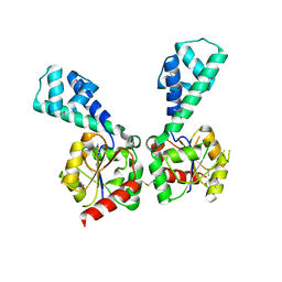
Loading
PDBj
MenuPDBj@FacebookPDBj@TwitterPDBj@YouTubewwPDB FoundationwwPDB
RCSB PDBPDBeBMRBAdv. SearchSearch help
Search by PDB author
4N3Z
DownloadVisualize
BU of 4n3z by Molmil
Crystal structure of Rabex-5delta and Rabaptin-5C21 complex
Descriptor: PHOSPHATE ION, Rab GTPase-binding effector protein 1, Rab5 GDP/GTP exchange factor
Authors:Zhang, Z, Zhang, T, Ding, J.
Deposit date:2013-10-08
Release date:2014-07-23
Last modified:2023-11-08
Method:X-RAY DIFFRACTION (3.1 Å)
Cite:Molecular mechanism for Rabex-5 GEF activation by Rabaptin-5
Elife, 3, 2014
3NMW
DownloadVisualize
BU of 3nmw by Molmil
Crystal structure of armadillo repeats domain of APC
Descriptor: APC variant protein, SULFATE ION
Authors:Zhang, Z, Chen, L, Gao, L, Lin, K, Wu, G.
Deposit date:2010-06-22
Release date:2011-07-06
Last modified:2023-12-27
Method:X-RAY DIFFRACTION (1.6 Å)
Cite:Structural basis for the recognition of Asef by adenomatous polyposis coli.
Cell Res., 22, 2012
4N3X
DownloadVisualize
BU of 4n3x by Molmil
Crystal structure of Rabex-5 CC domain
Descriptor: Rab5 GDP/GTP exchange factor
Authors:Zhang, Z, Zhang, T, Ding, J.
Deposit date:2013-10-08
Release date:2014-07-23
Last modified:2023-11-08
Method:X-RAY DIFFRACTION (2 Å)
Cite:Molecular mechanism for Rabex-5 GEF activation by Rabaptin-5
Elife, 3, 2014
4N3Y
DownloadVisualize
BU of 4n3y by Molmil
Crystal structure of Rabex-5CC and Rabaptin-5C21 complex
Descriptor: Rab GTPase-binding effector protein 1, Rab5 GDP/GTP exchange factor
Authors:Zhang, Z, Zhang, T, Ding, J.
Deposit date:2013-10-08
Release date:2014-07-23
Last modified:2023-11-08
Method:X-RAY DIFFRACTION (2.2 Å)
Cite:Molecular mechanism for Rabex-5 GEF activation by Rabaptin-5
Elife, 3, 2014
8BDP
DownloadVisualize
BU of 8bdp by Molmil
A GH20 family sulfoglycosidase Bt4394 in complex with NAG-thiazoline and sulfite
Descriptor: Beta-N-acetylhexosaminidase, CHLORIDE ION
Authors:Zhang, Z, He, Y, Jin, Y.
Deposit date:2022-10-19
Release date:2023-01-11
Last modified:2024-01-31
Method:X-RAY DIFFRACTION (1.47 Å)
Cite:Mechanistic and Structural Insights into the Specificity and Biological Functions of Bacterial Sulfoglycosidases
Acs Catalysis, 2022
1JJR
DownloadVisualize
BU of 1jjr by Molmil
The Three-Dimensional Structure of the C-terminal DNA Binding Domain of Human Ku70
Descriptor: THYROID AUTOANTIGEN
Authors:Zhang, Z, Chen, Y.
Deposit date:2001-07-09
Release date:2001-10-03
Last modified:2024-05-22
Method:SOLUTION NMR
Cite:The three-dimensional structure of the C-terminal DNA-binding domain of human Ku70.
J.Biol.Chem., 276, 2001
1M4Z
DownloadVisualize
BU of 1m4z by Molmil
Crystal structure of the N-terminal BAH domain of Orc1p
Descriptor: MANGANESE (II) ION, ORIGIN RECOGNITION COMPLEX SUBUNIT 1
Authors:Zhang, Z, Hayashi, M.K, Merkel, O, Stillman, B, Xu, R.-M.
Deposit date:2002-07-05
Release date:2002-09-11
Last modified:2024-02-14
Method:X-RAY DIFFRACTION (2.2 Å)
Cite:Structure and function of the BAH-containing domain of Orc1p in epigenetic silencing.
EMBO J., 21, 2002
1RW2
DownloadVisualize
BU of 1rw2 by Molmil
Three-dimensional structure of Ku80 CTD
Descriptor: ATP-dependent DNA helicase II, 80 kDa subunit
Authors:Zhang, Z, Hu, W, Cano, L, Lee, T.D, Chen, D.J, Chen, Y.
Deposit date:2003-12-15
Release date:2003-12-30
Last modified:2024-05-22
Method:SOLUTION NMR
Cite:Solution structure of the C-terminal domain of Ku80 suggests important sites for protein-protein interactions.
STRUCTURE, 12, 2004
1TPH
DownloadVisualize
BU of 1tph by Molmil
1.8 ANGSTROMS CRYSTAL STRUCTURE OF WILD TYPE CHICKEN TRIOSEPHOSPHATE ISOMERASE-PHOSPHOGLYCOLOHYDROXAMATE COMPLEX
Descriptor: PHOSPHOGLYCOLOHYDROXAMIC ACID, TRIOSEPHOSPHATE ISOMERASE
Authors:Zhang, Z, Sugio, S, Komives, E.A, Liu, K.D, Knowles, J.R, Petsko, G.A, Ringe, D.
Deposit date:1993-12-22
Release date:1994-04-30
Last modified:2024-02-14
Method:X-RAY DIFFRACTION (1.8 Å)
Cite:Crystal structure of recombinant chicken triosephosphate isomerase-phosphoglycolohydroxamate complex at 1.8-A resolution.
Biochemistry, 33, 1994
1TPW
DownloadVisualize
BU of 1tpw by Molmil
TRIOSEPHOSPHATE ISOMERASE DRINKS WATER TO KEEP HEALTHY
Descriptor: PHOSPHOGLYCOLOHYDROXAMIC ACID, TRIOSEPHOSPHATE ISOMERASE
Authors:Zhang, Z, Sugio, S, Komives, E.A, Liu, K.D, Stock, A.M, Narayana, N, Xuong, Ng.H, Knowles, J.R, Petsko, G.A, Ringe, D.
Deposit date:1994-11-07
Release date:1995-04-20
Last modified:2024-02-14
Method:X-RAY DIFFRACTION (1.9 Å)
Cite:The role of water in the catalytic efficiency of triosephosphate isomerase.
Biochemistry, 38, 1999
1W9Y
DownloadVisualize
BU of 1w9y by Molmil
The structure of ACC oxidase
Descriptor: 1-AMINOCYCLOPROPANE-1-CARBOXYLATE OXIDASE 1, SULFATE ION
Authors:Zhang, Z, Ren, J.-S, Clifton, I.J, Schofield, C.J.
Deposit date:2004-10-20
Release date:2005-10-26
Last modified:2011-07-13
Method:X-RAY DIFFRACTION (2.1 Å)
Cite:Crystal Structure and Mechanistic Implications of 1-Aminocyclopropane-1-Carboxylic Acid Oxidase (the Ethyling Forming Enzyme)
Chem.Biol., 11, 2004
7YTP
DownloadVisualize
BU of 7ytp by Molmil
TLR7 in complex with an inhibitor
Descriptor: (2R,6R)-4-(8-cyanoquinolin-5-yl)-N-[(3S,4R)-4-fluoranylpyrrolidin-3-yl]-6-methyl-morpholine-2-carboxamide, 2-acetamido-2-deoxy-beta-D-glucopyranose, 2-acetamido-2-deoxy-beta-D-glucopyranose-(1-4)-2-acetamido-2-deoxy-beta-D-glucopyranose, ...
Authors:Zhang, Z, Ohto, U, Shimizu, T.
Deposit date:2022-08-15
Release date:2023-11-15
Method:ELECTRON MICROSCOPY (2.77 Å)
Cite:TLR7 in complex with an inhibitor
To Be Published
2KKA
DownloadVisualize
BU of 2kka by Molmil
Human telomere DNA two-tetrad quadruplex structure in K+ solution
Descriptor: 5'-D(*AP*GP*GP*GP*TP*TP*AP*GP*GP*GP*TP*TP*AP*IP*GP*GP*TP*TP*AP*GP*GP*GP*T)-3'
Authors:Zhang, Z, Dai, J, Yang, D.
Deposit date:2009-06-16
Release date:2009-12-15
Last modified:2024-05-01
Method:SOLUTION NMR
Cite:Structure of a two-G-tetrad intramolecular G-quadruplex formed by a variant human telomeric sequence in K+ solution: insights into the interconversion of human telomeric G-quadruplex structures.
Nucleic Acids Res., 38, 2010
4OV4
DownloadVisualize
BU of 4ov4 by Molmil
Isopropylmalate synthase binding with ketoisovalerate
Descriptor: 2-isopropylmalate synthase, 3-METHYL-2-OXOBUTANOIC ACID, ZINC ION
Authors:Zhang, Z, Wu, J, Wang, C, Zhang, P.
Deposit date:2014-02-20
Release date:2014-08-20
Last modified:2024-03-20
Method:X-RAY DIFFRACTION (2 Å)
Cite:Subdomain II of alpha-isopropylmalate synthase is essential for activity: inferring a mechanism of feedback inhibition.
J.Biol.Chem., 289, 2014
4OV9
DownloadVisualize
BU of 4ov9 by Molmil
Structure of isopropylmalate synthase binding with alpha-isopropylmalate
Descriptor: (2S)-2-hydroxy-2-(propan-2-yl)butanedioic acid, ZINC ION, isopropylmalate synthase
Authors:Zhang, Z, Wu, J, Wang, C, Zhang, P.
Deposit date:2014-02-20
Release date:2014-08-20
Last modified:2024-03-20
Method:X-RAY DIFFRACTION (2.2 Å)
Cite:Subdomain II of alpha-isopropylmalate synthase is essential for activity: inferring a mechanism of feedback inhibition.
J.Biol.Chem., 289, 2014
4QXA
DownloadVisualize
BU of 4qxa by Molmil
Crystal structure of the Rab9A-RUTBC2 RBD complex
Descriptor: GUANOSINE-5'-TRIPHOSPHATE, MAGNESIUM ION, Ras-related protein Rab-9A, ...
Authors:Zhang, Z, Wang, S, Ding, J.
Deposit date:2014-07-19
Release date:2014-09-10
Last modified:2023-11-08
Method:X-RAY DIFFRACTION (2.3 Å)
Cite:Crystal structure of the Rab9A-RUTBC2 RBD complex reveals the molecular basis for the binding specificity of Rab9A with RUTBC2.
Structure, 22, 2014
3NND
DownloadVisualize
BU of 3nnd by Molmil
The crystal structure of ABC transporter from Rhodopseudomonas palustris
Descriptor: Possible substrate binding protein of ABC transporter system
Authors:Zhang, Z, Burley, S.K, Swaminathan, S, New York SGX Research Center for Structural Genomics (NYSGXRC)
Deposit date:2010-06-23
Release date:2010-10-13
Last modified:2023-12-27
Method:X-RAY DIFFRACTION (2.8 Å)
Cite:The crystal structure of ABC transporter from Rhodopseudomonas palustris
To be Published
3NPK
DownloadVisualize
BU of 3npk by Molmil
The crystal structure of geranyltranstransferase from Campylobacter jejuni
Descriptor: GLYCEROL, Geranyltranstransferase, PYROPHOSPHATE
Authors:Zhang, Z, Burley, S.K, Swaminathan, S, New York SGX Research Center for Structural Genomics (NYSGXRC)
Deposit date:2010-06-28
Release date:2010-07-14
Last modified:2023-12-27
Method:X-RAY DIFFRACTION (1.5 Å)
Cite:The crystal structure of geranyltranstransferase from Campylobacter jejuni
To be Published
3PAN
DownloadVisualize
BU of 3pan by Molmil
The crystal structure of adenosine deaminase with hypoxanthine bound from Pseudomonas aeruginosa
Descriptor: Adenosine deaminase, HYPOXANTHINE, ZINC ION
Authors:Zhang, Z, Burley, S.K, Swaminathan, S, New York SGX Research Center for Structural Genomics (NYSGXRC)
Deposit date:2010-10-19
Release date:2010-12-22
Last modified:2023-12-06
Method:X-RAY DIFFRACTION (2.627 Å)
Cite:The crystal structure of adenosine deaminase with hypoxanthine bound from Pseudomonas aeruginosa
TO BE PUBLISHED
3OPN
DownloadVisualize
BU of 3opn by Molmil
The crystal structure of a putative hemolysin from Lactococcus lactis
Descriptor: GLYCEROL, SULFATE ION, putative hemolysin
Authors:Zhang, Z, Burley, S.K, Swaminathan, S, New York SGX Research Center for Structural Genomics (NYSGXRC)
Deposit date:2010-09-01
Release date:2010-10-13
Last modified:2021-02-10
Method:X-RAY DIFFRACTION (2.05 Å)
Cite:The crystal structure of a putative hemolysin from Lactococcus lactis
To be Published
3NAS
DownloadVisualize
BU of 3nas by Molmil
The crystal structure of beta-phosphoglucomutase from Bacillus subtilis
Descriptor: beta-phosphoglucomutase
Authors:Zhang, Z, Burley, S.K, Swaminathan, S, New York SGX Research Center for Structural Genomics (NYSGXRC)
Deposit date:2010-06-02
Release date:2010-07-28
Last modified:2021-02-10
Method:X-RAY DIFFRACTION (3 Å)
Cite:The crystal structure of beta-phosphoglucomutase from Bacillus subtilis
To be Published
3NIV
DownloadVisualize
BU of 3niv by Molmil
The crystal structure of Glutathione S-transferase from Legionella pneumophila
Descriptor: Glutathione S-transferase
Authors:Zhang, Z, Burley, S.K, Swaminathan, S, New York SGX Research Center for Structural Genomics (NYSGXRC)
Deposit date:2010-06-16
Release date:2010-07-14
Last modified:2021-02-10
Method:X-RAY DIFFRACTION (2.3 Å)
Cite:The crystal structure of Glutathione S-transferase from Legionella pneumophila
To be Published
3PAO
DownloadVisualize
BU of 3pao by Molmil
The crystal structure of adenosine deaminase with adenine bound from Pseudomonas aeruginosa
Descriptor: ADENINE, Adenosine deaminase, ZINC ION
Authors:Zhang, Z, Burley, S.K, Swaminathan, S, New York SGX Research Center for Structural Genomics (NYSGXRC)
Deposit date:2010-10-19
Release date:2010-12-22
Last modified:2023-12-06
Method:X-RAY DIFFRACTION (2.493 Å)
Cite:The crystal structure of adenosine deaminase with adenine bound from Pseudomonas aeruginosa
To be Published
3MPO
DownloadVisualize
BU of 3mpo by Molmil
The crystal structure of a hydrolase from Lactobacillus brevis
Descriptor: Predicted hydrolase of the HAD superfamily
Authors:Zhang, Z, Burley, S.K, Swaminathan, S, New York SGX Research Center for Structural Genomics (NYSGXRC)
Deposit date:2010-04-27
Release date:2010-05-12
Last modified:2021-02-10
Method:X-RAY DIFFRACTION (2.9 Å)
Cite:The crystal structure of a hydrolase from Lactobacillus brevis
To be Published
3MSR
DownloadVisualize
BU of 3msr by Molmil
The crystal structure of an amidohydrolase from Mycoplasma synoviae
Descriptor: GLYCEROL, PHOSPHATE ION, amidohydrolases
Authors:Zhang, Z, Burley, S.K, Swaminathan, S, New York SGX Research Center for Structural Genomics (NYSGXRC)
Deposit date:2010-04-29
Release date:2010-05-12
Last modified:2021-02-10
Method:X-RAY DIFFRACTION (2.162 Å)
Cite:The crystal structure of an amidohydrolase from Mycoplasma synoviae
To be Published

222624

数据于2024-07-17公开中

PDB statisticsPDBj update infoContact PDBjnumon