Loading
PDBj
MenuPDBj@FacebookPDBj@TwitterPDBj@YouTubewwPDB FoundationwwPDB
RCSB PDBPDBeBMRBAdv. SearchSearch help
Search by PDB author
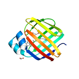
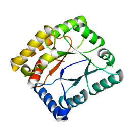
4QGV
DownloadVisualize
BU of 4qgv by Molmil
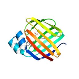
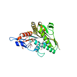
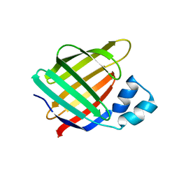
Crystal structure of the R132K:R111L mutant of Cellular Retinoic Acid Binding ProteinII complexed with a synthetic ligand (Merocyanine) at 1.73 angstrom resolution.
Descriptor: (2E,4E,6E)-3-methyl-6-(1,3,3-trimethyl-1,3-dihydro-2H-indol-2-ylidene)hexa-2,4-dienal, Cellular retinoic acid-binding protein 2
Authors:Nosrati, M, Yapici, I, Geiger, J.H.
Deposit date:2014-05-25
Release date:2015-01-28
Last modified:2023-09-20
Method:X-RAY DIFFRACTION (1.73 Å)
Cite:"Turn-on" protein fluorescence: in situ formation of cyanine dyes.
J.Am.Chem.Soc., 137, 2015
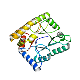
4QZU
DownloadVisualize
BU of 4qzu by Molmil
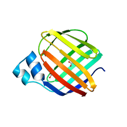
Crystal Structure of wild type Human Cellular Retinol Binding Protein II (hCRBPII) bound to retinol at 11 KeV beam energy
Descriptor: ACETATE ION, GLYCEROL, RETINOL, ...
Authors:Assar, Z, Geiger, J.H.
Deposit date:2014-07-28
Release date:2014-12-10
Last modified:2023-09-20
Method:X-RAY DIFFRACTION (1.5 Å)
Cite:Structures of holo wild-type human cellular retinol-binding protein II (hCRBPII) bound to retinol and retinal.
Acta Crystallogr.,Sect.D, 70, 2014
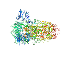
8H3E
DownloadVisualize
BU of 8h3e by Molmil
Complex structure of a small molecule (SPC-14) bound SARS-CoV-2 spike protein, closed state
Descriptor: 2-acetamido-2-deoxy-beta-D-glucopyranose, 7-(6-nitro-2,3-dihydroindol-1-yl)-7-oxidanylidene-heptanoic acid, Spike glycoprotein,Fibritin
Authors:Meng, F, Wang, Q, Xie, Y, Ni, X, Huang, N.
Deposit date:2022-10-08
Release date:2023-03-22
Last modified:2024-11-06
Method:ELECTRON MICROSCOPY (3.06 Å)
Cite:In Silico Discovery of Small Molecule Modulators Targeting the Achilles' Heel of SARS-CoV-2 Spike Protein.
Acs Cent.Sci., 9, 2023
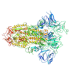
8H3D
DownloadVisualize
BU of 8h3d by Molmil
Structure of apo SARS-CoV-2 spike protein with one RBD up
Descriptor: 2-acetamido-2-deoxy-beta-D-glucopyranose, Spike glycoprotein,Fibritin
Authors:Meng, F, Wang, Q, Xie, Y, Ni, X, Huang, N.
Deposit date:2022-10-08
Release date:2023-03-22
Method:ELECTRON MICROSCOPY (3.27 Å)
Cite:In Silico Discovery of Small Molecule Modulators Targeting the Achilles' Heel of SARS-CoV-2 Spike Protein.
Acs Cent.Sci., 9, 2023
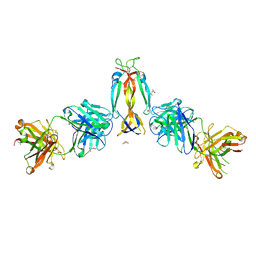
7WKX
DownloadVisualize
BU of 7wkx by Molmil
IL-17A in complex with the humanized antibody HB0017
Descriptor: ACETIC ACID, Heavy chain of HB0017 Fab, Interleukin-17A, ...
Authors:Xu, J, Zhu, X, He, Y.
Deposit date:2022-01-12
Release date:2022-03-23
Last modified:2024-10-16
Method:X-RAY DIFFRACTION (2.81 Å)
Cite:Structural and functional insights into a novel pre-clinical-stage antibody targeting IL-17A for treatment of autoimmune diseases.
Int.J.Biol.Macromol., 202, 2022
4R0U
DownloadVisualize
BU of 4r0u by Molmil
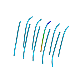
Tgvtava, an amyloid forming segment from alpha synuclein, residues 72-78
Descriptor: Alpha-synuclein
Authors:Ivanova, M.I, Eisenberg, D.S, Sawaya, M.R.
Deposit date:2014-08-01
Release date:2014-12-17
Last modified:2024-04-03
Method:X-RAY DIFFRACTION (1.38 Å)
Cite:Structure-based design of functional amyloid materials.
J.Am.Chem.Soc., 136, 2014
4R0P
DownloadVisualize
BU of 4r0p by Molmil
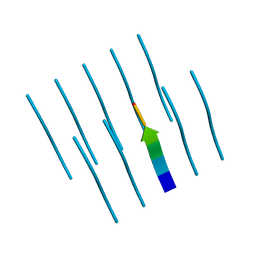
Ifqins, an amyloid forming segment from human lysozyme spanning residues 56-61
Descriptor: Lysozyme C
Authors:Sievers, S, Eisenberg, D.S, Sawaya, M.R.
Deposit date:2014-08-01
Release date:2014-12-17
Last modified:2024-02-28
Method:X-RAY DIFFRACTION (1.52 Å)
Cite:Structure-based design of functional amyloid materials.
J.Am.Chem.Soc., 136, 2014
4R0W
DownloadVisualize
BU of 4r0w by Molmil
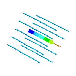
Vvtgvta, an amyloid forming segment from alpha synuclein, residues 70-76
Descriptor: Alpha-synuclein
Authors:Ivanova, M.I, Eisenberg, D.S, Sawaya, M.R.
Deposit date:2014-08-01
Release date:2014-12-17
Last modified:2024-04-03
Method:X-RAY DIFFRACTION (1.5 Å)
Cite:Structure-based design of functional amyloid materials.
J.Am.Chem.Soc., 136, 2014
4Z9P
DownloadVisualize
BU of 4z9p by Molmil
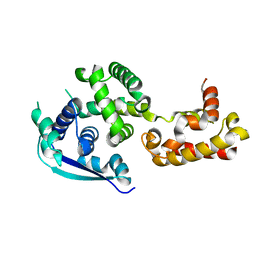
Crystal structure of Ebola virus nucleoprotein core domain at 1.8A resolution
Descriptor: Nucleoprotein
Authors:Guo, Y, Dong, S.S, Yang, P, Li, G.B, Liu, B.C, Yang, C, Rao, Z.H.
Deposit date:2015-04-11
Release date:2015-05-20
Last modified:2024-03-20
Method:X-RAY DIFFRACTION (1.792 Å)
Cite:Insight into the Ebola virus nucleocapsid assembly mechanism: crystal structure of Ebola virus nucleoprotein core domain at 1.8 A resolution.
Protein Cell, 6, 2015
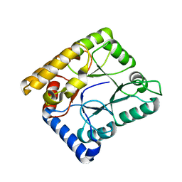
4QZT
DownloadVisualize
BU of 4qzt by Molmil
Crystal Structure of wild type Human Cellular Retinol Binding Protein II (hCRBPII) bound to retinol at 7 KeV beam energy
Descriptor: ACETATE ION, RETINOL, Retinol-binding protein 2
Authors:Assar, Z, Geiger, J.H.
Deposit date:2014-07-28
Release date:2014-12-10
Last modified:2023-09-20
Method:X-RAY DIFFRACTION (1.9 Å)
Cite:Structures of holo wild-type human cellular retinol-binding protein II (hCRBPII) bound to retinol and retinal.
Acta Crystallogr.,Sect.D, 70, 2014
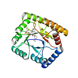
4ZGU
DownloadVisualize
BU of 4zgu by Molmil
Crystal structure of monomer Y60W hCRBPII
Descriptor: ACETATE ION, Retinol-binding protein 2
Authors:Nossoni, Z, Geiger, J.
Deposit date:2015-04-24
Release date:2016-06-08
Last modified:2024-03-06
Method:X-RAY DIFFRACTION (1.49 Å)
Cite:Domain-Swapped Dimers of Intracellular Lipid-Binding Proteins: Evidence for Ordered Folding Intermediates.
Structure, 24, 2016
2FSA
DownloadVisualize
BU of 2fsa by Molmil
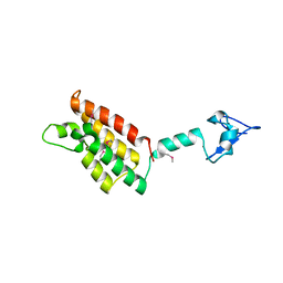
Crystal structure of PHD finger-linker-bromodomain fragment of human BPTF in the H3(1-15)K4ME2 bound state
Descriptor: Histone H3(1-15)K4me2 peptide, ZINC ION, bromodomain PHD finger transcription factor
Authors:Li, H, Patel, D.J.
Deposit date:2006-01-21
Release date:2006-07-11
Last modified:2023-11-15
Method:X-RAY DIFFRACTION (1.9 Å)
Cite:Molecular basis for site-specific read-out of histone H3K4me3 by the BPTF PHD finger of NURF.
Nature, 442, 2006
2F6J
DownloadVisualize
BU of 2f6j by Molmil
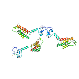
Crystal structure of PHD finger-linker-bromodomain fragment of human BPTF in the H3(1-15)K4me3 bound state
Descriptor: ZINC ION, bromodomain PHD finger transcription factor, histone H3, ...
Authors:Li, H, Patel, D.J.
Deposit date:2005-11-29
Release date:2006-07-11
Last modified:2011-07-13
Method:X-RAY DIFFRACTION (2 Å)
Cite:Molecular basis for site-specific read-out of histone H3K4me3 by the BPTF PHD finger of NURF.
Nature, 442, 2006
3K3B
DownloadVisualize
BU of 3k3b by Molmil
Co-crystal structure of the human kinesin Eg5 with a novel tetrahydro-beta-carboline
Descriptor: 3-[(1R)-2-acetyl-6-methyl-2,3,4,9-tetrahydro-1H-beta-carbolin-1-yl]phenol, ADENOSINE-5'-DIPHOSPHATE, CHLORIDE ION, ...
Authors:Bussiere, D.E, Bellamacina, C, Le, V.
Deposit date:2009-10-02
Release date:2009-12-15
Last modified:2024-02-21
Method:X-RAY DIFFRACTION (2.4 Å)
Cite:The discovery of tetrahydro-beta-carbolines as inhibitors of the kinesin Eg5.
Bioorg.Med.Chem.Lett., 20, 2010
2FUI
DownloadVisualize
BU of 2fui by Molmil
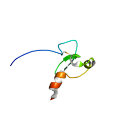
NMR solution structure of PHD finger fragment of human BPTF in free state
Descriptor: bromodomain PHD finger transcription factor
Authors:Ilin, S, Patel, D.J.
Deposit date:2006-01-26
Release date:2006-07-11
Last modified:2022-03-09
Method:SOLUTION NMR
Cite:Molecular basis for site-specific read-out of histone H3K4me3 by the BPTF PHD finger of NURF.
Nature, 442, 2006
2FUU
DownloadVisualize
BU of 2fuu by Molmil
NMR solution structure of the PHD domain from the human BPTF in complex with H3(1-15)K4me3 peptide
Descriptor: Histone H3, ZINC ION, bromodomain PHD finger transcription factor
Authors:Ilin, S, Patel, D.J.
Deposit date:2006-01-27
Release date:2006-07-11
Last modified:2022-03-09
Method:SOLUTION NMR
Cite:Molecular basis for site-specific read-out of histone H3K4me3 by the BPTF PHD finger of NURF.
Nature, 442, 2006
7EFT
DownloadVisualize
BU of 7eft by Molmil
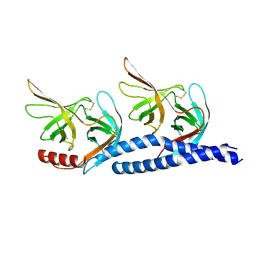
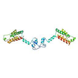
Crystal structure of cell shape-determining protein MreC
Descriptor: CHLORIDE ION, Cell shape protein MreC
Authors:Xu, Q, Xiao, Q.J, Sun, B.
Deposit date:2021-03-23
Release date:2021-10-20
Last modified:2024-10-23
Method:X-RAY DIFFRACTION (2.1 Å)
Cite:The crystal structure of MreC provides insights into polymer formation.
Febs Open Bio, 12, 2022
5ZBF
DownloadVisualize
BU of 5zbf by Molmil
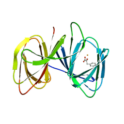
Crystal structure of 4-hydroxyphenylpyruvic acid bound AerE from Microcystis aeruginosa
Descriptor: 3-(4-HYDROXY-PHENYL)PYRUVIC ACID, Cupin domain protein, FE (II) ION
Authors:Qiu, X, Zhu, W.
Deposit date:2018-02-11
Release date:2019-02-13
Last modified:2024-03-27
Method:X-RAY DIFFRACTION (1.6 Å)
Cite:Structural and functional insights into the role of a cupin superfamily isomerase in the biosynthesis of Choi moiety of aeruginosin.
J. Struct. Biol., 205, 2019
7WMW
DownloadVisualize
BU of 7wmw by Molmil
Crystal structure of methylenetetrahydrofolate reductase MSMEG_6649 from Mycobacterium smegmatis
Descriptor: Methylenetetrahydrofolate reductase
Authors:Lin, W, Wang, W.
Deposit date:2022-01-17
Release date:2023-01-25
Last modified:2024-05-29
Method:X-RAY DIFFRACTION (2.053 Å)
Cite:Structural and functional characterization of a mycobacterial methylenetetrahydrofolate reductase utilizing NADH as the exclusive cofactor.
Biochem.J., 480, 2023
7WMY
DownloadVisualize
BU of 7wmy by Molmil
Crystal structure of methylenetetrahydrofolate reductase MSMEG_6649 from Mycobacterium smegmatis with 5-methyltetrahydrofolate
Descriptor: methylenetetrahydrofolate reductase
Authors:Lin, W, Wang, W.
Deposit date:2022-01-17
Release date:2023-01-25
Last modified:2024-05-29
Method:X-RAY DIFFRACTION (2.266 Å)
Cite:Structural and functional characterization of a mycobacterial methylenetetrahydrofolate reductase utilizing NADH as the exclusive cofactor.
Biochem.J., 480, 2023
7WMX
DownloadVisualize
BU of 7wmx by Molmil
Crystal structure of methylenetetrahydrofolate reductase MSMEG_6649 from Mycobacterium smegmatis with 5,10-methylenetetrahydrofolate
Descriptor: Methylenetetrahydrofolate reductase
Authors:Lin, W, Wang, W.
Deposit date:2022-01-17
Release date:2023-01-25
Last modified:2024-05-29
Method:X-RAY DIFFRACTION (2.264 Å)
Cite:Structural and functional characterization of a mycobacterial methylenetetrahydrofolate reductase utilizing NADH as the exclusive cofactor.
Biochem.J., 480, 2023
7WMZ
DownloadVisualize
BU of 7wmz by Molmil
Crystal structure of methylenetetrahydrofolate reductase MSMEG_6649 from Mycobacterium smegmatis with NADH
Descriptor: 1,4-DIHYDRONICOTINAMIDE ADENINE DINUCLEOTIDE, Methylenetetrahydrofolate reductase
Authors:Lin, W, Wang, W.
Deposit date:2022-01-17
Release date:2023-01-25
Last modified:2024-05-29
Method:X-RAY DIFFRACTION (1.916 Å)
Cite:Structural and functional characterization of a mycobacterial methylenetetrahydrofolate reductase utilizing NADH as the exclusive cofactor.
Biochem.J., 480, 2023
6KLR
DownloadVisualize
BU of 6klr by Molmil
Crystal structure of human WIPI3 in complex with the WIR-peptide from ATG2A
Descriptor: chimera ATG2A and WIPI3
Authors:Ren, J.Q, Liang, R.B, Feng, W.
Deposit date:2019-07-30
Release date:2020-04-29
Last modified:2023-11-22
Method:X-RAY DIFFRACTION (2.21 Å)
Cite:Multi-site-mediated entwining of the linear WIR-motif around WIPI beta-propellers for autophagy.
Nat Commun, 11, 2020
2F6N
DownloadVisualize
BU of 2f6n by Molmil
Crystal structure of PHD finger-linker-bromodomain fragment of human BPTF in the free form
Descriptor: ZINC ION, bromodomain PHD finger transcription factor
Authors:Li, H, Patel, D.J.
Deposit date:2005-11-29
Release date:2006-07-11
Last modified:2024-10-09
Method:X-RAY DIFFRACTION (2 Å)
Cite:Molecular basis for site-specific read-out of histone H3K4me3 by the BPTF PHD finger of NURF.
Nature, 442, 2006
5XS4
DownloadVisualize
BU of 5xs4 by Molmil
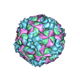
Structure of Coxsackievirus A6 (CVA6) virus A-particle
Descriptor: Genome polyprotein
Authors:Zheng, Q.B, He, M.Z, Xu, L.F, Yu, H, Li, S.W, Cheng, T.
Deposit date:2017-06-12
Release date:2017-09-27
Last modified:2024-03-27
Method:ELECTRON MICROSCOPY (3.1 Å)
Cite:Atomic structures of Coxsackievirus A6 and its complex with a neutralizing antibody
Nat Commun, 8, 2017

227111

건을2024-11-06부터공개중

PDB statisticsPDBj update infoContact PDBjnumon