Loading
PDBj
MenuPDBj@FacebookPDBj@TwitterPDBj@YouTubewwPDB FoundationwwPDB
RCSB PDBPDBeBMRBAdv. SearchSearch help
Search by PDB author
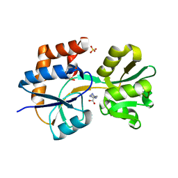
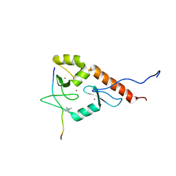
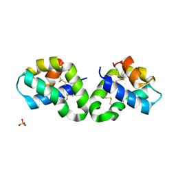
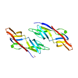
2R0R
DownloadVisualize
BU of 2r0r by Molmil
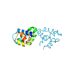
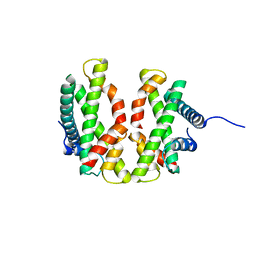
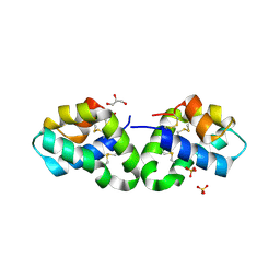
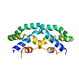
Crystal Structure of Human Saposin D variant SapD K9E
Descriptor: Proactivator polypeptide, SULFATE ION
Authors:Rossmann, M, Saenger, W, Maier, T.
Deposit date:2007-08-21
Release date:2008-04-29
Last modified:2024-11-06
Method:X-RAY DIFFRACTION (2.5 Å)
Cite:Crystal structures of human saposins C and d: implications for lipid recognition and membrane interactions.
Structure, 16, 2008
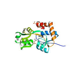
1ZSO
DownloadVisualize
BU of 1zso by Molmil
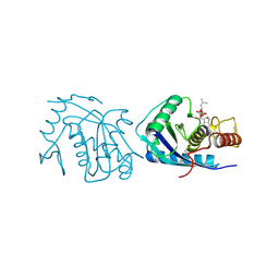
Hypothetical protein from plasmodium falciparum
Descriptor: hypothetical protein
Authors:Holmes, M.A, Merritt, E.A, Structural Genomics of Pathogenic Protozoa Consortium (SGPP)
Deposit date:2005-05-24
Release date:2005-06-07
Last modified:2024-10-30
Method:X-RAY DIFFRACTION (2.17 Å)
Cite:Structure of the conserved hypothetical protein MAL13P1.257 from Plasmodium falciparum.
Acta Crystallogr.,Sect.F, 62, 2006
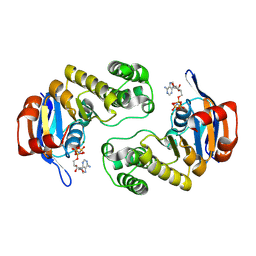
2R1Q
DownloadVisualize
BU of 2r1q by Molmil
Crystal Structure of Iodinated Human Saposin D in Space Group C2221
Descriptor: Proactivator polypeptide
Authors:Maier, T, Rossman, M, Saenger, W.
Deposit date:2007-08-23
Release date:2008-04-29
Last modified:2024-10-30
Method:X-RAY DIFFRACTION (2.5 Å)
Cite:Crystal structures of human saposins C and d: implications for lipid recognition and membrane interactions.
Structure, 16, 2008
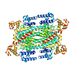
3CUJ
DownloadVisualize
BU of 3cuj by Molmil
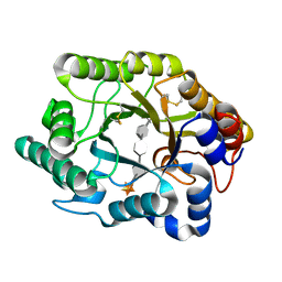
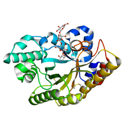
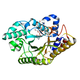
Cellulomonas fimi Xylanase/Cellulase Cex (Cf Xyn10A) in complex with sulfur substituted beta-1,4 xylopentaose.
Descriptor: Exo-beta-1,4-glucanase, beta-D-xylopyranose-(1-4)-4-thio-beta-D-xylopyranose-(1-4)-4-thio-beta-D-xylopyranose-(1-4)-4-thio-beta-D-xylopyranose-(1-4)-4-thio-beta-D-xylopyranose
Authors:Kuntz, D.A, Saul, M, Rose, D.R.
Deposit date:2008-04-16
Release date:2009-04-21
Last modified:2024-10-30
Method:X-RAY DIFFRACTION (1.7 Å)
Cite:Probing the binding sites of Family 10 and 11 Xylanases with extended Oligosaccharides
to be published
1YZV
DownloadVisualize
BU of 1yzv by Molmil
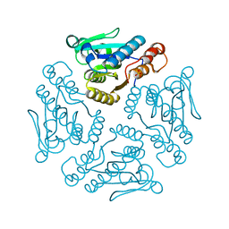
HYPOTHETICAL PROTEIN FROM TRYPANOSOMA CRUZI
Descriptor: HYPOTHETICAL PROTEIN, SULFATE ION
Authors:Caruthers, J, Merritt, E.A, Structural Genomics of Pathogenic Protozoa Consortium (SGPP)
Deposit date:2005-02-28
Release date:2005-03-08
Last modified:2024-02-14
Method:X-RAY DIFFRACTION (2.001 Å)
Cite:Crystal structures and proposed structural/functional classification of three protozoan proteins from the isochorismatase superfamily.
Protein Sci., 14, 2005
3CUI
DownloadVisualize
BU of 3cui by Molmil
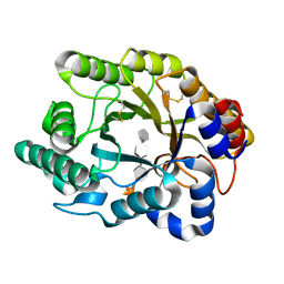
Cellulomonas fimi Xylanase/Cellulase Cex (Cf Xyn10A) in complex with sulfur substituted beta-1,4 xylotetraose
Descriptor: Exo-beta-1,4-glucanase, beta-D-xylopyranose-(1-4)-4-thio-beta-D-xylopyranose-(1-4)-4-thio-beta-D-xylopyranose-(1-4)-4-thio-beta-D-xylopyranose
Authors:Kuntz, D.A, Saul, M, Rose, D.R.
Deposit date:2008-04-16
Release date:2009-04-21
Last modified:2024-10-16
Method:X-RAY DIFFRACTION (1.5 Å)
Cite:Probing the binding sites of Family 10 and 11 Xylanases with extended Oligosaccharides
to be published
2POX
DownloadVisualize
BU of 2pox by Molmil
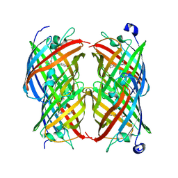
Dark state structure of the reversibly switchable fluorescent protein Dronpa
Descriptor: Fluorescent protein Dronpa
Authors:Trowitzsch, S, Weber, G, Wahl, M.C.
Deposit date:2007-04-27
Release date:2007-07-17
Last modified:2024-10-30
Method:X-RAY DIFFRACTION (1.946 Å)
Cite:Structural basis for reversible photoswitching in Dronpa
Proc.Natl.Acad.Sci.Usa, 104, 2007
3CUG
DownloadVisualize
BU of 3cug by Molmil
Cellulomonas fimi Xylanase/Cellulase Cex (Cf Xyn10A) in complex with cellotetraose-like isofagomine
Descriptor: (3R,4R,5R)-3-hydroxy-5-(hydroxymethyl)piperidin-4-yl beta-D-glucopyranosyl-(1->4)-beta-D-glucopyranosyl-(1->4)-beta-D-glucopyranoside, Exo-beta-1,4-glucanase
Authors:Kuntz, D.A, Saul, M, Rose, D.R.
Deposit date:2008-04-16
Release date:2009-04-21
Last modified:2019-07-24
Method:X-RAY DIFFRACTION (1.68 Å)
Cite:Probing the binding sites of Family 10 and 11 Xylanases with extended Oligosaccharides
to be published
1AVO
DownloadVisualize
BU of 1avo by Molmil
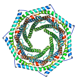
PROTEASOME ACTIVATOR REG(ALPHA)
Descriptor: 11S REGULATOR
Authors:Hill, C.P, Knowlton, J.R.
Deposit date:1997-09-18
Release date:1997-12-31
Last modified:2024-02-07
Method:X-RAY DIFFRACTION (2.8 Å)
Cite:Structure of the proteasome activator REGalpha (PA28alpha).
Nature, 390, 1997
4E44
DownloadVisualize
BU of 4e44 by Molmil
Crystal structure of the hMHF1/hMHF2 Histone-Fold Tetramer
Descriptor: Centromere protein S, Centromere protein X
Authors:Fox III, D, Zhao, Y, Yang, W, Weidong, W.
Deposit date:2012-03-12
Release date:2013-03-20
Last modified:2023-09-13
Method:X-RAY DIFFRACTION (2.1 Å)
Cite:Crystal Structures Reveal that FANCM remodels the MHF Tetramer in favor of binding Branched DNA
To be Published
2Q2C
DownloadVisualize
BU of 2q2c by Molmil
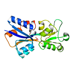
Crystal structures of the arginine-, lysine-, histidine-binding protein ArtJ from the thermophilic bacterium Geobacillus stearothermophilus
Descriptor: ArtJ, GLYCEROL, HISTIDINE, ...
Authors:Vahedi-Faridi, A, Scheffel, F, Eckey, V, Saenger, W, Schneider, E.
Deposit date:2007-05-28
Release date:2008-01-15
Last modified:2024-02-21
Method:X-RAY DIFFRACTION (2.35 Å)
Cite:Crystal structures and mutational analysis of the arginine-, lysine-, histidine-binding protein ArtJ from Geobacillus stearothermophilus. Implications for interactions of ArtJ with its cognate ATP-binding cassette transporter, Art(MP)2
J.Mol.Biol., 375, 2008
2PVU
DownloadVisualize
BU of 2pvu by Molmil
Crystal structures of the arginine-, lysine-, histidine-binding protein ArtJ from the thermophilic bacterium Geobacillus stearothermophilus
Descriptor: ArtJ, LYSINE, SULFATE ION
Authors:Vahedi-Faridi, A, Scheffel, F, Eckey, V, Saenger, W, Schneider, E.
Deposit date:2007-05-10
Release date:2008-01-15
Last modified:2024-02-21
Method:X-RAY DIFFRACTION (1.79 Å)
Cite:Crystal structures and mutational analysis of the arginine-, lysine-, histidine-binding protein ArtJ from Geobacillus stearothermophilus. Implications for interactions of ArtJ with its cognate ATP-binding cassette transporter, Art(MP)2
J.Mol.Biol., 375, 2008
3C41
DownloadVisualize
BU of 3c41 by Molmil
ABC protein ArtP in complex with AMP-PNP/Mg2+
Descriptor: Amino acid ABC transporter (ArtP), MAGNESIUM ION, PHOSPHOAMINOPHOSPHONIC ACID-ADENYLATE ESTER
Authors:Thaben, P.F, Eckey, V, Scheffel, F, Saenger, W, Schneider, E, Vahedi-Faridi, A.
Deposit date:2008-01-29
Release date:2009-02-03
Last modified:2023-11-01
Method:X-RAY DIFFRACTION (2.25 Å)
Cite:Crystal Structures of the ATP-binding cassette (ABC) Protein ArtP from Geobacillus stearothermophilus in complexes with nucleotides and nucleotide analogs reveal an intermediate semiclosed dimer in the post hydrolyses state and an asymmetry in the dimerisation region
To be Published
1AUW
DownloadVisualize
BU of 1auw by Molmil
H91N DELTA 2 CRYSTALLIN FROM DUCK
Descriptor: DELTA 2 CRYSTALLIN
Authors:Abu-Abed, M, Vallee, F, Howell, P.L.
Deposit date:1997-09-03
Release date:1998-03-18
Last modified:2024-04-03
Method:X-RAY DIFFRACTION (2.5 Å)
Cite:Structural comparison of the enzymatically active and inactive forms of delta crystallin and the role of histidine 91.
Biochemistry, 36, 1997
3CUH
DownloadVisualize
BU of 3cuh by Molmil
Cellulomonas fimi Xylanase/Cellulase Cex (Cf Xyn10A) in complex with cellotriose-like isofagomine
Descriptor: (3R,4R,5R)-3-hydroxy-5-(hydroxymethyl)piperidin-4-yl 4-O-beta-D-glucopyranosyl-beta-D-glucopyranoside, Exo-beta-1,4-glucanase
Authors:Kuntz, D.A, Saul, M, Rose, D.R.
Deposit date:2008-04-16
Release date:2009-04-21
Last modified:2024-10-30
Method:X-RAY DIFFRACTION (1.89 Å)
Cite:Probing the binding sites of Family 10 and 11 Xylanases with extended Oligosaccharides
to be published
2RB3
DownloadVisualize
BU of 2rb3 by Molmil
Crystal Structure of Human Saposin D
Descriptor: GLYCEROL, Proactivator polypeptide, SULFATE ION
Authors:Maier, T, Rossman, M, Saenger, W.
Deposit date:2007-09-18
Release date:2008-04-29
Last modified:2024-10-30
Method:X-RAY DIFFRACTION (2.1 Å)
Cite:Crystal structures of human saposins C and d: implications for lipid recognition and membrane interactions.
Structure, 16, 2008
4AIG
DownloadVisualize
BU of 4aig by Molmil
ADAMALYSIN II WITH PHOSPHONATE INHIBITOR
Descriptor: ADAMALYSIN II, CALCIUM ION, N-[(FURAN-2-YL)CARBONYL]-(S)-LEUCYL-(R)-[1-AMINO-2(1H-INDOL-3-YL)ETHYL]-PHOSPHONIC ACID, ...
Authors:Pochetti, G, Mazza, F, Gavuzzo, E, Cirilli, M.
Deposit date:1997-10-16
Release date:1998-11-11
Last modified:2024-10-16
Method:X-RAY DIFFRACTION (2 Å)
Cite:2 angstrom X-ray structure of adamalysin II complexed with a peptide phosphonate inhibitor adopting a retro-binding mode.
FEBS Lett., 418, 1997
3BO8
DownloadVisualize
BU of 3bo8 by Molmil
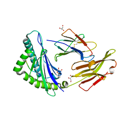
The High Resolution Crystal Structure of HLA-A1 Complexed with the MAGE-A1 Peptide
Descriptor: Beta-2-microglobulin, GLYCEROL, HLA class I histocompatibility antigen, ...
Authors:Kumar, P, Vahedi-Faridi, A, Saenger, W, Ziegler, A, Uchanska-Ziegler, B.
Deposit date:2007-12-17
Release date:2008-12-23
Last modified:2023-11-01
Method:X-RAY DIFFRACTION (1.8 Å)
Cite:Conformational changes within the HLA-A1:MAGE-A1 complex induced by binding of a recombinant antibody fragment with TCR-like specificity
Protein Sci., 18, 2009
2Z9A
DownloadVisualize
BU of 2z9a by Molmil
Crystal Structure of Human Saposin C Dimer in Open Conformation
Descriptor: GLYCEROL, Proactivator polypeptide
Authors:Rossmann, M, Saenger, W, Maier, T.
Deposit date:2007-09-18
Release date:2008-04-29
Last modified:2023-08-30
Method:X-RAY DIFFRACTION (2.5 Å)
Cite:Crystal structures of human saposins C and d: implications for lipid recognition and membrane interactions.
Structure, 16, 2008
1AMF
DownloadVisualize
BU of 1amf by Molmil
CRYSTAL STRUCTURE OF MODA, A MOLYBDATE TRANSPORT PROTEIN, COMPLEXED WITH MOLYBDATE
Descriptor: MOLYBDATE ION, MOLYBDATE TRANSPORT PROTEIN MODA
Authors:Hu, Y, Rech, S, Gunsalus, R.P, Rees, D.C.
Deposit date:1997-06-13
Release date:1997-12-24
Last modified:2024-02-07
Method:X-RAY DIFFRACTION (1.75 Å)
Cite:Crystal structure of the molybdate binding protein ModA.
Nat.Struct.Biol., 4, 1997
2LDL
DownloadVisualize
BU of 2ldl by Molmil
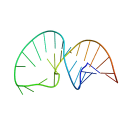
Solution NMR Structure of the HIV-1 Exon Splicing Silencer 3
Descriptor: RNA (27-MER)
Authors:Mishler, C, Levengood, J.D, Johnson, C.A, Rajan, P, Znosko, B.M.
Deposit date:2011-05-27
Release date:2011-12-28
Last modified:2024-05-01
Method:SOLUTION NMR
Cite:Solution Structure of the HIV-1 Exon Splicing Silencer 3.
J.Mol.Biol., 415, 2012
2MQH
DownloadVisualize
BU of 2mqh by Molmil
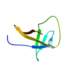
Solution structure of the Chlamydomonas reinhardtii NAB1 cold shock domain, CSD1
Descriptor: Nucleic acid binding protein
Authors:Sawyer, A, Mobli, M.
Deposit date:2014-06-20
Release date:2015-05-06
Last modified:2024-05-15
Method:SOLUTION NMR
Cite:Solution structure of the RNA-binding cold-shock domain of the Chlamydomonas reinhardtii NAB1 protein and insights into RNA recognition.
Biochem.J., 469, 2015
2LBM
DownloadVisualize
BU of 2lbm by Molmil
Solution structure of the ADD domain of ATRX complexed with histone tail H3 1-15 K9me3
Descriptor: Transcriptional regulator ATRX, ZINC ION, histone tail H3 K9me3
Authors:Eustermann, S, Yang, J, Neuhaus, D.
Deposit date:2011-04-08
Release date:2011-06-29
Last modified:2011-07-13
Method:SOLUTION NMR
Cite:Combinatorial readout of histone H3 modifications specifies localization of ATRX to heterochromatin
Nat.Struct.Mol.Biol., 2011

227111

건을2024-11-06부터공개중

PDB statisticsPDBj update infoContact PDBjnumon