Loading
PDBj
MenuPDBj@FacebookPDBj@TwitterPDBj@YouTubewwPDB FoundationwwPDB
RCSB PDBPDBeBMRBAdv. SearchSearch help
Search by PDB author
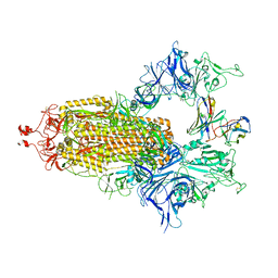
7UZ8
DownloadVisualize
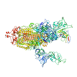
BU of 7uz8 by Molmil
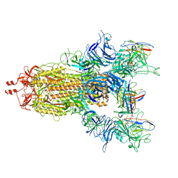
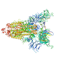
Structure of the SARS-CoV-2 Omicron BA.1 S 6P trimer in complex with the mouse antibody Fab fragment, M8a-31
Descriptor: 2-acetamido-2-deoxy-beta-D-glucopyranose, 2-acetamido-2-deoxy-beta-D-glucopyranose-(1-4)-2-acetamido-2-deoxy-beta-D-glucopyranose, M8a-31 Fab heavy chain, ...
Authors:Fan, C, Bjorkman, P.J.
Deposit date:2022-05-08
Release date:2022-12-07
Last modified:2022-12-28
Method:ELECTRON MICROSCOPY (3.1 Å)
Cite:Neutralizing monoclonal antibodies elicited by mosaic RBD nanoparticles bind conserved sarbecovirus epitopes.
Immunity, 55, 2022
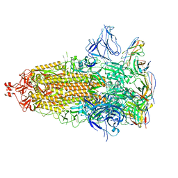
7UZA
DownloadVisualize
BU of 7uza by Molmil
Structure of the SARS-CoV-2 S 6P trimer in complex with the mouse antibody Fab fragment, HSW-1
Descriptor: 2-acetamido-2-deoxy-beta-D-glucopyranose, 2-acetamido-2-deoxy-beta-D-glucopyranose-(1-4)-2-acetamido-2-deoxy-beta-D-glucopyranose, HSW-1 Fab heavy chain, ...
Authors:Fan, C, Bjorkman, P.J.
Deposit date:2022-05-08
Release date:2022-12-07
Last modified:2022-12-28
Method:ELECTRON MICROSCOPY (3.1 Å)
Cite:Neutralizing monoclonal antibodies elicited by mosaic RBD nanoparticles bind conserved sarbecovirus epitopes.
Immunity, 55, 2022
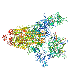
7UZ5
DownloadVisualize
BU of 7uz5 by Molmil
Structure of the SARS-CoV-2 S 6P trimer in complex with the mouse antibody Fab fragment, M8a-6
Descriptor: 2-acetamido-2-deoxy-beta-D-glucopyranose, M8a-6 Fab heavy chain, M8a-6 Fab light chain, ...
Authors:Fan, C, Bjorkman, P.J.
Deposit date:2022-05-08
Release date:2022-12-07
Last modified:2022-12-28
Method:ELECTRON MICROSCOPY (3.2 Å)
Cite:Neutralizing monoclonal antibodies elicited by mosaic RBD nanoparticles bind conserved sarbecovirus epitopes.
Immunity, 55, 2022
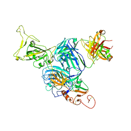
7UZ6
DownloadVisualize
BU of 7uz6 by Molmil
Structure of the SARS-CoV-2 S 6P trimer in complex with the mouse antibody Fab fragment, M8a-28
Descriptor: 2-acetamido-2-deoxy-beta-D-glucopyranose, M8a-28 Fab heavy chain, M8a-28 Fab light chain, ...
Authors:Fan, C, Bjorkman, P.J.
Deposit date:2022-05-08
Release date:2022-12-07
Last modified:2022-12-28
Method:ELECTRON MICROSCOPY (2.8 Å)
Cite:Neutralizing monoclonal antibodies elicited by mosaic RBD nanoparticles bind conserved sarbecovirus epitopes.
Immunity, 55, 2022
7UZ4
DownloadVisualize
BU of 7uz4 by Molmil
Structure of the SARS-CoV-2 S 6P trimer in complex with the mouse antibody Fab fragment, M8a-3
Descriptor: 2-acetamido-2-deoxy-beta-D-glucopyranose, 2-acetamido-2-deoxy-beta-D-glucopyranose-(1-4)-2-acetamido-2-deoxy-beta-D-glucopyranose, M8a-3 Fab heavy chain, ...
Authors:Fan, C, Bjorkman, P.J.
Deposit date:2022-05-08
Release date:2022-12-07
Last modified:2022-12-28
Method:ELECTRON MICROSCOPY (3.1 Å)
Cite:Neutralizing monoclonal antibodies elicited by mosaic RBD nanoparticles bind conserved sarbecovirus epitopes.
Immunity, 55, 2022
7UZB
DownloadVisualize
BU of 7uzb by Molmil
Structure of the SARS-CoV-2 S S1 doamin in complex with the mouse antibody Fab fragment, HSW-2
Descriptor: 2-acetamido-2-deoxy-beta-D-glucopyranose, HSW-2 Fab heavy chain, HSW-2 Fab light chain, ...
Authors:Fan, C, Bjorkman, P.J.
Deposit date:2022-05-08
Release date:2022-12-07
Last modified:2022-12-28
Method:ELECTRON MICROSCOPY (4.1 Å)
Cite:Neutralizing monoclonal antibodies elicited by mosaic RBD nanoparticles bind conserved sarbecovirus epitopes.
Immunity, 55, 2022
7UZ7
DownloadVisualize
BU of 7uz7 by Molmil
Structure of the SARS-CoV-2 S 6P trimer in complex with the mouse antibody Fab fragment, M8a-31
Descriptor: 2-acetamido-2-deoxy-beta-D-glucopyranose, 2-acetamido-2-deoxy-beta-D-glucopyranose-(1-4)-2-acetamido-2-deoxy-beta-D-glucopyranose, M8a-31 Fab heavy chain, ...
Authors:Fan, C, Bjorkman, P.J.
Deposit date:2022-05-08
Release date:2022-12-07
Last modified:2022-12-28
Method:ELECTRON MICROSCOPY (2.9 Å)
Cite:Neutralizing monoclonal antibodies elicited by mosaic RBD nanoparticles bind conserved sarbecovirus epitopes.
Immunity, 55, 2022
7UZC
DownloadVisualize
BU of 7uzc by Molmil
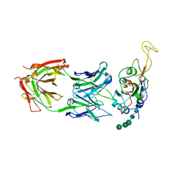
Structure of the SARS-CoV-2 RBD in complex with the mouse antibody Fab fragment, M8a-34
Descriptor: M8a-34 Fab heavy chain, M8a-34 Fab light chain, Spike protein S1, ...
Authors:Fan, C, Bjorkman, P.J.
Deposit date:2022-05-08
Release date:2023-01-11
Last modified:2023-10-25
Method:X-RAY DIFFRACTION (2.2 Å)
Cite:Neutralizing monoclonal antibodies elicited by mosaic RBD nanoparticles bind conserved sarbecovirus epitopes.
Immunity, 55, 2022
7UZD
DownloadVisualize
BU of 7uzd by Molmil
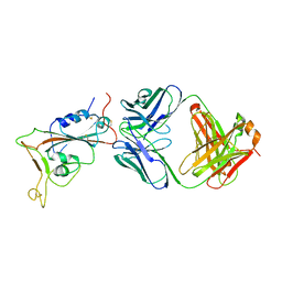
Structure of the SARS-CoV-2 RBD in complex with the mouse antibody Fab fragment, HSW-2
Descriptor: 2-acetamido-2-deoxy-beta-D-glucopyranose-(1-4)-2-acetamido-2-deoxy-beta-D-glucopyranose, HSW-2 Fab heavy chain, HSW-2 Fab light chain, ...
Authors:Fan, C, Bjorkman, P.J.
Deposit date:2022-05-08
Release date:2023-01-11
Last modified:2023-10-25
Method:X-RAY DIFFRACTION (3 Å)
Cite:Neutralizing monoclonal antibodies elicited by mosaic RBD nanoparticles bind conserved sarbecovirus epitopes.
Immunity, 55, 2022
2OO9
DownloadVisualize
BU of 2oo9 by Molmil
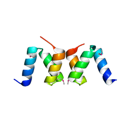
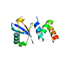
crystal structure of the UBA domain from human c-Cbl ubiquitin ligase
Descriptor: E3 ubiquitin-protein ligase CBL
Authors:Kozlov, G, Gehring, K.
Deposit date:2007-01-25
Release date:2007-02-06
Last modified:2023-12-27
Method:X-RAY DIFFRACTION (2.1 Å)
Cite:Structural basis for UBA-mediated dimerization of c-Cbl ubiquitin ligase.
J.Biol.Chem., 282, 2007
2OOB
DownloadVisualize
BU of 2oob by Molmil
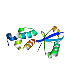
crystal structure of the UBA domain from Cbl-b ubiquitin ligase in complex with ubiquitin
Descriptor: E3 ubiquitin-protein ligase CBL-B, Ubiquitin
Authors:Kozlov, G, Gehring, K.
Deposit date:2007-01-25
Release date:2007-02-06
Last modified:2023-08-30
Method:X-RAY DIFFRACTION (1.9 Å)
Cite:Structural basis for ubiquitin-mediated dimerization and activation of the ubiquitin protein ligase Cbl-b.
Mol.Cell, 27, 2007
2QHO
DownloadVisualize
BU of 2qho by Molmil
Crystal structure of the UBA domain from EDD ubiquitin ligase in complex with ubiquitin
Descriptor: E3 ubiquitin-protein ligase EDD1, Ubiquitin
Authors:Kozlov, G, Gehring, K.
Deposit date:2007-07-02
Release date:2007-09-25
Last modified:2023-08-30
Method:X-RAY DIFFRACTION (1.85 Å)
Cite:Structural basis of ubiquitin recognition by the ubiquitin-associated (UBA) domain of the ubiquitin ligase EDD.
J.Biol.Chem., 282, 2007
5WU7
DownloadVisualize
BU of 5wu7 by Molmil
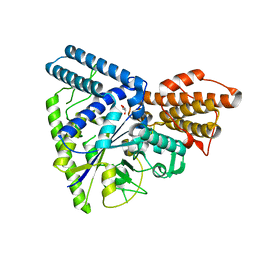
Crystal structure of GH57-type branching enzyme from hyperthermophilic archaeon Pyrococcus horikoshii
Descriptor: GLYCEROL, Uncharacterized protein
Authors:Na, S, Jo, I, Ha, N.-C.
Deposit date:2016-12-16
Release date:2017-03-08
Last modified:2023-11-08
Method:X-RAY DIFFRACTION (2.31 Å)
Cite:Structural basis for the transglycosylase activity of a GH57-type glycogen branching enzyme from Pyrococcus horikoshii.
Biochem. Biophys. Res. Commun., 484, 2017
2OOA
DownloadVisualize
BU of 2ooa by Molmil
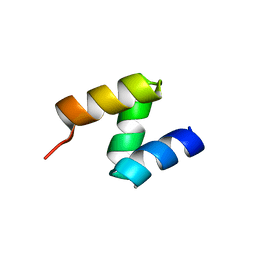
crystal structure of the UBA domain from Cbl-b ubiquitin ligase
Descriptor: E3 ubiquitin-protein ligase CBL-B
Authors:Kozlov, G, Gehring, K.
Deposit date:2007-01-25
Release date:2007-02-06
Last modified:2023-12-27
Method:X-RAY DIFFRACTION (1.56 Å)
Cite:Structural basis for ubiquitin-mediated dimerization and activation of the ubiquitin protein ligase Cbl-b.
Mol.Cell, 27, 2007
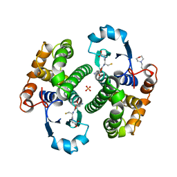
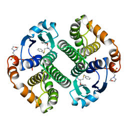
8GSS
DownloadVisualize
BU of 8gss by Molmil
Human glutathione S-transferase P1-1, complex with glutathione
Descriptor: 2-(N-MORPHOLINO)-ETHANESULFONIC ACID, GLUTATHIONE, GLUTATHIONE S-TRANSFERASE P1-1, ...
Authors:Oakley, A, Parker, M.
Deposit date:1997-08-14
Release date:1998-09-16
Last modified:2023-08-09
Method:X-RAY DIFFRACTION (1.9 Å)
Cite:The structures of human glutathione transferase P1-1 in complex with glutathione and various inhibitors at high resolution.
J.Mol.Biol., 274, 1997
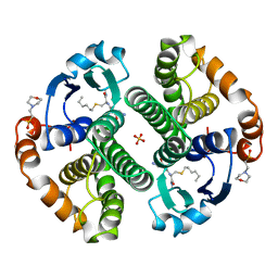
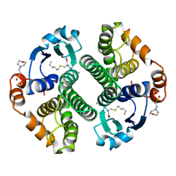
9GSS
DownloadVisualize
BU of 9gss by Molmil
HUMAN GLUTATHIONE S-TRANSFERASE P1-1, COMPLEX WITH S-HEXYL GLUTATHIONE
Descriptor: 2-(N-MORPHOLINO)-ETHANESULFONIC ACID, GLUTATHIONE S-TRANSFERASE P1-1, S-HEXYLGLUTATHIONE, ...
Authors:Oakley, A, Parker, M.
Deposit date:1997-08-14
Release date:1998-09-16
Last modified:2024-05-22
Method:X-RAY DIFFRACTION (1.97 Å)
Cite:The structures of human glutathione transferase P1-1 in complex with glutathione and various inhibitors at high resolution.
J.Mol.Biol., 274, 1997
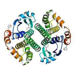
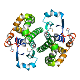
6GSS
DownloadVisualize
BU of 6gss by Molmil
HUMAN GLUTATHIONE S-TRANSFERASE P1-1, COMPLEX WITH GLUTATHIONE
Descriptor: 2-(N-MORPHOLINO)-ETHANESULFONIC ACID, GLUTATHIONE, GLUTATHIONE S-TRANSFERASE P1-1
Authors:Oakley, A, Parker, M.
Deposit date:1997-08-13
Release date:1998-09-16
Last modified:2024-05-22
Method:X-RAY DIFFRACTION (1.9 Å)
Cite:The structures of human glutathione transferase P1-1 in complex with glutathione and various inhibitors at high resolution.
J.Mol.Biol., 274, 1997
6WLF
DownloadVisualize
BU of 6wlf by Molmil
Phosphoethanolamine Methyltransferase from the Pine Wilt Nematode Bursaphelenchus xylophilus
Descriptor: PHOSPHORIC ACID MONO-(2-AMINO-ETHYL) ESTER, Phosphoethanolamine N-methyltransferase 1, S-ADENOSYL-L-HOMOCYSTEINE
Authors:Lee, S.G, Jez, J.M.
Deposit date:2020-04-20
Release date:2020-06-17
Last modified:2023-10-18
Method:X-RAY DIFFRACTION (2.05 Å)
Cite:Structural and biochemical analysis of phosphoethanolamine methyltransferase from the pine wilt nematode Bursaphelenchus xylophilus.
Mol.Biochem.Parasitol., 238, 2020
10GS
DownloadVisualize
BU of 10gs by Molmil
HUMAN GLUTATHIONE S-TRANSFERASE P1-1, COMPLEX WITH TER117
Descriptor: 2-(N-MORPHOLINO)-ETHANESULFONIC ACID, GLUTATHIONE S-TRANSFERASE P1-1, L-gamma-glutamyl-S-benzyl-N-[(S)-carboxy(phenyl)methyl]-L-cysteinamide
Authors:Oakley, A, Parker, M.
Deposit date:1997-08-14
Release date:1998-09-16
Last modified:2024-05-22
Method:X-RAY DIFFRACTION (2.2 Å)
Cite:The structures of human glutathione transferase P1-1 in complex with glutathione and various inhibitors at high resolution.
J.Mol.Biol., 274, 1997
7GSS
DownloadVisualize
BU of 7gss by Molmil
Human glutathione S-transferase P1-1, complex with glutathione
Descriptor: 2-(N-MORPHOLINO)-ETHANESULFONIC ACID, GLUTATHIONE, GLUTATHIONE S-TRANSFERASE P1-1
Authors:Oakley, A, Parker, M.
Deposit date:1997-08-13
Release date:1998-09-16
Last modified:2024-05-22
Method:X-RAY DIFFRACTION (2.2 Å)
Cite:The structures of human glutathione transferase P1-1 in complex with glutathione and various inhibitors at high resolution.
J.Mol.Biol., 274, 1997
4GSS
DownloadVisualize
BU of 4gss by Molmil
HUMAN GLUTATHIONE S-TRANSFERASE P1-1 Y108F MUTANT
Descriptor: 2-(N-MORPHOLINO)-ETHANESULFONIC ACID, GLUTATHIONE S-TRANSFERASE, S-HEXYLGLUTATHIONE
Authors:Oakley, A, Rossjohn, J, Parker, M.
Deposit date:1997-01-20
Release date:1998-01-28
Last modified:2024-05-22
Method:X-RAY DIFFRACTION (2.5 Å)
Cite:Multifunctional role of Tyr 108 in the catalytic mechanism of human glutathione transferase P1-1. Crystallographic and kinetic studies on the Y108F mutant enzyme.
Biochemistry, 36, 1997
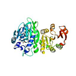
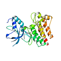
3ZXZ
DownloadVisualize
BU of 3zxz by Molmil
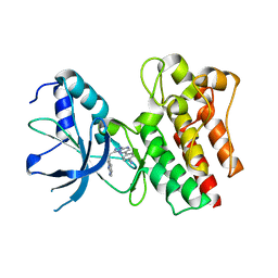
X-ray Structure of PF-04217903 bound to the kinase domain of c-Met
Descriptor: 2-{4-[1-(QUINOLIN-6-YLMETHYL)-1H-[1,2,3]TRIAZOLO[4,5-B]PYRAZIN-6-YL]-1H-PYRAZOL-1-YL}ETHANOL, HEPATOCYTE GROWTH FACTOR RECEPTOR
Authors:McTigue, M, Grodsky, N, Ryan, K, Cui, J.J.
Deposit date:2011-08-16
Release date:2011-08-31
Last modified:2023-12-20
Method:X-RAY DIFFRACTION (1.8 Å)
Cite:Discovery of a Novel Class of Exquisitely Selective Mesenchymal-Epithelial Transition Factor (C-met) Protein Kinase Inhibitors and Identification of the Clinical Candidate 2-(4-(1-(Quinolin-6-Ylmethyl)-1H-[1,2, 3]Triazolo[4,5-B]Pyrazin-6-Yl)-1H-Pyrazol-1-Yl)Ethanol (Pf-04217903) for the Treatment of Cancer.
J.Med.Chem., 55, 2012
4AOI
DownloadVisualize
BU of 4aoi by Molmil
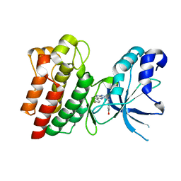
Crystal structure of C-MET kinase domain in complex with 4-(3-((1H- pyrrolo(2,3-b)pyridin-3-yl)methyl)-(1,2,4)triazolo(4,3-b)(1,2,4) triazin-6-yl)benzonitrile
Descriptor: 4-[3-(1H-pyrrolo[2,3-b]pyridin-3-ylmethyl)-[1,2,4]triazolo[4,3-b][1,2,4]triazin-6-yl]benzenecarbonitrile, HEPATOCYTE GROWTH FACTOR RECEPTOR
Authors:McTigue, M, Grodsky, N, Ryan, K, Cui, J.J.
Deposit date:2012-03-27
Release date:2012-09-26
Last modified:2023-12-20
Method:X-RAY DIFFRACTION (1.9 Å)
Cite:Discovery of a Novel Class of Exquisitely Selective Mesenchymal-Epithelial Transition Factor (C-met) Protein Kinase Inhibitors and Identification of the Clinical Candidate 2-(4-(1-(Quinolin-6-Ylmethyl)-1H-[1,2, 3]Triazolo[4,5-B]Pyrazin-6-Yl)-1H-Pyrazol-1-Yl)Ethanol (Pf-04217903) for the Treatment of Cancer.
J.Med.Chem., 55, 2012
4AP7
DownloadVisualize
BU of 4ap7 by Molmil
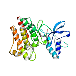
Crystal structure of C-MET kinase domain in complex with 4-((6-(4- fluorophenyl)-(1,2,4)triazolo(4,3-b)(1,2,4)triazin-3-yl)methyl)phenol
Descriptor: 4-[[6-(4-fluorophenyl)-[1,2,4]triazolo[4,3-b][1,2,4]triazin-3-yl]methyl]phenol, HEPATOCYTE GROWTH FACTOR RECEPTOR
Authors:McTigue, M, Wickersham, J.
Deposit date:2012-03-30
Release date:2012-09-26
Last modified:2024-05-08
Method:X-RAY DIFFRACTION (1.8 Å)
Cite:Discovery of a Novel Class of Exquisitely Selective Mesenchymal-Epithelial Transition Factor (C-met) Protein Kinase Inhibitors and Identification of the Clinical Candidate 2-(4-(1-(Quinolin-6-Ylmethyl)-1H-[1,2, 3]Triazolo[4,5-B]Pyrazin-6-Yl)-1H-Pyrazol-1-Yl)Ethanol (Pf-04217903) for the Treatment of Cancer.
J.Med.Chem., 55, 2012
3ZZE
DownloadVisualize
BU of 3zze by Molmil
Crystal structure of C-MET kinase domain in complex with N'-((3Z)-4- chloro-7-methyl-2-oxo-1,2-dihydro-3H-indol-3-ylidene)-2-(4- hydroxyphenyl)propanohydrazide
Descriptor: (2S)-N'-[(3R)-4-chloro-7-methyl-2-oxo-2,3-dihydro-1H-indol-3-yl]-2-(4-hydroxyphenyl)propanehydrazide, HEPATOCYTE GROWTH FACTOR RECEPTOR
Authors:McTigue, M, Deng, Y, Ryan, K, Cui, J.J.
Deposit date:2011-08-31
Release date:2011-09-14
Last modified:2023-12-20
Method:X-RAY DIFFRACTION (1.87 Å)
Cite:Discovery of a Novel Class of Exquisitely Selective Mesenchymal-Epithelial Transition Factor (C-met) Protein Kinase Inhibitors and Identification of the Clinical Candidate 2-(4-(1-(Quinolin-6-Ylmethyl)-1H-[1,2, 3]Triazolo[4,5-B]Pyrazin-6-Yl)-1H-Pyrazol-1-Yl)Ethanol (Pf-04217903) for the Treatment of Cancer.
J.Med.Chem., 55, 2012

222926

건을2024-07-24부터공개중

PDB statisticsPDBj update infoContact PDBjnumon