Loading
PDBj
MenuPDBj@FacebookPDBj@TwitterPDBj@YouTubewwPDB FoundationwwPDB
RCSB PDBPDBeBMRBAdv. SearchSearch help
Search by PDB author
1J20
DownloadVisualize
BU of 1j20 by Molmil
Crystal Structure of Thermus thermophilus HB8 Argininosuccinate Synthetase in complex with product
Descriptor: ADENOSINE MONOPHOSPHATE, ARGININOSUCCINATE, Argininosuccinate Synthetase, ...
Authors:Goto, M, Hirotsu, K, RIKEN Structural Genomics/Proteomics Initiative (RSGI)
Deposit date:2002-12-24
Release date:2003-04-22
Last modified:2023-12-27
Method:X-RAY DIFFRACTION (2 Å)
Cite:Structures of argininosuccinate synthetase in enzyme-ATP-substrates and enzyme-AMP-product forms
J.Biol.Chem., 278, 2003
2YW9
DownloadVisualize
BU of 2yw9 by Molmil
Crystal structure of TT0143 from Thermus thermophilus HB8
Descriptor: Enoyl-[acyl carrier protein] reductase, NADP NICOTINAMIDE-ADENINE-DINUCLEOTIDE PHOSPHATE
Authors:Miyano, M, Ago, H, RIKEN Structural Genomics/Proteomics Initiative (RSGI)
Deposit date:2007-04-20
Release date:2007-05-15
Last modified:2023-10-25
Method:X-RAY DIFFRACTION (2.5 Å)
Cite:Crystal structure of TT0143 from Thermus thermophilus HB8
To be Published
5WR3
DownloadVisualize
BU of 5wr3 by Molmil
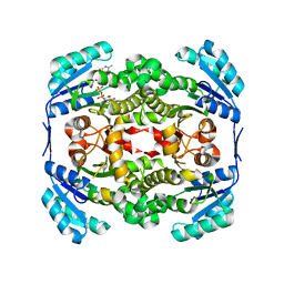
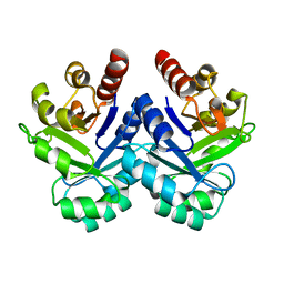
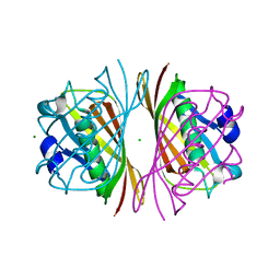
Thermolysin, SFX liganded form with water-based carrier
Descriptor: CALCIUM ION, N-[(benzyloxy)carbonyl]-L-aspartic acid, Thermolysin, ...
Authors:Kunishima, N, Naitow, H, Matsuura, Y.
Deposit date:2016-11-29
Release date:2017-08-16
Last modified:2023-11-08
Method:X-RAY DIFFRACTION (2.1 Å)
Cite:Protein-ligand complex structure from serial femtosecond crystallography using soaked thermolysin microcrystals and comparison with structures from synchrotron radiation
Acta Crystallogr D Struct Biol, 73, 2017
5WR6
DownloadVisualize
BU of 5wr6 by Molmil
Thermolysin, liganded form with cryo condition 2
Descriptor: CALCIUM ION, N-[(benzyloxy)carbonyl]-L-aspartic acid, Thermolysin, ...
Authors:Kunishima, N, Naitow, H, Matsuura, Y.
Deposit date:2016-11-29
Release date:2017-08-16
Last modified:2023-11-08
Method:X-RAY DIFFRACTION (2.3 Å)
Cite:Protein-ligand complex structure from serial femtosecond crystallography using soaked thermolysin microcrystals and comparison with structures from synchrotron radiation
Acta Crystallogr D Struct Biol, 73, 2017
5WR4
DownloadVisualize
BU of 5wr4 by Molmil
Thermolysin, SFX unliganded form with oil-based carrier
Descriptor: CALCIUM ION, Thermolysin, ZINC ION
Authors:Kunishima, N, Naitow, H, Matsuura, Y.
Deposit date:2016-11-29
Release date:2017-08-16
Last modified:2023-11-08
Method:X-RAY DIFFRACTION (2.1 Å)
Cite:Protein-ligand complex structure from serial femtosecond crystallography using soaked thermolysin microcrystals and comparison with structures from synchrotron radiation
Acta Crystallogr D Struct Biol, 73, 2017
5WR5
DownloadVisualize
BU of 5wr5 by Molmil
Thermolysin, liganded form with cryo condition 1
Descriptor: CALCIUM ION, N-[(benzyloxy)carbonyl]-L-aspartic acid, TETRAETHYLENE GLYCOL, ...
Authors:Kunishima, N, Naitow, H, Matsuura, Y.
Deposit date:2016-11-29
Release date:2017-08-16
Last modified:2023-11-08
Method:X-RAY DIFFRACTION (1.9 Å)
Cite:Protein-ligand complex structure from serial femtosecond crystallography using soaked thermolysin microcrystals and comparison with structures from synchrotron radiation
Acta Crystallogr D Struct Biol, 73, 2017
6JZH
DownloadVisualize
BU of 6jzh by Molmil
Structure of human A2A adenosine receptor in complex with ZM241385 obtained from SFX experiments under atmospheric pressure
Descriptor: 4-{2-[(7-amino-2-furan-2-yl[1,2,4]triazolo[1,5-a][1,3,5]triazin-5-yl)amino]ethyl}phenol, Adenosine receptor A2a,Soluble cytochrome b562,Adenosine receptor A2a, CHOLESTEROL, ...
Authors:Nango, E, Shimamura, T, Nakane, T, Yamanaka, Y, Mori, C, Kimura, K.T, Fujiwara, T, Tanaka, T, Iwata, S.
Deposit date:2019-05-02
Release date:2019-10-30
Last modified:2023-11-22
Method:X-RAY DIFFRACTION (2.25 Å)
Cite:High-viscosity sample-injection device for serial femtosecond crystallography at atmospheric pressure.
J.Appl.Crystallogr., 52, 2019
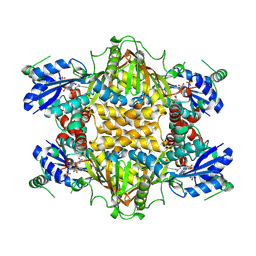
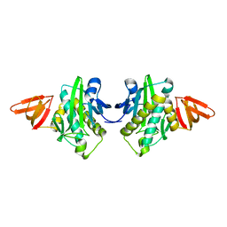
1UB3
DownloadVisualize
BU of 1ub3 by Molmil
Crystal Structure of Tetrameric Structure of Aldolase from thermus thermophilus HB8
Descriptor: 1-HYDROXY-PENTANE-3,4-DIOL-5-PHOSPHATE, Aldolase protein
Authors:Lokanath, N.K, Miyano, M, Yokoyama, S, Kuramitsu, S, Kunishima, N, RIKEN Structural Genomics/Proteomics Initiative (RSGI)
Deposit date:2003-03-28
Release date:2003-04-08
Last modified:2023-12-27
Method:X-RAY DIFFRACTION (1.4 Å)
Cite:Structure of aldolase from Thermus thermophilus HB8 showing the contribution of oligomeric state to thermostability.
Acta Crystallogr.,Sect.D, 60, 2004
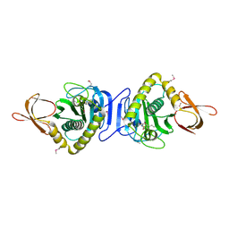
2P6D
DownloadVisualize
BU of 2p6d by Molmil
Crystal structure of PH0725 from Pyrococcus horikoshii OT3
Descriptor: S-ADENOSYL-L-HOMOCYSTEINE, diphthine synthase
Authors:Yamamoto, H, Taketa, M, Morikawa, Y, Matsuura, Y, Kunishima, N, RIKEN Structural Genomics/Proteomics Initiative (RSGI)
Deposit date:2007-03-17
Release date:2007-09-18
Last modified:2023-10-25
Method:X-RAY DIFFRACTION (2.4 Å)
Cite:Crystal structure of PH0725 from Pyrococcus horikoshii OT3
To be Published
2P5C
DownloadVisualize
BU of 2p5c by Molmil
Crystal structure of PH0725 from Pyrococcus horikoshii OT3
Descriptor: GLYCEROL, S-ADENOSYL-L-HOMOCYSTEINE, diphthine synthase
Authors:Yamamoto, H, Taketa, M, Ono, N, Matsuura, Y, Kunishima, N, RIKEN Structural Genomics/Proteomics Initiative (RSGI)
Deposit date:2007-03-15
Release date:2007-09-18
Last modified:2023-10-25
Method:X-RAY DIFFRACTION (2.4 Å)
Cite:Crystal structure of PH0725 from Pyrococcus horikoshii OT3
To be Published
1WR8
DownloadVisualize
BU of 1wr8 by Molmil
Crystal structure of hypothetical protein PH1421 from Pyrococcus horikoshii.
Descriptor: ACETATE ION, Phosphoglycolate phosphatase
Authors:Yamamoto, H, Kunishima, N, RIKEN Structural Genomics/Proteomics Initiative (RSGI)
Deposit date:2004-10-13
Release date:2004-10-26
Last modified:2024-03-13
Method:X-RAY DIFFRACTION (1.6 Å)
Cite:Structure of a haloacid dehalogenase superfamily phosphatase PH1421 from Pyrococcus horikoshii OT3: oligomeric state and thermoadaptation mechanism.
Acta Crystallogr.,Sect.D, 64, 2008
1WNL
DownloadVisualize
BU of 1wnl by Molmil
Crystal Structure Of Biotin-(Acetyl-CoA-Carboxylase) ligase From Pyrococcus Horikoshii Ot3 in complex with ADP
Descriptor: ADENOSINE-5'-DIPHOSPHATE, GLYCEROL, biotin--[acetyl-CoA-carboxylase] ligase
Authors:Bagautdinov, B, Kunishima, N, RIKEN Structural Genomics/Proteomics Initiative (RSGI)
Deposit date:2004-08-05
Release date:2005-08-16
Last modified:2024-04-03
Method:X-RAY DIFFRACTION (1.6 Å)
Cite:Crystal Structures of Biotin Protein Ligase from Pyrococcus horikoshii OT3 and its Complexes: Structural Basis of Biotin Activation
J.Mol.Biol., 353, 2005
1WQW
DownloadVisualize
BU of 1wqw by Molmil
Crystal Structure Of Biotin Protein Ligase From Pyrococcus Horikoshii Ot3 in complex with Biotinyl-5-AMP
Descriptor: BIOTINYL-5-AMP, biotin--[acetyl-CoA-carboxylase] ligase
Authors:Bagautdinov, B, Kunishima, N, RIKEN Structural Genomics/Proteomics Initiative (RSGI)
Deposit date:2004-10-04
Release date:2005-10-04
Last modified:2023-10-25
Method:X-RAY DIFFRACTION (1.45 Å)
Cite:Crystal Structures of Biotin Protein Ligase from Pyrococcus horikoshii OT3 and its Complexes: Structural Basis of Biotin Activation
J.Mol.Biol., 353, 2005
1WN2
DownloadVisualize
BU of 1wn2 by Molmil
Crystal structure of project ID PH1539 from Pyrococcus horikoshii OT3
Descriptor: Peptidyl-tRNA hydrolase, SODIUM ION
Authors:Shimizu, K, Kunishima, N, RIKEN Structural Genomics/Proteomics Initiative (RSGI)
Deposit date:2004-07-26
Release date:2005-07-19
Last modified:2023-10-25
Method:X-RAY DIFFRACTION (1.2 Å)
Cite:Structure of peptidyl-tRNA hydrolase 2 from Pyrococcus horikoshii OT3: insight into the functional role of its dimeric state.
Acta Crystallogr.,Sect.D, 64, 2008
1WQ7
DownloadVisualize
BU of 1wq7 by Molmil
Crystal Structure Of Biotin-(Acetyl-CoA-Carboxylase) ligase From Pyrococcus Horikoshii Ot3
Descriptor: biotin--[acetyl-CoA-carboxylase] ligase
Authors:Bagautdinov, B, Kunishima, N, RIKEN Structural Genomics/Proteomics Initiative (RSGI)
Deposit date:2004-09-23
Release date:2005-10-04
Last modified:2024-03-13
Method:X-RAY DIFFRACTION (1.6 Å)
Cite:Crystal Structures of Biotin Protein Ligase from Pyrococcus horikoshii OT3 and its Complexes: Structural Basis of Biotin Activation
J.Mol.Biol., 353, 2005
2BEC
DownloadVisualize
BU of 2bec by Molmil
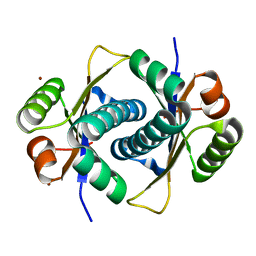
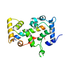
Crystal structure of CHP2 in complex with its binding region in NHE1 and insights into the mechanism of pH regulation
Descriptor: Calcineurin B homologous protein 2, Sodium/hydrogen exchanger 1, YTTRIUM (III) ION
Authors:Ben Ammar, Y, Takeda, S, Hisamitsu, T, Mori, H, Wakabayashi, S.
Deposit date:2005-10-24
Release date:2006-06-27
Last modified:2024-03-13
Method:X-RAY DIFFRACTION (2.7 Å)
Cite:Crystal structure of CHP2 complexed with NHE1-cytosolic region and an implication for pH regulation
Embo J., 25, 2006
1WPY
DownloadVisualize
BU of 1wpy by Molmil
Crystal Structure Of Biotin-(Acetyl-CoA-Carboxylase) ligase From Pyrococcus Horikoshii Ot3 in complex with biotin
Descriptor: BIOTIN, biotin--[acetyl-CoA-carboxylase] ligase
Authors:Bagautdinov, B, Kunishima, N, RIKEN Structural Genomics/Proteomics Initiative (RSGI)
Deposit date:2004-09-17
Release date:2005-10-04
Last modified:2023-11-15
Method:X-RAY DIFFRACTION (1.6 Å)
Cite:Crystal Structures of Biotin Protein Ligase from Pyrococcus horikoshii OT3 and its Complexes: Structural Basis of Biotin Activation
J.Mol.Biol., 353, 2005
1V5X
DownloadVisualize
BU of 1v5x by Molmil
Crystal structure of Phosphoribosyl anthranilate isomerase from Thermus Thermophilus
Descriptor: Phosphoribosylanthranilate isomerase
Authors:Taka, J, Kunishima, N, Yutani, K, RIKEN Structural Genomics/Proteomics Initiative (RSGI)
Deposit date:2003-11-26
Release date:2003-12-23
Last modified:2023-10-25
Method:X-RAY DIFFRACTION (2 Å)
Cite:Stabilization due to dimer formation of phosphoribosyl anthranilate isomerase from Thermus thermophilus HB8: X-ray Analysis and DSC experiments.
J.Biochem.(Tokyo), 137, 2005
2DSL
DownloadVisualize
BU of 2dsl by Molmil
Mutant N33D structure of phenylacetic acid degradation protein PaaI from Thermus thermophilus HB8
Descriptor: CHLORIDE ION, MAGNESIUM ION, Phenylacetic acid degradation protein PaaI
Authors:Shimizu, K, RIKEN Structural Genomics/Proteomics Initiative (RSGI)
Deposit date:2006-06-30
Release date:2006-12-30
Last modified:2023-10-25
Method:X-RAY DIFFRACTION (1.7 Å)
Cite:Mutant N33D structure of phenylacetic acid degradation protein PaaI from Thermus thermophilus HB8
To be Published
2D3K
DownloadVisualize
BU of 2d3k by Molmil
Structural study on Project ID PH1539 from Pyrococcus horikoshii OT3
Descriptor: Peptidyl-tRNA hydrolase, ZINC ION
Authors:Shimizu, K, Kunishima, N, RIKEN Structural Genomics/Proteomics Initiative (RSGI)
Deposit date:2005-09-29
Release date:2006-03-29
Last modified:2023-10-25
Method:X-RAY DIFFRACTION (1.9 Å)
Cite:Structure of peptidyl-tRNA hydrolase 2 from Pyrococcus horikoshii OT3: insight into the functional role of its dimeric state.
Acta Crystallogr.,Sect.D, 64, 2008

222624

건을2024-07-17부터공개중

PDB statisticsPDBj update infoContact PDBjnumon