Loading
PDBj
MenuPDBj@FacebookPDBj@TwitterPDBj@YouTubewwPDB FoundationwwPDB
RCSB PDBPDBeBMRBAdv. SearchSearch help
Search by PDB author
3JS4
DownloadVisualize
BU of 3js4 by Molmil
Crystal structure of iron superoxide dismutase from Anaplasma phagocytophilum
Descriptor: FE (III) ION, SODIUM ION, Superoxide dismutase
Authors:Seattle Structural Genomics Center for Infectious Disease (SSGCID)
Deposit date:2009-09-09
Release date:2009-09-22
Last modified:2023-09-06
Method:X-RAY DIFFRACTION (1.95 Å)
Cite:Crystal structure of Iron superoxide dismutase from Anaplasma phagocytophilum
To be Published
3P85
DownloadVisualize
BU of 3p85 by Molmil
Crystal structure enoyl-coa hydratase from mycobacterium avium
Descriptor: 1,2-ETHANEDIOL, ACETATE ION, CACODYLATE ION, ...
Authors:Seattle Structural Genomics Center for Infectious Disease (SSGCID)
Deposit date:2010-10-13
Release date:2010-11-03
Last modified:2023-09-06
Method:X-RAY DIFFRACTION (1.9 Å)
Cite:Increasing the structural coverage of tuberculosis drug targets.
Tuberculosis (Edinb), 95, 2015
3K2H
DownloadVisualize
BU of 3k2h by Molmil
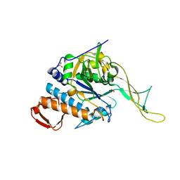
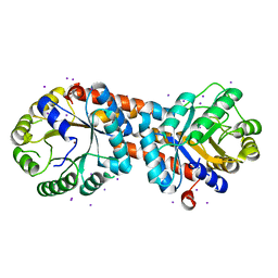
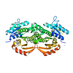
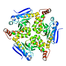
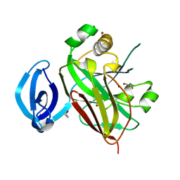
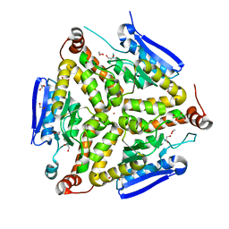
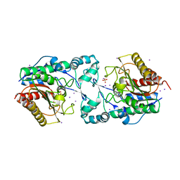
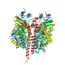
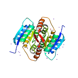
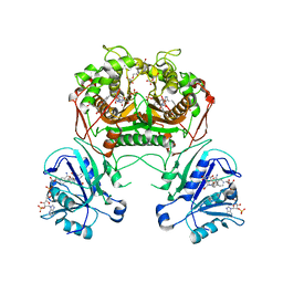
Co-crystal structure of dihydrofolate reductase/thymidylate synthase from Babesia bovis with dUMP, Pemetrexed and NADP
Descriptor: 1,2-ETHANEDIOL, 2'-DEOXYURIDINE 5'-MONOPHOSPHATE, 2-{4-[2-(2-AMINO-4-OXO-4,7-DIHYDRO-3H-PYRROLO[2,3-D]PYRIMIDIN-5-YL)-ETHYL]-BENZOYLAMINO}-PENTANEDIOIC ACID, ...
Authors:Seattle Structural Genomics Center for Infectious Disease (SSGCID)
Deposit date:2009-09-30
Release date:2009-10-13
Last modified:2023-09-06
Method:X-RAY DIFFRACTION (2.2 Å)
Cite:Inhibitor-bound complexes of dihydrofolate reductase-thymidylate synthase from Babesia bovis.
Acta Crystallogr.,Sect.F, 67, 2011
3P4I
DownloadVisualize
BU of 3p4i by Molmil
Crystal structure of acetate kinase from Mycobacterium avium
Descriptor: 1,2-ETHANEDIOL, Acetate kinase
Authors:Seattle Structural Genomics Center for Infectious Disease (SSGCID)
Deposit date:2010-10-06
Release date:2010-11-10
Last modified:2023-09-06
Method:X-RAY DIFFRACTION (2.35 Å)
Cite:Increasing the structural coverage of tuberculosis drug targets.
Tuberculosis (Edinb), 95, 2015
3OC9
DownloadVisualize
BU of 3oc9 by Molmil
Crystal structure of putative UDP-N-acetylglucosamine pyrophosphorylase from Entamoeba histolytica
Descriptor: 1,2-ETHANEDIOL, SULFATE ION, UDP-N-acetylglucosamine pyrophosphorylase
Authors:Seattle Structural Genomics Center for Infectious Disease (SSGCID)
Deposit date:2010-08-09
Release date:2010-08-18
Last modified:2023-09-06
Method:X-RAY DIFFRACTION (1.8 Å)
Cite:Structure of uridine diphosphate N-acetylglucosamine pyrophosphorylase from Entamoeba histolytica.
Acta Crystallogr F Struct Biol Commun, 71, 2015
3O38
DownloadVisualize
BU of 3o38 by Molmil
Crystal structure of a short chain dehydrogenase from Mycobacterium smegmatis
Descriptor: SODIUM ION, Short chain dehydrogenase
Authors:Seattle Structural Genomics Center for Infectious Disease (SSGCID)
Deposit date:2010-07-23
Release date:2010-08-11
Last modified:2023-09-06
Method:X-RAY DIFFRACTION (1.95 Å)
Cite:Increasing the structural coverage of tuberculosis drug targets.
Tuberculosis (Edinb), 95, 2015
3JS5
DownloadVisualize
BU of 3js5 by Molmil
Crystal structure of protein tyrosine phosphatase from Entamoeba histolytica with Hepes in the active site. High resolution, alternative crystal form with 1 molecule in asymmetric unit
Descriptor: 4-(2-HYDROXYETHYL)-1-PIPERAZINE ETHANESULFONIC ACID, Protein tyrosine phosphatase, SODIUM ION
Authors:Seattle Structural Genomics Center for Infectious Disease (SSGCID)
Deposit date:2009-09-09
Release date:2009-09-22
Last modified:2023-09-06
Method:X-RAY DIFFRACTION (1.94 Å)
Cite:Crystal structure and putative substrate identification for the Entamoeba histolytica low molecular weight tyrosine phosphatase.
Mol.Biochem.Parasitol., 193, 2014
3JVI
DownloadVisualize
BU of 3jvi by Molmil
Product state mimic crystal structure of protein tyrosine phosphatase from Entamoeba histolytica
Descriptor: Protein tyrosine phosphatase, SULFATE ION
Authors:Seattle Structural Genomics Center for Infectious Disease (SSGCID)
Deposit date:2009-09-16
Release date:2009-09-22
Last modified:2023-09-06
Method:X-RAY DIFFRACTION (1.8 Å)
Cite:Crystal structure and putative substrate identification for the Entamoeba histolytica low molecular weight tyrosine phosphatase.
Mol.Biochem.Parasitol., 193, 2014
3OXK
DownloadVisualize
BU of 3oxk by Molmil
Crystal structure of a histidine triad family protein from Entamoeba histolytica, bound to GMP
Descriptor: ETHANOL, GUANOSINE-5'-MONOPHOSPHATE, Putative histidine triad family protein, ...
Authors:Seattle Structural Genomics Center for Infectious Disease (SSGCID)
Deposit date:2010-09-21
Release date:2010-10-06
Last modified:2023-09-06
Method:X-RAY DIFFRACTION (1.55 Å)
Cite:Structures of a histidine triad family protein from Entamoeba histolytica bound to sulfate, AMP and GMP.
Acta Crystallogr F Struct Biol Commun, 71, 2015
3P08
DownloadVisualize
BU of 3p08 by Molmil
Crystal structure of the human BTK kinase domain
Descriptor: Tyrosine-protein kinase BTK
Authors:Yu, C.L, Hymowitz, S.G.
Deposit date:2010-09-27
Release date:2010-11-03
Last modified:2023-09-06
Method:X-RAY DIFFRACTION (2.3 Å)
Cite:A novel, specific BTK inhibitor antagonizes BCR and FcgR signaling and suppresses inflammatory arthritis
To be Published
3PZY
DownloadVisualize
BU of 3pzy by Molmil
Crystal structure of Molybdopterin biosynthesis mog protein from Mycobacterium paratuberculosis
Descriptor: Mog, PHOSPHATE ION
Authors:Seattle Structural Genomics Center for Infectious Disease (SSGCID)
Deposit date:2010-12-14
Release date:2011-01-05
Last modified:2023-09-13
Method:X-RAY DIFFRACTION (1.8 Å)
Cite:Increasing the structural coverage of tuberculosis drug targets.
Tuberculosis (Edinb), 95, 2015
3JS9
DownloadVisualize
BU of 3js9 by Molmil
Crystal structure of nucleoside diphosphate kinase family protein from Babesia bovis
Descriptor: Nucleoside diphosphate kinase family protein, SULFATE ION
Authors:Seattle Structural Genomics Center for Infectious Disease (SSGCID)
Deposit date:2009-09-09
Release date:2009-09-22
Last modified:2023-09-06
Method:X-RAY DIFFRACTION (2.5 Å)
Cite:Crystal structure of nucleoside diphosphate kinase family protein from Babesia bovis
To be Published
3PM6
DownloadVisualize
BU of 3pm6 by Molmil
Crystal structure of a putative fructose-1,6-biphosphate aldolase from Coccidioides immitis solved by combined SAD MR
Descriptor: IODIDE ION, Putative fructose-bisphosphate aldolase, ZINC ION
Authors:Seattle Structural Genomics Center for Infectious Disease (SSGCID)
Deposit date:2010-11-16
Release date:2010-12-15
Last modified:2023-09-06
Method:X-RAY DIFFRACTION (2.2 Å)
Cite:SAD phasing using iodide ions in a high-throughput structural genomics environment.
J Struct Funct Genomics, 12, 2011
3QLJ
DownloadVisualize
BU of 3qlj by Molmil
Crystal structure of a short chain dehydrogenase from Mycobacterium avium
Descriptor: 1,2-ETHANEDIOL, CHLORIDE ION, SODIUM ION, ...
Authors:Seattle Structural Genomics Center for Infectious Disease (SSGCID)
Deposit date:2011-02-02
Release date:2011-02-16
Last modified:2023-09-13
Method:X-RAY DIFFRACTION (1.8 Å)
Cite:Increasing the structural coverage of tuberculosis drug targets.
Tuberculosis (Edinb), 95, 2015
3QXI
DownloadVisualize
BU of 3qxi by Molmil
Crystal structure of enoyl-CoA hydratase EchA1 from Mycobacterium marinum
Descriptor: Enoyl-CoA hydratase EchA1
Authors:Seattle Structural Genomics Center for Infectious Disease (SSGCID)
Deposit date:2011-03-01
Release date:2011-03-16
Last modified:2023-09-13
Method:X-RAY DIFFRACTION (2.2 Å)
Cite:Increasing the structural coverage of tuberculosis drug targets.
Tuberculosis (Edinb), 95, 2015
3QDF
DownloadVisualize
BU of 3qdf by Molmil
Crystal structure of 2-hydroxyhepta-2,4-diene-1,7-dioate isomerase from Mycobacterium marinum
Descriptor: 1,2-ETHANEDIOL, 2-hydroxyhepta-2,4-diene-1,7-dioate isomerase, ZINC ION
Authors:Seattle Structural Genomics Center for Infectious Disease (SSGCID)
Deposit date:2011-01-18
Release date:2011-02-02
Last modified:2023-09-13
Method:X-RAY DIFFRACTION (2.05 Å)
Cite:Increasing the structural coverage of tuberculosis drug targets.
Tuberculosis (Edinb), 95, 2015
3QXZ
DownloadVisualize
BU of 3qxz by Molmil
Crystal structure of a probable enoyl-CoA hydratase/isomerase from Mycobacterium abscessus
Descriptor: 1,2-ETHANEDIOL, GLYCEROL, POTASSIUM ION, ...
Authors:Seattle Structural Genomics Center for Infectious Disease (SSGCID)
Deposit date:2011-03-02
Release date:2011-03-16
Last modified:2023-09-13
Method:X-RAY DIFFRACTION (1.35 Å)
Cite:Increasing the structural coverage of tuberculosis drug targets.
Tuberculosis (Edinb), 95, 2015
3MEN
DownloadVisualize
BU of 3men by Molmil
Crystal structure of acetylpolyamine aminohydrolase from Burkholderia pseudomallei, iodide soak
Descriptor: Acetylpolyamine aminohydrolase, IODIDE ION, POTASSIUM ION, ...
Authors:Seattle Structural Genomics Center for Infectious Disease (SSGCID)
Deposit date:2010-03-31
Release date:2010-04-21
Last modified:2024-02-21
Method:X-RAY DIFFRACTION (2.2 Å)
Cite:SAD phasing using iodide ions in a high-throughput structural genomics environment.
J Struct Funct Genomics, 12, 2011
3PY5
DownloadVisualize
BU of 3py5 by Molmil
Crystal structure of a beta-lactamase-like protein from brucella melitensis bound to AMP
Descriptor: ADENOSINE MONOPHOSPHATE, Beta-lactamase-like, L(+)-TARTARIC ACID, ...
Authors:Seattle Structural Genomics Center for Infectious Disease (SSGCID)
Deposit date:2010-12-11
Release date:2010-12-29
Last modified:2023-09-13
Method:X-RAY DIFFRACTION (1.7 Å)
Cite:BrabA.11339.a: anomalous diffraction and ligand binding guide towards the elucidation of the function of a `putative beta-lactamase-like protein from Brucella melitensis.
Acta Crystallogr.,Sect.F, 67, 2011
3MOY
DownloadVisualize
BU of 3moy by Molmil
Crystal structure of probable enoyl-CoA hydratase from Mycobacterium smegmatis
Descriptor: 1,2-ETHANEDIOL, 2-AMINO-2-HYDROXYMETHYL-PROPANE-1,3-DIOL, GLYCEROL, ...
Authors:Seattle Structural Genomics Center for Infectious Disease (SSGCID)
Deposit date:2010-04-23
Release date:2010-05-12
Last modified:2023-09-06
Method:X-RAY DIFFRACTION (1.5 Å)
Cite:Increasing the structural coverage of tuberculosis drug targets.
Tuberculosis (Edinb), 95, 2015
3QKA
DownloadVisualize
BU of 3qka by Molmil
Crystal structure of enoyl-CoA hydratase EchA5 from Mycobacterium marinum
Descriptor: Enoyl-CoA hydratase, EchA5
Authors:Seattle Structural Genomics Center for Infectious Disease (SSGCID)
Deposit date:2011-01-31
Release date:2011-02-09
Last modified:2023-09-13
Method:X-RAY DIFFRACTION (2.15 Å)
Cite:Increasing the structural coverage of tuberculosis drug targets.
Tuberculosis (Edinb), 95, 2015
3QBP
DownloadVisualize
BU of 3qbp by Molmil
Crystal structure of fumarase Fum from Mycobacterium marinum
Descriptor: Fumarase Fum
Authors:Seattle Structural Genomics Center for Infectious Disease (SSGCID)
Deposit date:2011-01-13
Release date:2011-01-26
Last modified:2023-09-13
Method:X-RAY DIFFRACTION (1.85 Å)
Cite:Increasing the structural coverage of tuberculosis drug targets.
Tuberculosis (Edinb), 95, 2015
3QMJ
DownloadVisualize
BU of 3qmj by Molmil
Crystal structure of Enoyl-CoA hydratase EchA8_6 from Mycobacterium marinum
Descriptor: Enoyl-CoA hydratase, EchA8_6, FORMIC ACID
Authors:Seattle Structural Genomics Center for Infectious Disease (SSGCID)
Deposit date:2011-02-04
Release date:2011-03-09
Last modified:2023-09-13
Method:X-RAY DIFFRACTION (2.2 Å)
Cite:Increasing the structural coverage of tuberculosis drug targets.
Tuberculosis (Edinb), 95, 2015
3QH8
DownloadVisualize
BU of 3qh8 by Molmil
Crystal structure of a beta-lactamase-like protein bound to AMP from brucella melitensis, long wavelength synchrotron data
Descriptor: 1,2-ETHANEDIOL, ADENOSINE MONOPHOSPHATE, Beta-lactamase-like, ...
Authors:Seattle Structural Genomics Center for Infectious Disease (SSGCID)
Deposit date:2011-01-25
Release date:2011-02-09
Last modified:2023-09-13
Method:X-RAY DIFFRACTION (1.6 Å)
Cite:Crystal structure of a Beta-Lactamase-Like Protein bound to AMP from Brucella Melitensis, long wavelength synchrotron data
Acta Crystallogr.,Sect.F, 67, 2011
3QD5
DownloadVisualize
BU of 3qd5 by Molmil
Crystal structure of a putative ribose-5-phosphate isomerase from Coccidioides immitis solved by combined iodide ion SAD and MR
Descriptor: 1,2-ETHANEDIOL, IODIDE ION, putative ribose-5-phosphate isomerase
Authors:Seattle Structural Genomics Center for Infectious Disease (SSGCID)
Deposit date:2011-01-17
Release date:2011-02-09
Last modified:2017-09-27
Method:X-RAY DIFFRACTION (1.9 Å)
Cite:Structural characterization of a ribose-5-phosphate isomerase B from the pathogenic fungus Coccidioides immitis.
Bmc Struct.Biol., 11, 2011

222926

건을2024-07-24부터공개중

PDB statisticsPDBj update infoContact PDBjnumon