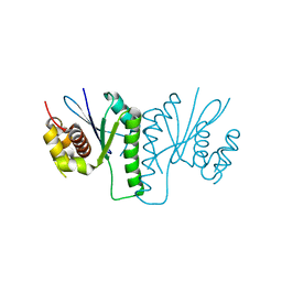
1T6W
 
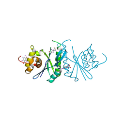
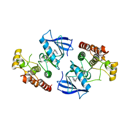
 | | RATIONAL DESIGN OF A CALCIUM-BINDING ADHESION PROTEIN NMR, 20 STRUCTURES | | 分子名称: | CALCIUM ION, hypothetical protein XP_346638 | | 著者 | Yang, W, Wilkins, A.L, Ye, Y, Liu, Z.-R, Urbauer, J.L, Kearney, A, van der Merwe, P.A, Yang, J.J. | | 登録日 | 2004-05-07 | | 公開日 | 2005-02-15 | | 最終更新日 | 2024-05-22 | | 実験手法 | SOLUTION NMR | | 主引用文献 | Design of a calcium-binding protein with desired structure in a cell adhesion molecule.
J.Am.Chem.Soc., 127, 2005
|
|
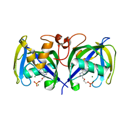
5FBH
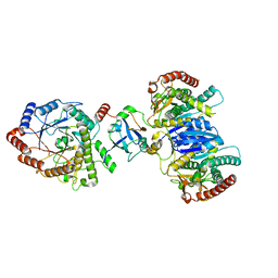
 | | Crystal structure of the extracellular domain of human calcium sensing receptor with bound Gd3+ | | 分子名称: | 2-acetamido-2-deoxy-beta-D-glucopyranose, BICARBONATE ION, CHLORIDE ION, ... | | 著者 | Zhang, T, Zhang, C, Miller, C.L, Zou, J, Moremen, K.W, Brown, E.M, Yang, J.J, Hu, J. | | 登録日 | 2015-12-14 | | 公開日 | 2016-06-22 | | 最終更新日 | 2020-07-29 | | 実験手法 | X-RAY DIFFRACTION (2.7 Å) | | 主引用文献 | Structural basis for regulation of human calcium-sensing receptor by magnesium ions and an unexpected tryptophan derivative co-agonist.
Sci Adv, 2, 2016
|
|
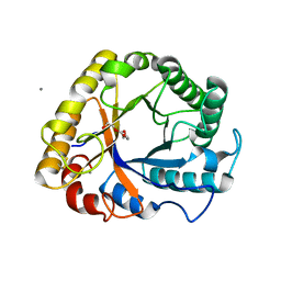
5FBK
 | | Crystal structure of the extracellular domain of human calcium sensing receptor | | 分子名称: | 2-acetamido-2-deoxy-beta-D-glucopyranose, BICARBONATE ION, CHLORIDE ION, ... | | 著者 | Zhang, T, Zhang, C, Miller, C.L, Zou, J, Moremen, K.W, Brown, E.M, Yang, J.J, Hu, J. | | 登録日 | 2015-12-14 | | 公開日 | 2016-06-22 | | 最終更新日 | 2020-07-29 | | 実験手法 | X-RAY DIFFRACTION (2.1 Å) | | 主引用文献 | Structural basis for regulation of human calcium-sensing receptor by magnesium ions and an unexpected tryptophan derivative co-agonist.
Sci Adv, 2, 2016
|
|
7YI9
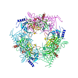
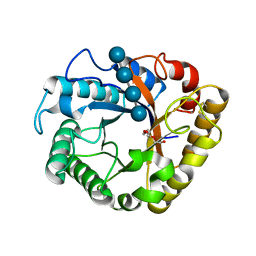
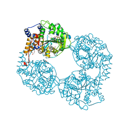
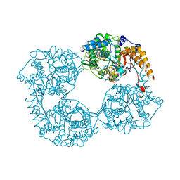
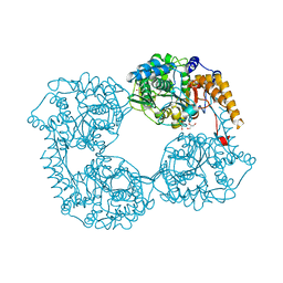
 | | Cryo-EM structure of SAM-bound MTA1-MTA9-p1-p2 complex | | 分子名称: | MT-a70 family protein, MTA9, P1, ... | | 著者 | Yan, J.J, Guan, Z.Y, Liu, F.Q, Yan, X.H, Hou, M.J, Yin, P. | | 登録日 | 2022-07-15 | | 公開日 | 2023-01-18 | | 最終更新日 | 2024-07-03 | | 実験手法 | ELECTRON MICROSCOPY (2.6 Å) | | 主引用文献 | Structural insights into DNA N 6 -adenine methylation by the MTA1 complex.
Cell Discov, 9, 2023
|
|
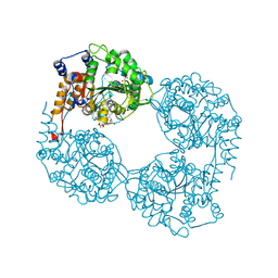
7YI8
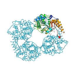
 | | Cryo-EM structure of SAH-bound MTA1-MTA9-p1-p2 complex | | 分子名称: | MT-a70 family protein, MTA9, P1, ... | | 著者 | Yan, J.J, Guan, Z.Y, Liu, F.Q, Yan, X.H, Hou, M.J, Yin, P. | | 登録日 | 2022-07-15 | | 公開日 | 2023-01-18 | | 最終更新日 | 2024-07-03 | | 実験手法 | ELECTRON MICROSCOPY (2.7 Å) | | 主引用文献 | Structural insights into DNA N 6 -adenine methylation by the MTA1 complex.
Cell Discov, 9, 2023
|
|
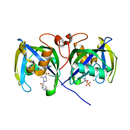
7Y77
 | | Crystal structure of rice NAL1 | | 分子名称: | ADENOSINE-5'-TRIPHOSPHATE, Protein NARROW LEAF 1 | | 著者 | Yan, J.J, Guan, Z.Y, Yin, P, Xiong, L.Z. | | 登録日 | 2022-06-21 | | 公開日 | 2023-07-12 | | 最終更新日 | 2024-05-29 | | 実験手法 | X-RAY DIFFRACTION (2.6 Å) | | 主引用文献 | Serine protease NAL1 exerts pleiotropic functions through degradation of TOPLESS-related corepressor in rice.
Nat.Plants, 9, 2023
|
|
6KVN
 | |
6LCT
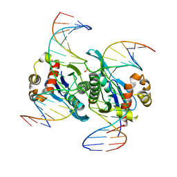 | | Crystal structure of catalytic inactive chloroplast resolvase NtMOC1 in complex with Holliday junction | | 分子名称: | DNA (5'-D(*AP*AP*GP*AP*TP*GP*TP*CP*CP*CP*TP*CP*TP*GP*TP*TP*GP*T)-3'), DNA (5'-D(*AP*CP*AP*AP*CP*AP*GP*AP*GP*GP*AP*TP*GP*GP*AP*GP*CP*T)-3'), DNA (5'-D(*GP*CP*CP*TP*TP*GP*CP*TP*GP*GP*GP*AP*CP*AP*TP*CP*TP*T)-3'), ... | | 著者 | Yan, J.J, Hong, S.X, Guan, Z.Y, Yin, P. | | 登録日 | 2019-11-19 | | 公開日 | 2020-04-08 | | 最終更新日 | 2023-11-22 | | 実験手法 | X-RAY DIFFRACTION (2.55 Å) | | 主引用文献 | Structural insights into sequence-dependent Holliday junction resolution by the chloroplast resolvase MOC1.
Nat Commun, 11, 2020
|
|
6KVO
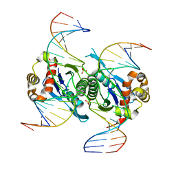 | | Crystal structure of chloroplast resolvase in complex with Holliday junction | | 分子名称: | DNA (5'-D(*AP*CP*AP*AP*CP*AP*GP*AP*TP*GP*AP*TP*GP*GP*AP*GP*CP*T)-3'), DNA (5'-D(*GP*CP*CP*TP*TP*GP*CP*TP*TP*GP*GP*AP*CP*AP*TP*CP*TP*T)-3'), DNA (5'-D(P*AP*AP*GP*AP*TP*GP*TP*CP*CP*AP*TP*CP*TP*GP*TP*TP*GP*T)-3'), ... | | 著者 | Yan, J.J, Hong, S.X, Guan, Z.Y, Yin, P. | | 登録日 | 2019-09-05 | | 公開日 | 2020-04-08 | | 最終更新日 | 2024-03-27 | | 実験手法 | X-RAY DIFFRACTION (2.5 Å) | | 主引用文献 | Structural insights into sequence-dependent Holliday junction resolution by the chloroplast resolvase MOC1.
Nat Commun, 11, 2020
|
|
6LCM
 | | Crystal structure of chloroplast resolvase ZmMOC1 with the magic triangle I3C | | 分子名称: | 5-amino-2,4,6-triiodobenzene-1,3-dicarboxylic acid, ZmMoc1 | | 著者 | Yan, J.J, Hong, S.X, Guan, Z.Y, Yin, P. | | 登録日 | 2019-11-19 | | 公開日 | 2020-04-08 | | 最終更新日 | 2024-03-27 | | 実験手法 | X-RAY DIFFRACTION (2.5 Å) | | 主引用文献 | Structural insights into sequence-dependent Holliday junction resolution by the chloroplast resolvase MOC1.
Nat Commun, 11, 2020
|
|
8J5D
 | | Cryo-EM structure of starch degradation complex of BAM1-LSF1-MDH | | 分子名称: | Beta-amylase 1, chloroplastic, Malate dehydrogenase, ... | | 著者 | Guan, Z.Y, Liu, J, Yan, J.J. | | 登録日 | 2023-04-21 | | 公開日 | 2024-01-10 | | 実験手法 | ELECTRON MICROSCOPY (3 Å) | | 主引用文献 | The LIKE SEX FOUR 1-malate dehydrogenase complex functions as a scaffold to recruit beta-amylase to promote starch degradation.
Plant Cell, 36, 2023
|
|
5I78
 | | Crystal structure of a beta-1,4-endoglucanase from Aspergillus niger | | 分子名称: | 2-acetamido-2-deoxy-beta-D-glucopyranose, CALCIUM ION, Endo-beta-1, ... | | 著者 | Liu, W.D, Yan, J.J, Li, Y.J, Zheng, Y.Y, Chen, C.C, Guo, R.T. | | 登録日 | 2016-02-17 | | 公開日 | 2016-12-21 | | 最終更新日 | 2023-11-08 | | 実験手法 | X-RAY DIFFRACTION (1.58 Å) | | 主引用文献 | Functional and structural analysis of Pichia pastoris-expressed Aspergillus niger 1,4-beta-endoglucanase
Biochem. Biophys. Res. Commun., 475, 2016
|
|
5I79
 | | Crystal structure of a beta-1,4-endoglucanase mutant from Aspergillus niger in complex with sugar | | 分子名称: | 2-acetamido-2-deoxy-beta-D-glucopyranose, CALCIUM ION, Endo-beta-1, ... | | 著者 | Liu, W.D, Yan, J.J, Li, Y.J, Zheng, Y.Y, Chen, C.C, Guo, R.T. | | 登録日 | 2016-02-17 | | 公開日 | 2016-12-21 | | 最終更新日 | 2023-11-08 | | 実験手法 | X-RAY DIFFRACTION (2.35 Å) | | 主引用文献 | Functional and structural analysis of Pichia pastoris-expressed Aspergillus niger 1,4-beta-endoglucanase
Biochem. Biophys. Res. Commun., 475, 2016
|
|
3Q96
 | | B-Raf kinase domain in complex with a tetrahydronaphthalene inhibitor | | 分子名称: | (2S)-N-[3-(2-aminopropan-2-yl)-5-(trifluoromethyl)phenyl]-7-[(7-oxo-5,6,7,8-tetrahydro-1,8-naphthyridin-4-yl)oxy]-1,2,3,4-tetrahydronaphthalene-2-carboxamide, Serine/threonine-protein kinase B-raf | | 著者 | Sintchak, M.D, Aertgeerts, K, Yano, J. | | 登録日 | 2011-01-07 | | 公開日 | 2011-03-23 | | 最終更新日 | 2024-02-21 | | 実験手法 | X-RAY DIFFRACTION (3.1 Å) | | 主引用文献 | Design and optimization of potent and orally bioavailable tetrahydronaphthalene raf inhibitors.
J.Med.Chem., 54, 2011
|
|
7B7V
 | |
5OPL
 | | Crystal structure of K25E cN-II mutant | | 分子名称: | Cytosolic purine 5'-nucleotidase, GLYCEROL, MAGNESIUM ION | | 著者 | Kugler, M, Hnizda, A, Pachl, P, Rezacova, P. | | 登録日 | 2017-08-10 | | 公開日 | 2018-06-13 | | 最終更新日 | 2024-05-08 | | 実験手法 | X-RAY DIFFRACTION (1.8 Å) | | 主引用文献 | Relapsed acute lymphoblastic leukemia-specific mutations in NT5C2 cluster into hotspots driving intersubunit stimulation.
Leukemia, 32, 2018
|
|
5OPM
 | | Crystal structure of D52N/R238W cN-II mutant bound to dATP and free phosphate | | 分子名称: | 2'-DEOXYADENOSINE 5'-TRIPHOSPHATE, Cytosolic purine 5'-nucleotidase, GLYCEROL, ... | | 著者 | Hnizda, A, Pachl, P, Rezacova, P. | | 登録日 | 2017-08-10 | | 公開日 | 2018-06-13 | | 最終更新日 | 2024-01-17 | | 実験手法 | X-RAY DIFFRACTION (1.68 Å) | | 主引用文献 | Relapsed acute lymphoblastic leukemia-specific mutations in NT5C2 cluster into hotspots driving intersubunit stimulation.
Leukemia, 32, 2018
|
|
5OPK
 | | Crystal structure of D52N/R367Q cN-II mutant bound to dATP and free phosphate | | 分子名称: | 2'-DEOXYADENOSINE 5'-TRIPHOSPHATE, Cytosolic purine 5'-nucleotidase, GLYCEROL, ... | | 著者 | Hnizda, A, Pachl, P, Rezacova, P. | | 登録日 | 2017-08-10 | | 公開日 | 2018-06-13 | | 最終更新日 | 2024-01-17 | | 実験手法 | X-RAY DIFFRACTION (1.74 Å) | | 主引用文献 | Relapsed acute lymphoblastic leukemia-specific mutations in NT5C2 cluster into hotspots driving intersubunit stimulation.
Leukemia, 32, 2018
|
|
5OPP
 | | Crystal structure of S408R cN-II mutant | | 分子名称: | BROMIDE ION, Cytosolic purine 5'-nucleotidase, GLYCEROL, ... | | 著者 | Hnizda, A, Pachl, P, Rezacova, P. | | 登録日 | 2017-08-10 | | 公開日 | 2018-06-13 | | 最終更新日 | 2024-01-17 | | 実験手法 | X-RAY DIFFRACTION (1.7 Å) | | 主引用文献 | Relapsed acute lymphoblastic leukemia-specific mutations in NT5C2 cluster into hotspots driving intersubunit stimulation.
Leukemia, 32, 2018
|
|
5OPN
 | | Crystal structure of R39Q cN-II mutant | | 分子名称: | Cytosolic purine 5'-nucleotidase, GLYCEROL | | 著者 | Hnizda, A, Pachl, P, Rezacova, P. | | 登録日 | 2017-08-10 | | 公開日 | 2018-06-13 | | 最終更新日 | 2024-01-17 | | 実験手法 | X-RAY DIFFRACTION (1.77 Å) | | 主引用文献 | Relapsed acute lymphoblastic leukemia-specific mutations in NT5C2 cluster into hotspots driving intersubunit stimulation.
Leukemia, 32, 2018
|
|
5OPO
 | | Crystal structure of R238G cN-II mutant | | 分子名称: | Cytosolic purine 5'-nucleotidase, GLYCEROL, MAGNESIUM ION | | 著者 | Hnizda, A, Pachl, P, Rezacova, P. | | 登録日 | 2017-08-10 | | 公開日 | 2018-06-13 | | 最終更新日 | 2024-01-17 | | 実験手法 | X-RAY DIFFRACTION (2 Å) | | 主引用文献 | Relapsed acute lymphoblastic leukemia-specific mutations in NT5C2 cluster into hotspots driving intersubunit stimulation.
Leukemia, 32, 2018
|
|
7B67
 | | Structure of NUDT15 V18_V19insGV Mutant in complex with TH7755 | | 分子名称: | (R)-6-((2-methyl-4-(1-methyl-1H-indole-5-carbonyl)piperazin-1-yl)sulfonyl)benzo[d]oxazol-2(3H)-one, MAGNESIUM ION, Nucleotide triphosphate diphosphatase NUDT15, ... | | 著者 | Rehling, D, Stenmark, P. | | 登録日 | 2020-12-07 | | 公開日 | 2021-03-24 | | 最終更新日 | 2024-01-31 | | 実験手法 | X-RAY DIFFRACTION (1.45 Å) | | 主引用文献 | Crystal structures of NUDT15 variants enabled by a potent inhibitor reveal the structural basis for thiopurine sensitivity.
J.Biol.Chem., 296, 2021
|
|
7B65
 | | Structure of NUDT15 R139C Mutant in complex with TH7755 | | 分子名称: | (R)-6-((2-methyl-4-(1-methyl-1H-indole-5-carbonyl)piperazin-1-yl)sulfonyl)benzo[d]oxazol-2(3H)-one, Nucleotide triphosphate diphosphatase NUDT15 | | 著者 | Rehling, D, Stenmark, P. | | 登録日 | 2020-12-07 | | 公開日 | 2021-03-24 | | 最終更新日 | 2024-01-31 | | 実験手法 | X-RAY DIFFRACTION (1.6 Å) | | 主引用文献 | Crystal structures of NUDT15 variants enabled by a potent inhibitor reveal the structural basis for thiopurine sensitivity.
J.Biol.Chem., 296, 2021
|
|
7B63
 | | Structure of NUDT15 in complex with TH7755 | | 分子名称: | (R)-6-((2-methyl-4-(1-methyl-1H-indole-5-carbonyl)piperazin-1-yl)sulfonyl)benzo[d]oxazol-2(3H)-one, MAGNESIUM ION, Probable 8-oxo-dGTP diphosphatase NUDT15 | | 著者 | Rehling, D, Stenmark, P. | | 登録日 | 2020-12-07 | | 公開日 | 2021-03-24 | | 最終更新日 | 2024-01-31 | | 実験手法 | X-RAY DIFFRACTION (1.6 Å) | | 主引用文献 | Crystal structures of NUDT15 variants enabled by a potent inhibitor reveal the structural basis for thiopurine sensitivity.
J.Biol.Chem., 296, 2021
|
|
7B66
 | | Structure of NUDT15 R139H Mutant in complex with TH7755 | | 分子名称: | (R)-6-((2-methyl-4-(1-methyl-1H-indole-5-carbonyl)piperazin-1-yl)sulfonyl)benzo[d]oxazol-2(3H)-one, Nucleotide triphosphate diphosphatase NUDT15 | | 著者 | Rehling, D, Stenmark, P. | | 登録日 | 2020-12-07 | | 公開日 | 2021-03-24 | | 最終更新日 | 2024-01-31 | | 実験手法 | X-RAY DIFFRACTION (1.6 Å) | | 主引用文献 | Crystal structures of NUDT15 variants enabled by a potent inhibitor reveal the structural basis for thiopurine sensitivity.
J.Biol.Chem., 296, 2021
|
|


