6HYS
 
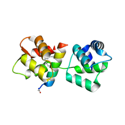
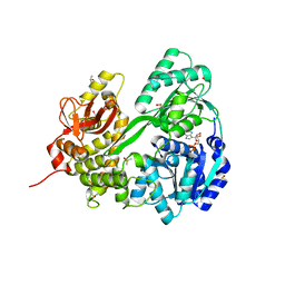 | | Crystal structure of DHX8 helicase domain bound to ADP at 2.6 angstrom | | 分子名称: | 1,2-ETHANEDIOL, ACETATE ION, ADENOSINE-5'-DIPHOSPHATE, ... | | 著者 | Felisberto-Rodrigues, C, Thomas, J.C, McAndrew, P.C, Le Bihan, Y.V, Burke, R, Workman, P, van Montfort, R.L.M. | | 登録日 | 2018-10-22 | | 公開日 | 2019-08-28 | | 最終更新日 | 2024-01-24 | | 実験手法 | X-RAY DIFFRACTION (2.6 Å) | | 主引用文献 | Structural and functional characterisation of human RNA helicase DHX8 provides insights into the mechanism of RNA-stimulated ADP release.
Biochem.J., 476, 2019
|
|
6HYT
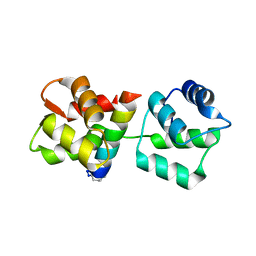
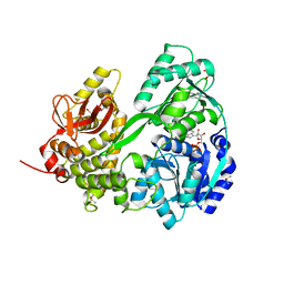 | | Crystal structure of DHX8 helicase domain bound to ADP at 2.3 Angstrom | | 分子名称: | 1,2-ETHANEDIOL, ACETATE ION, ADENOSINE-5'-DIPHOSPHATE, ... | | 著者 | Felisberto-Rodrigues, C, Thomas, J.C, McAndrew, P.C, Le Bihan, Y.V, Burke, R, Workman, P, van Montfort, R.L.M. | | 登録日 | 2018-10-22 | | 公開日 | 2019-08-28 | | 最終更新日 | 2024-05-15 | | 実験手法 | X-RAY DIFFRACTION (2.33 Å) | | 主引用文献 | Structural and functional characterisation of human RNA helicase DHX8 provides insights into the mechanism of RNA-stimulated ADP release.
Biochem.J., 476, 2019
|
|
6HYU
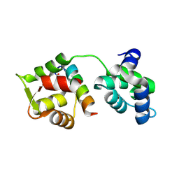
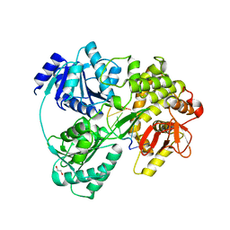 | | Crystal structure of DHX8 helicase bound to single stranded poly-adenine RNA | | 分子名称: | 1,2-ETHANEDIOL, ATP-dependent RNA helicase DHX8, RNA (5'-R(*A*AP*A)-3'), ... | | 著者 | Felisberto-Rodrigues, C, Thomas, J.C, McAndrew, P.C, Le Bihan, Y.V, Burke, R, Workman, P, van Montfort, R.L.M. | | 登録日 | 2018-10-22 | | 公開日 | 2019-08-28 | | 最終更新日 | 2024-05-15 | | 実験手法 | X-RAY DIFFRACTION (3.22 Å) | | 主引用文献 | Structural and functional characterisation of human RNA helicase DHX8 provides insights into the mechanism of RNA-stimulated ADP release.
Biochem.J., 476, 2019
|
|
4AOM
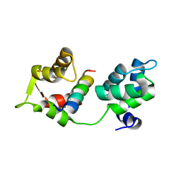 | | MTIP and MyoA complex | | 分子名称: | MYOSIN A TAIL DOMAIN INTERACTING PROTEIN, MYOSIN-A | | 著者 | Salgado, P.S, Douse, C.H, Simpson, P.J, Thomas, J.C, Holder, A.A, Tate, E.W, Cota, E. | | 登録日 | 2012-03-29 | | 公開日 | 2012-09-05 | | 最終更新日 | 2023-12-20 | | 実験手法 | X-RAY DIFFRACTION (1.939 Å) | | 主引用文献 | Regulation of the Plasmodium Motor Complex: Phosphorylation of Myosin a Tail Interacting Protein (Mtip) Loosens its Grip on Myoa
J.Biol.Chem., 287, 2012
|
|
3ZNG
 | | Ankyrin repeat and SOCS-box protein 9 (ASB9) in complex with ElonginB and ElonginC | | 分子名称: | 1,2-ETHANEDIOL, ANKYRIN REPEAT AND SOCS BOX PROTEIN 9, TRANSCRIPTION ELONGATION FACTOR B POLYPEPTIDE 1, ... | | 著者 | Thomas, J, Van Molle, I, Ciulli, A. | | 登録日 | 2013-02-14 | | 公開日 | 2013-12-04 | | 最終更新日 | 2023-12-20 | | 実験手法 | X-RAY DIFFRACTION (2.85 Å) | | 主引用文献 | Multimeric Complexes Among Ankyrin-Repeat and Socs-Box Protein 9 (Asb9), Elonginbc, and Cullin 5: Insights Into the Structure and Assembly of Ecs-Type Cullin-Ring E3 Ubiquitin Ligases.
Biochemistry, 52, 2013
|
|
4MZK
 | | Crystal Structure of MTIP from Plasmodium falciparum in complex with pGly[807,811], a stapled myoA tail peptide | | 分子名称: | Myosin A tail domain interacting protein, pGly[807,811], a stapled myoA tail peptide | | 著者 | Douse, C.H, Garnett, J.A, Maas, S.J, Cota, E, Tate, E.W. | | 登録日 | 2013-09-30 | | 公開日 | 2013-11-06 | | 最終更新日 | 2023-12-06 | | 実験手法 | X-RAY DIFFRACTION (1.82 Å) | | 主引用文献 | Crystal Structures of Stapled and Hydrogen Bond Surrogate Peptides Targeting a Fully Buried Protein-Helix Interaction.
Acs Chem.Biol., 8, 2014
|
|
4MZL
 | | Crystal Structure of MTIP from Plasmodium falciparum in complex with HBS myoA, a hydrogen bond surrogate myoA helix mimetic | | 分子名称: | Myosin A tail domain interacting protein, hydrogen bond surrogate (HBS) myoA helix mimetic | | 著者 | Douse, C.H, Garnett, J.A, Maas, S.J, Cota, E, Tate, E.W. | | 登録日 | 2013-09-30 | | 公開日 | 2013-11-06 | | 最終更新日 | 2023-06-07 | | 実験手法 | X-RAY DIFFRACTION (2.01 Å) | | 主引用文献 | Crystal Structures of Stapled and Hydrogen Bond Surrogate Peptides Targeting a Fully Buried Protein-Helix Interaction.
Acs Chem.Biol., 8, 2014
|
|
4MZJ
 | | Crystal Structure of MTIP from Plasmodium falciparum in complex with pGly[801,805], a stapled myoA tail peptide | | 分子名称: | Myosin A tail domain interacting protein, Myosin-A | | 著者 | Douse, C.H, Garnett, J.A, Maas, S.J, Cota, E, Tate, E.W. | | 登録日 | 2013-09-30 | | 公開日 | 2013-11-06 | | 最終更新日 | 2023-12-06 | | 実験手法 | X-RAY DIFFRACTION (1.474 Å) | | 主引用文献 | Crystal Structures of Stapled and Hydrogen Bond Surrogate Peptides Targeting a Fully Buried Protein-Helix Interaction.
Acs Chem.Biol., 8, 2014
|
|


