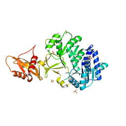
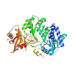
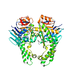
7SX5
 
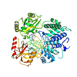 | | Crystal structure of ligase I with nick duplexes containing mismatch A:C | | 分子名称: | ADENOSINE MONOPHOSPHATE, DNA chain 1, DNA chain 2, ... | | 著者 | Tang, Q, Gulkis, M, McKenna, R, Caglayan, M. | | 登録日 | 2021-11-22 | | 公開日 | 2022-07-13 | | 最終更新日 | 2023-10-18 | | 実験手法 | X-RAY DIFFRACTION (2.8 Å) | | 主引用文献 | Structures of LIG1 that engage with mutagenic mismatches inserted by pol beta in base excision repair.
Nat Commun, 13, 2022
|
|
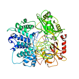
7SXE
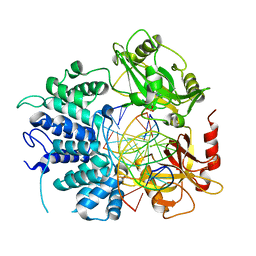 | | Crystal structure of ligase I with nick duplexes containing cognate G:T | | 分子名称: | ADENOSINE MONOPHOSPHATE, DNA chain 1, DNA chain 2, ... | | 著者 | Tang, Q, Gulkis, M, McKenna, R, Caglayan, M. | | 登録日 | 2021-11-22 | | 公開日 | 2022-07-13 | | 最終更新日 | 2023-10-18 | | 実験手法 | X-RAY DIFFRACTION (3 Å) | | 主引用文献 | Structures of LIG1 that engage with mutagenic mismatches inserted by pol beta in base excision repair.
Nat Commun, 13, 2022
|
|
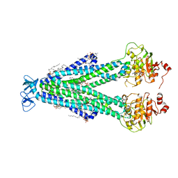
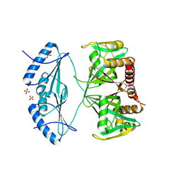
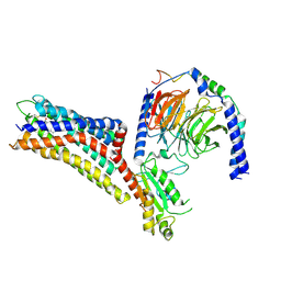
4MBS
 | | Crystal Structure of the CCR5 Chemokine Receptor | | 分子名称: | (2R)-2,3-dihydroxypropyl (9Z)-octadec-9-enoate, 4,4-difluoro-N-[(1S)-3-{(3-exo)-3-[3-methyl-5-(propan-2-yl)-4H-1,2,4-triazol-4-yl]-8-azabicyclo[3.2.1]oct-8-yl}-1-phenylpropyl]cyclohexanecarboxamide, Chimera protein of C-C chemokine receptor type 5 and Rubredoxin, ... | | 著者 | Tan, Q, Zhu, Y, Han, G.W, Li, J, Fenalti, G, Liu, H, Cherezov, V, Stevens, R.C, GPCR Network (GPCR), Zhao, Q, Wu, B. | | 登録日 | 2013-08-19 | | 公開日 | 2013-09-11 | | 最終更新日 | 2023-09-20 | | 実験手法 | X-RAY DIFFRACTION (2.71 Å) | | 主引用文献 | Structure of the CCR5 chemokine receptor-HIV entry inhibitor maraviroc complex.
Science, 341, 2013
|
|
8FMV
 | |
8FHK
 | |
8PYY
 | |
8T1P
 | |
8SZC
 | |
8T3K
 | |
8PYX
 | |
8PPP
 | |
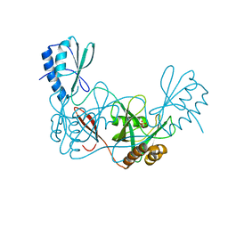
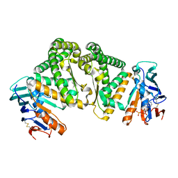
7SUM
 | | Crystal structure of human ligase I with nick duplexes containing cognate A:T | | 分子名称: | ADENOSINE MONOPHOSPHATE, DNA ligase 1, DNA(5'-*GP*CP*TP*GP*AP*TP*GP*CP*GP*TP*A-3'), ... | | 著者 | Tang, Q, Gulkis, M, McKenna, R, Caglayan, M. | | 登録日 | 2021-11-17 | | 公開日 | 2022-07-13 | | 最終更新日 | 2023-10-18 | | 実験手法 | X-RAY DIFFRACTION (2.9 Å) | | 主引用文献 | Structures of LIG1 that engage with mutagenic mismatches inserted by pol beta in base excision repair.
Nat Commun, 13, 2022
|
|
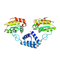
4ZBP
 | | Crystal structure of the AMPCPR-bound AtNUDT7 | | 分子名称: | ALPHA-BETA METHYLENE ADP-RIBOSE, Nudix hydrolase 7, SULFATE ION | | 著者 | Tang, Q, Liu, C, Zhong, C, Ding, J. | | 登録日 | 2015-04-15 | | 公開日 | 2015-09-09 | | 最終更新日 | 2023-11-08 | | 実験手法 | X-RAY DIFFRACTION (2.6 Å) | | 主引用文献 | Crystal Structures of Arabidopsis thaliana Nudix Hydrolase NUDT7 Reveal a Previously Unobserved Conformation.
Mol Plant, 8, 2015
|
|
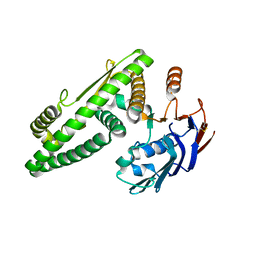
4ZB3
 | | Crystal structure of the apo AtNUDT7 | | 分子名称: | Nudix hydrolase 7, SULFATE ION | | 著者 | Tang, Q, Liu, C, Zhong, C, Ding, J. | | 登録日 | 2015-04-14 | | 公開日 | 2015-09-09 | | 最終更新日 | 2024-03-20 | | 実験手法 | X-RAY DIFFRACTION (2.3 Å) | | 主引用文献 | Crystal Structures of Arabidopsis thaliana Nudix Hydrolase NUDT7 Reveal a Previously Unobserved Conformation.
Mol Plant, 8, 2015
|
|
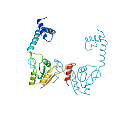
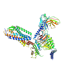
5Z68
 | | Structure of the recombination mediator protein RecF-ATP in RecFOR pathway | | 分子名称: | ADENOSINE-5'-TRIPHOSPHATE, DNA replication and repair protein RecF, IMIDAZOLE, ... | | 著者 | Tang, Q, Liu, Y.-P, Yan, X.-X. | | 登録日 | 2018-01-22 | | 公開日 | 2018-04-18 | | 実験手法 | X-RAY DIFFRACTION (3 Å) | | 主引用文献 | ATP-dependent conformational change in ABC-ATPase RecF serves as a switch in DNA repair.
Sci Rep, 8, 2018
|
|
5Z69
 | |
5Z67
 | |
3VE5
 | |
3VDU
 | |
3VDP
 | |
4O6P
 | | Structural and functional studies the characterization of C58G/C70G mutant in Cys4 Zinc-finger motif in the recombination mediator protein RecR | | 分子名称: | Recombination protein RecR, ZINC ION | | 著者 | Tang, Q, Liu, Y.P, Yan, X.X, Liang, D.C. | | 登録日 | 2013-12-23 | | 公開日 | 2014-12-10 | | 最終更新日 | 2023-11-08 | | 実験手法 | X-RAY DIFFRACTION (3 Å) | | 主引用文献 | Structural and functional characterization of Cys4 zinc finger motif in the recombination mediator protein RecR.
DNA Repair (Amst.), 24, 2014
|
|
4O6O
 | | Structural and functional studies the characterization of Cys4 Zinc-finger motif in the recombination mediator protein RecR | | 分子名称: | IMIDAZOLE, Recombination protein RecR, ZINC ION | | 著者 | Tang, Q, Liu, Y.P, Yan, X.X, Liang, D.C. | | 登録日 | 2013-12-23 | | 公開日 | 2014-12-10 | | 最終更新日 | 2023-11-08 | | 実験手法 | X-RAY DIFFRACTION (3 Å) | | 主引用文献 | Structural and functional characterization of Cys4 zinc finger motif in the recombination mediator protein RecR.
DNA Repair (Amst.), 24, 2014
|
|
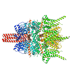
7XXI
 | | Cryo-EM structure of the purinergic receptor P2Y12R in complex with 2MeSADP and Gi | | 分子名称: | 2-(methylsulfanyl)adenosine 5'-(trihydrogen diphosphate), Guanine nucleotide-binding protein G(I)/G(S)/G(O) subunit gamma-2, Guanine nucleotide-binding protein G(I)/G(S)/G(T) subunit beta-1, ... | | 著者 | Tan, Q, Li, B, Han, S, Zhao, Q, Wu, B. | | 登録日 | 2022-05-30 | | 公開日 | 2023-06-07 | | 最終更新日 | 2023-09-20 | | 実験手法 | ELECTRON MICROSCOPY (3 Å) | | 主引用文献 | Structural insights into signal transduction of the purinergic receptors P2Y1R and P2Y12R.
Protein Cell, 14, 2023
|
|
7XXH
 | | Cryo-EM structure of the purinergic receptor P2Y1R in complex with 2MeSADP and G11 | | 分子名称: | 2-(methylsulfanyl)adenosine 5'-(trihydrogen diphosphate), Guanine nucleotide-binding protein G(11) subunit alpha, Guanine nucleotide-binding protein G(I)/G(S)/G(O) subunit gamma-2, ... | | 著者 | Tan, Q, Li, B, Han, S, Zhao, Q, Wu, B. | | 登録日 | 2022-05-30 | | 公開日 | 2023-06-07 | | 最終更新日 | 2023-09-20 | | 実験手法 | ELECTRON MICROSCOPY (2.9 Å) | | 主引用文献 | Structural insights into signal transduction of the purinergic receptors P2Y1R and P2Y12R.
Protein Cell, 14, 2023
|
|
5ZBG
 | |


