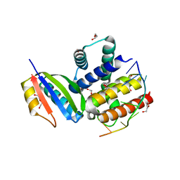
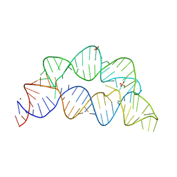
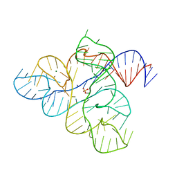
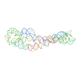
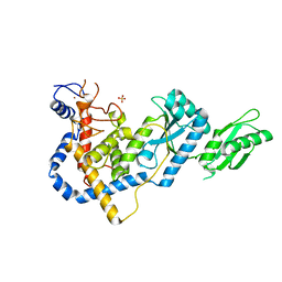
5E54
 
 | | Two apo structures of the adenine riboswitch aptamer domain determined using an X-ray free electron laser | | 分子名称: | MAGNESIUM ION, Vibrio vulnificus strain 93U204 chromosome II, adenine riboswitch aptamer domain | | 著者 | Stagno, J.R, Wang, Y.-X, Liu, Y, Bhandari, Y.R, Conrad, C.E, Nelson, G, Li, C, Wendel, D.R, White, T.A, Barty, A, Tuckey, R.A, Zatsepin, N.A, Grant, T.D, Fromme, P, Tan, K, Ji, X, Spence, J.C.H. | | 登録日 | 2015-10-07 | | 公開日 | 2016-11-23 | | 最終更新日 | 2023-08-30 | | 実験手法 | X-RAY DIFFRACTION (2.3 Å) | | 主引用文献 | Structures of riboswitch RNA reaction states by mix-and-inject XFEL serial crystallography.
Nature, 541, 2017
|
|
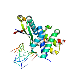
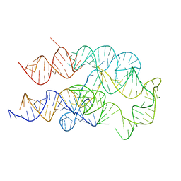
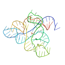
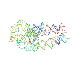
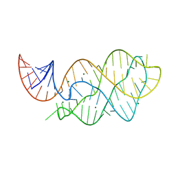
4EYA
 | | Crystal Structure of a Plectonemic RNA Supercoil | | 分子名称: | GLYCEROL, N utilization substance protein B homolog, RNA (5'-R(*GP*GP*CP*UP*CP*CP*UP*UP*GP*GP*CP*A)-3'), ... | | 著者 | Stagno, J.R, Ji, X. | | 登録日 | 2012-05-01 | | 公開日 | 2012-06-20 | | 最終更新日 | 2023-09-13 | | 実験手法 | X-RAY DIFFRACTION (3.2 Å) | | 主引用文献 | Crystal structure of a plectonemic RNA supercoil.
Nat Commun, 3, 2012
|
|
3R2D
 | |
3R2C
 | |
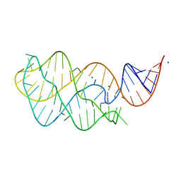
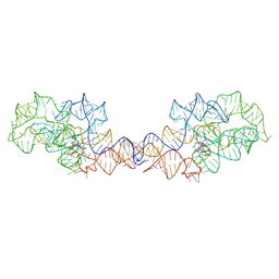
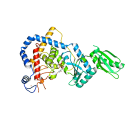
5SWD
 | | Structure of the adenine riboswitch aptamer domain in an intermediate-bound state | | 分子名称: | ADENINE, MAGNESIUM ION, Vibrio vulnificus strain 93U204 chromosome II, ... | | 著者 | Stagno, J.R, Wang, Y.-X, Liu, Y, Bhandari, Y.R, Conrad, C.E, Nelson, G, Li, C, Wendel, D.R, White, T.A, Barty, A, Tuckey, R.A, Zatsepin, N.A, Grant, T.D, Fromme, P, Tan, K, Ji, X, Spence, J.C.H. | | 登録日 | 2016-08-08 | | 公開日 | 2016-11-23 | | 最終更新日 | 2023-10-04 | | 実験手法 | X-RAY DIFFRACTION (2.5 Å) | | 主引用文献 | Structures of riboswitch RNA reaction states by mix-and-inject XFEL serial crystallography.
Nature, 541, 2017
|
|
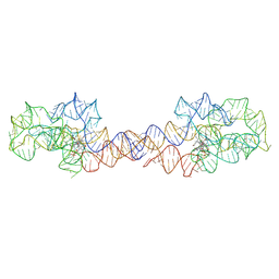
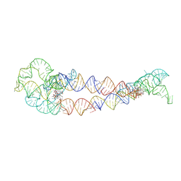
5SWE
 | | Ligand-bound structure of adenine riboswitch aptamer domain converted in crystal from its ligand-free state using ligand mixing serial femtosecond crystallography | | 分子名称: | ADENINE, Vibrio vulnificus strain 93U204 chromosome II, adenine riboswitch aptamer domain | | 著者 | Stagno, J.R, Wang, Y.-X, Liu, Y, Bhandari, Y.R, Conrad, C.E, Nelson, G, Li, C, Wendel, D.R, White, T.A, Barty, A, Tuckey, R.A, Zatsepin, N.A, Grant, T.D, Fromme, P, Tan, K, Ji, X, Spence, J.C.H. | | 登録日 | 2016-08-08 | | 公開日 | 2016-11-23 | | 最終更新日 | 2023-10-04 | | 実験手法 | X-RAY DIFFRACTION (3 Å) | | 主引用文献 | Structures of riboswitch RNA reaction states by mix-and-inject XFEL serial crystallography.
Nature, 541, 2017
|
|
6VWT
 | |
4U5T
 | |
8FW4
 | |
6VWV
 | |
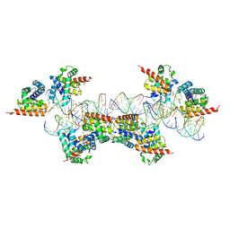
5KAL
 | | Terminal uridylyl transferase 4 from Trypanosoma brucei with bound UTP and UpU | | 分子名称: | MAGNESIUM ION, RNA (5'-R(*UP*U)-3'), RNA uridylyltransferase 4, ... | | 著者 | Stagno, J.R, Luecke, H, Afasizhev, R. | | 登録日 | 2016-06-01 | | 公開日 | 2016-10-26 | | 最終更新日 | 2023-09-27 | | 実験手法 | X-RAY DIFFRACTION (2.75 Å) | | 主引用文献 | RNA Editing TUTase 1: structural foundation of substrate recognition, complex interactions and drug targeting.
Nucleic Acids Res., 44, 2016
|
|
8F4O
 | | Apo structure of the TPP riboswitch aptamer domain | | 分子名称: | IRIDIUM HEXAMMINE ION, TETRAETHYLENE GLYCOL, TPP riboswitch aptamer domain, ... | | 著者 | Lee, H.-K, Wang, Y.-X, Stagno, J.R. | | 登録日 | 2022-11-11 | | 公開日 | 2023-05-17 | | 最終更新日 | 2023-10-25 | | 実験手法 | X-RAY DIFFRACTION (3.1 Å) | | 主引用文献 | Crystal structure of Escherichia coli thiamine pyrophosphate-sensing riboswitch in the apo state.
Structure, 31, 2023
|
|
7KD1
 | |
6WJR
 | |
6WJS
 | |
5UZA
 | | Adenine riboswitch aptamer domain labelled with iodo-uridine by position-selective labelling of RNA (PLOR) | | 分子名称: | ADENINE, MAGNESIUM ION, RNA (71-MER) | | 著者 | Liu, Y, Stagno, J.R, Wang, Y.-X. | | 登録日 | 2017-02-25 | | 公開日 | 2018-02-28 | | 最終更新日 | 2024-03-06 | | 実験手法 | X-RAY DIFFRACTION (2.22 Å) | | 主引用文献 | Incorporation of isotopic, fluorescent, and heavy-atom-modified nucleotides into RNAs by position-selective labeling of RNA.
Nat Protoc, 13, 2018
|
|
8SA2
 | | Adenosylcobalamin-bound riboswitch dimer, form 1 | | 分子名称: | Adenosylcobalamin, adenosylcobalamin riboswitch form 1 | | 著者 | Ding, J, Deme, J.C, Stagno, J.R, Yu, P, Lea, S.M, Wang, Y.X. | | 登録日 | 2023-03-31 | | 公開日 | 2023-07-26 | | 最終更新日 | 2023-10-25 | | 実験手法 | ELECTRON MICROSCOPY (3.1 Å) | | 主引用文献 | Capturing heterogeneous conformers of cobalamin riboswitch by cryo-EM.
Nucleic Acids Res., 51, 2023
|
|
8SA4
 | | Adenosylcobalamin-bound riboswitch dimer, form 3 | | 分子名称: | Adenosylcobalamin, adenosylcobalamin riboswitch form 3 | | 著者 | Ding, J, Deme, J.C, Stagno, J.R, Yu, P, Lea, S.M, Wang, Y.X. | | 登録日 | 2023-03-31 | | 公開日 | 2023-07-26 | | 最終更新日 | 2023-10-25 | | 実験手法 | ELECTRON MICROSCOPY (3.1 Å) | | 主引用文献 | Capturing heterogeneous conformers of cobalamin riboswitch by cryo-EM.
Nucleic Acids Res., 51, 2023
|
|
8SA3
 | | Adenosylcobalamin-bound riboswitch dimer, form 2 | | 分子名称: | Adenosylcobalamin, adenosylcobalamin riboswitch form 2 | | 著者 | Ding, J, Deme, J.C, Stagno, J.R, Yu, P, Lea, S.M, Wang, Y.X. | | 登録日 | 2023-03-31 | | 公開日 | 2023-07-26 | | 最終更新日 | 2023-10-25 | | 実験手法 | ELECTRON MICROSCOPY (3 Å) | | 主引用文献 | Capturing heterogeneous conformers of cobalamin riboswitch by cryo-EM.
Nucleic Acids Res., 51, 2023
|
|
8SA6
 | | apo form of adenosylcobalamin riboswitch dimer | | 分子名称: | apo form of adenosylcobalamin riboswitch dimer | | 著者 | Ding, J, Deme, J.C, Stagno, J.R, Yu, P, Lea, S.M, Wang, Y.X. | | 登録日 | 2023-03-31 | | 公開日 | 2023-07-26 | | 最終更新日 | 2023-10-25 | | 実験手法 | ELECTRON MICROSCOPY (5.3 Å) | | 主引用文献 | Capturing heterogeneous conformers of cobalamin riboswitch by cryo-EM.
Nucleic Acids Res., 51, 2023
|
|
8SA5
 | | Adenosylcobalamin-bound riboswitch dimer, form 4 | | 分子名称: | Adenosylcobalamin, adenosylcobalamin riboswitch form 4 | | 著者 | Ding, J, Deme, J.C, Stagno, J.R, Yu, P, Lea, S.M, Wang, Y.X. | | 登録日 | 2023-03-31 | | 公開日 | 2023-07-26 | | 最終更新日 | 2023-10-25 | | 実験手法 | ELECTRON MICROSCOPY (3.5 Å) | | 主引用文献 | Capturing heterogeneous conformers of cobalamin riboswitch by cryo-EM.
Nucleic Acids Res., 51, 2023
|
|
4XNR
 | | Vibrio Vulnificus Adenine Riboswitch Aptamer Domain, Synthesized by Position-selective Labeling of RNA (PLOR), in Complex with Adenine | | 分子名称: | ADENINE, MAGNESIUM ION, Vibrio Vulnificus Adenine Riboswitch | | 著者 | Zhang, J, Liu, Y, Wang, Y.-X, Ferre-D'Amare, A.R. | | 登録日 | 2015-01-16 | | 公開日 | 2015-05-06 | | 最終更新日 | 2023-09-27 | | 実験手法 | X-RAY DIFFRACTION (2.21 Å) | | 主引用文献 | Synthesis and applications of RNAs with position-selective labelling and mosaic composition.
Nature, 522, 2015
|
|
6PQ7
 | |
5HZD
 | | RNA Editing TUTase 1 from Trypanosoma brucei | | 分子名称: | 3' terminal uridylyl transferase, CHLORIDE ION, SULFATE ION, ... | | 著者 | Thore, S, Rajappa, L.T. | | 登録日 | 2016-02-02 | | 公開日 | 2016-10-26 | | 最終更新日 | 2024-01-10 | | 実験手法 | X-RAY DIFFRACTION (1.6 Å) | | 主引用文献 | RNA Editing TUTase 1: structural foundation of substrate recognition, complex interactions and drug targeting.
Nucleic Acids Res., 44, 2016
|
|
5I49
 | |


