1WCT
 
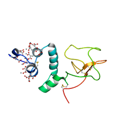
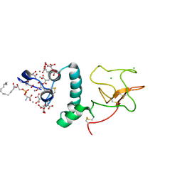
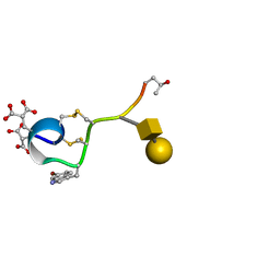 | | A NOVEL CONOTOXIN FROM CONUS TEXTILE WITH UNUSUAL POST-TRANSLATIONAL MODIFICATIONS REDUCES PRESYNAPTIC CALCIUM INFLUX, NMR, 1 STRUCTURE, GLYCOSYLATED PROTEIN | | 分子名称: | OMEGAC-TXIX, beta-D-galactopyranose-(1-3)-2-acetamido-2-deoxy-beta-D-galactopyranose | | 著者 | Rigby, A.C, Hambe, B, Czerwiec, E, Baleja, J.D, Furie, B.C, Furie, B, Stenflo, J. | | 登録日 | 1998-12-18 | | 公開日 | 1999-06-08 | | 最終更新日 | 2025-03-26 | | 実験手法 | SOLUTION NMR | | 主引用文献 | A conotoxin from Conus textile with unusual posttranslational modifications reduces presynaptic Ca2+ influx.
Proc.Natl.Acad.Sci.USA, 96, 1999
|
|
1AWY
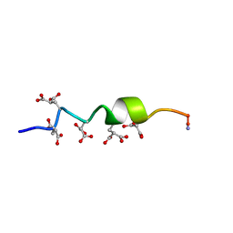 | | NMR STRUCTURE OF CALCIUM BOUND CONFORMER OF CONANTOKIN G, MINIMIZED AVERAGE STRUCTURE | | 分子名称: | CONANTOXIN G | | 著者 | Rigby, A.C, Baleja, J.D, Leping, L, Pedersen, L.G, Furie, B.C, Furie, B. | | 登録日 | 1997-10-06 | | 公開日 | 1998-04-08 | | 最終更新日 | 2022-02-16 | | 実験手法 | SOLUTION NMR | | 主引用文献 | Role of gamma-carboxyglutamic acid in the calcium-induced structural transition of conantokin G, a conotoxin from the marine snail Conus geographus.
Biochemistry, 36, 1997
|
|
1AD7
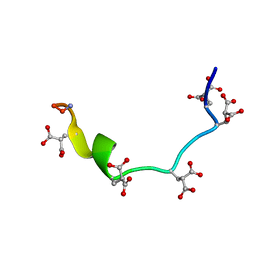 | | NMR STRUCTURE OF METAL-FREE CONANTOKIN G, 1 STRUCTURE | | 分子名称: | CONANTOXIN G | | 著者 | Rigby, A.C, Baleja, J.D, Furie, B.C, Furie, B. | | 登録日 | 1997-02-21 | | 公開日 | 1997-08-20 | | 最終更新日 | 2024-06-05 | | 実験手法 | SOLUTION NMR | | 主引用文献 | Three-dimensional structure of a gamma-carboxyglutamic acid-containing conotoxin, conantokin G, from the marine snail Conus geographus: the metal-free conformer.
Biochemistry, 36, 1997
|
|
1ZXA
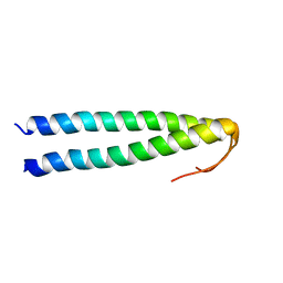
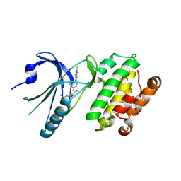
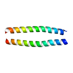 | | Solution Structure of the Coiled-Coil Domain of cGMP-dependent Protein Kinase Ia | | 分子名称: | cGMP-dependent protein kinase 1, alpha isozyme | | 著者 | Schnell, J.R, Zhou, G.P, Zweckstetter, M, Rigby, A.C, Chou, J.J. | | 登録日 | 2005-06-07 | | 公開日 | 2005-09-13 | | 最終更新日 | 2024-05-22 | | 実験手法 | SOLUTION NMR | | 主引用文献 | Rapid and accurate structure determination of coiled-coil domains using NMR dipolar couplings: Application to cGMP-dependent protein kinase I{alpha}
Protein Sci., 14, 2005
|
|
5HUZ
 | | Solution structure of coiled coil domain of myosin binding subunit of myosin light chain phosphatase | | 分子名称: | Protein phosphatase 1 regulatory subunit 12A | | 著者 | Sharma, A.K, Birrane, G, Anklin, C, Rigby, A.C, Pollak, M, Alper, S.L. | | 登録日 | 2016-01-27 | | 公開日 | 2016-03-02 | | 最終更新日 | 2024-05-15 | | 実験手法 | SOLUTION NMR | | 主引用文献 | To be published
To Be Published
|
|
2KLN
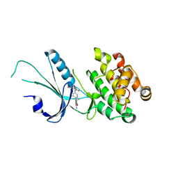
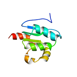 | | Solution Structure of STAS domain of RV1739c from M. tuberculosis | | 分子名称: | PROBABLE SULPHATE-TRANSPORT TRANSMEMBRANE PROTEIN, COG0659 | | 著者 | Sharma, A.K, Ye, L, Zolotarev, A.S, Alper, S.L, Rigby, A.C. | | 登録日 | 2009-07-06 | | 公開日 | 2010-12-15 | | 最終更新日 | 2024-05-01 | | 実験手法 | SOLUTION NMR | | 主引用文献 | Solution Structure of the Guanine Nucleotide-binding STAS Domain of SLC26-related SulP Protein Rv1739c from Mycobacterium tuberculosis.
J.Biol.Chem., 286, 2011
|
|
2ES3
 | |
8EQ9
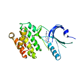 | | Co-crystal structure of PERK with compound 11 | | 分子名称: | (2R)-N-[(4M)-4-(4-amino-7-methyl-7H-pyrrolo[2,3-d]pyrimidin-5-yl)-3-methylphenyl]-2-(3-fluorophenyl)-2-hydroxyacetamide, Eukaryotic translation initiation factor 2-alpha kinase 3 | | 著者 | Zhu, G, Surman, M.D, Mulvihill, M.J. | | 登録日 | 2022-10-07 | | 公開日 | 2022-11-30 | | 最終更新日 | 2023-10-25 | | 実験手法 | X-RAY DIFFRACTION (2.86 Å) | | 主引用文献 | Optimization of a Novel Mandelamide-Derived Pyrrolopyrimidine Series of PERK Inhibitors.
Pharmaceutics, 14, 2022
|
|
8EQD
 | | Co-crystal structure of PERK with compound 24 | | 分子名称: | (2R)-N-[(4M)-4-(4-amino-2,7-dimethyl-7H-pyrrolo[2,3-d]pyrimidin-5-yl)-3-methylphenyl]-2-(3-fluorophenyl)-2-hydroxyacetamide, Eukaryotic translation initiation factor 2-alpha kinase 3 | | 著者 | Zhu, G, Surman, M.D, Mulvihill, M.J. | | 登録日 | 2022-10-07 | | 公開日 | 2022-11-30 | | 最終更新日 | 2023-10-25 | | 実験手法 | X-RAY DIFFRACTION (2.92 Å) | | 主引用文献 | Optimization of a Novel Mandelamide-Derived Pyrrolopyrimidine Series of PERK Inhibitors.
Pharmaceutics, 14, 2022
|
|
8EQE
 | | Co-crystal structure of PERK with compound 26 | | 分子名称: | (2R)-N-[(4M)-4-(4-amino-2,7-dimethyl-7H-pyrrolo[2,3-d]pyrimidin-5-yl)-3-methylphenyl]-2-hydroxy-2-[3-(trifluoromethyl)phenyl]acetamide, Eukaryotic translation initiation factor 2-alpha kinase 3 | | 著者 | Zhu, G, Surman, M.D, Mulvihill, M.J. | | 登録日 | 2022-10-07 | | 公開日 | 2022-11-30 | | 最終更新日 | 2023-10-25 | | 実験手法 | X-RAY DIFFRACTION (2.559 Å) | | 主引用文献 | Optimization of a Novel Mandelamide-Derived Pyrrolopyrimidine Series of PERK Inhibitors.
Pharmaceutics, 14, 2022
|
|
2OUH
 | | Crystal structure of the Thrombospondin-1 N-terminal domain in complex with fractionated Heparin DP10 | | 分子名称: | SULFATE ION, Thrombospondin-1 | | 著者 | Tan, K, Joachimiak, A, Wang, J, Lawler, J. | | 登録日 | 2007-02-11 | | 公開日 | 2008-01-08 | | 最終更新日 | 2024-10-09 | | 実験手法 | X-RAY DIFFRACTION (2.4 Å) | | 主引用文献 | Heparin-induced cis- and trans-Dimerization Modes of the Thrombospondin-1 N-terminal Domain.
J.Biol.Chem., 283, 2008
|
|
2OUJ
 | |
7MF0
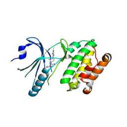
 | | Co-crystal structure of PERK with inhibitor (R)-2-amino-N-cyclopropyl-5-(4-(2-(3,5-difluorophenyl)-2-hydroxyacetamido)-2-methylphenyl)nicotinamide | | 分子名称: | 2-amino-N-cyclopropyl-5-(4-{[(2R)-2-(3,5-difluorophenyl)-2-hydroxyacetyl]amino}-2-methylphenyl)pyridine-3-carboxamide, Eukaryotic translation initiation factor 2-alpha kinase 3,Eukaryotic translation initiation factor 2-alpha kinase 3 | | 著者 | Wiens, B, Koszelak-Rosenblum, M, Surman, M.D, Zhu, G, Mulvihill, M.J. | | 登録日 | 2021-04-08 | | 公開日 | 2021-05-19 | | 最終更新日 | 2023-10-18 | | 実験手法 | X-RAY DIFFRACTION (2.809 Å) | | 主引用文献 | Discovery of 2-amino-3-amido-5-aryl-pyridines as highly potent, orally bioavailable, and efficacious PERK kinase inhibitors.
Bioorg.Med.Chem.Lett., 43, 2021
|
|
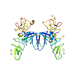
1NL1
 | | BOVINE PROTHROMBIN FRAGMENT 1 IN COMPLEX WITH CALCIUM ION | | 分子名称: | 2-acetamido-2-deoxy-beta-D-glucopyranose, 2-acetamido-2-deoxy-beta-D-glucopyranose-(1-4)-2-acetamido-2-deoxy-beta-D-glucopyranose, CALCIUM ION, ... | | 著者 | Huang, M, Huang, G, Furie, B, Seaton, B, Furie, B.C. | | 登録日 | 2003-01-06 | | 公開日 | 2003-09-16 | | 最終更新日 | 2023-11-15 | | 実験手法 | X-RAY DIFFRACTION (1.9 Å) | | 主引用文献 | Structural basis of membrane binding by Gla domains of vitamin K-dependent proteins.
Nat.Struct.Biol., 10, 2003
|
|
1NL2
 | | BOVINE PROTHROMBIN FRAGMENT 1 IN COMPLEX WITH CALCIUM AND LYSOPHOSPHOTIDYLSERINE | | 分子名称: | 2-acetamido-2-deoxy-beta-D-glucopyranose, 2-acetamido-2-deoxy-beta-D-glucopyranose-(1-4)-2-acetamido-2-deoxy-beta-D-glucopyranose, CALCIUM ION, ... | | 著者 | Huang, M, Huang, G, Furie, B, Seaton, B, Furie, B.C. | | 登録日 | 2003-01-06 | | 公開日 | 2003-09-16 | | 最終更新日 | 2023-11-15 | | 実験手法 | X-RAY DIFFRACTION (2.3 Å) | | 主引用文献 | Structural basis of membrane binding by Gla domains of vitamin K-dependent proteins.
Nat.Struct.Biol., 10, 2003
|
|
3SP8
 | | Crystal structure of NK2 in complex with fractionated Heparin DP10 | | 分子名称: | (4R)-2-METHYLPENTANE-2,4-DIOL, (4S)-2-METHYL-2,4-PENTANEDIOL, 2-(N-MORPHOLINO)-ETHANESULFONIC ACID, ... | | 著者 | Recacha, R, Mulloy, B, Gherardi, E. | | 登録日 | 2011-07-01 | | 公開日 | 2012-07-04 | | 最終更新日 | 2024-11-27 | | 実験手法 | X-RAY DIFFRACTION (1.86 Å) | | 主引用文献 | Crystal structure of NK2 in complex with fractionated Heparin DP10
TO BE PUBLISHED
|
|
4O03
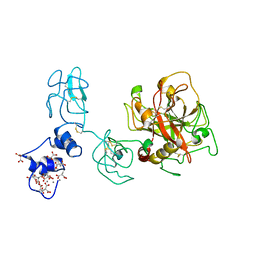 | | Crystal structure of Ca2+ bound prothrombin deletion mutant residues 146-167 | | 分子名称: | 2-acetamido-2-deoxy-beta-D-glucopyranose, CALCIUM ION, Prothrombin | | 著者 | Pozzi, N, Chen, Z, Shropshire, D.B, Pelc, L.A, Di Cera, E. | | 登録日 | 2013-12-13 | | 公開日 | 2014-05-21 | | 最終更新日 | 2023-12-06 | | 実験手法 | X-RAY DIFFRACTION (3.38 Å) | | 主引用文献 | The linker connecting the two kringles plays a key role in prothrombin activation.
Proc.Natl.Acad.Sci.USA, 111, 2014
|
|


