4LZ4
 
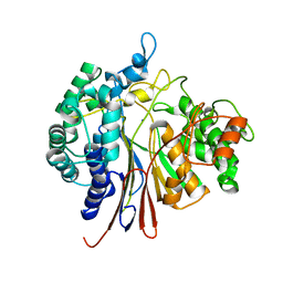
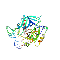
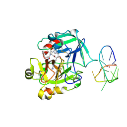 | | X-ray structure of the complex between human thrombin and the TBA deletion mutant lacking thymine 3 nucleobase | | 分子名称: | 2-acetamido-2-deoxy-beta-D-glucopyranose, D-phenylalanyl-N-[(2S,3S)-6-{[amino(iminio)methyl]amino}-1-chloro-2-hydroxyhexan-3-yl]-L-prolinamide, POTASSIUM ION, ... | | 著者 | Pica, A, Russo Krauss, I, Merlino, A, Sica, F. | | 登録日 | 2013-07-31 | | 公開日 | 2014-01-08 | | 最終更新日 | 2020-07-29 | | 実験手法 | X-RAY DIFFRACTION (2.56 Å) | | 主引用文献 | Dissecting the contribution of thrombin exosite I in the recognition of thrombin binding aptamer.
Febs J., 280, 2013
|
|
4LZ1
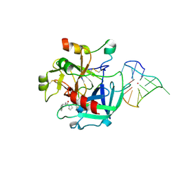 | | X-ray structure of the complex between human thrombin and the TBA deletion mutant lacking thymine 12 nucleobase | | 分子名称: | 2-acetamido-2-deoxy-beta-D-glucopyranose, D-phenylalanyl-N-[(2S,3S)-6-{[amino(iminio)methyl]amino}-1-chloro-2-hydroxyhexan-3-yl]-L-prolinamide, POTASSIUM ION, ... | | 著者 | Pica, A, Russo Krauss, I, Merlino, A, Sica, F. | | 登録日 | 2013-07-31 | | 公開日 | 2014-01-08 | | 最終更新日 | 2020-07-29 | | 実験手法 | X-RAY DIFFRACTION (1.65 Å) | | 主引用文献 | Dissecting the contribution of thrombin exosite I in the recognition of thrombin binding aptamer.
Febs J., 280, 2013
|
|
5EW1
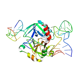 | | Human thrombin sandwiched between two DNA aptamers: HD22 and HD1-deltaT3 | | 分子名称: | 2-acetamido-2-deoxy-beta-D-glucopyranose, D-phenylalanyl-N-[(2S,3S)-6-{[amino(iminio)methyl]amino}-1-chloro-2-hydroxyhexan-3-yl]-L-prolinamide, HD1-deltaT3, ... | | 著者 | Pica, A, Russo Krauss, I, Parente, V, Sica, F. | | 登録日 | 2015-11-20 | | 公開日 | 2016-11-30 | | 最終更新日 | 2024-01-10 | | 実験手法 | X-RAY DIFFRACTION (2.95 Å) | | 主引用文献 | Through-bond effects in the ternary complexes of thrombin sandwiched by two DNA aptamers.
Nucleic Acids Res., 45, 2017
|
|
5EW2
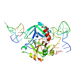 | | Human thrombin sandwiched between two DNA aptamers: HD22 and HD1-deltaT12 | | 分子名称: | 2-acetamido-2-deoxy-beta-D-glucopyranose, D-phenylalanyl-N-[(2S,3S)-6-{[amino(iminio)methyl]amino}-1-chloro-2-hydroxyhexan-3-yl]-L-prolinamide, HD1-deltaT12, ... | | 著者 | Pica, A, Russo Krauss, I, Parente, V, Sica, F. | | 登録日 | 2015-11-20 | | 公開日 | 2016-11-30 | | 最終更新日 | 2020-07-29 | | 実験手法 | X-RAY DIFFRACTION (3.59 Å) | | 主引用文献 | Through-bond effects in the ternary complexes of thrombin sandwiched by two DNA aptamers.
Nucleic Acids Res., 45, 2017
|
|
4KXH
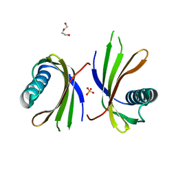
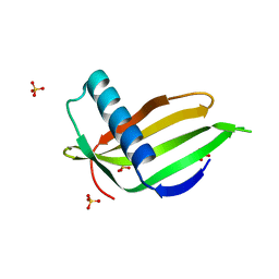
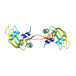
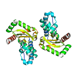
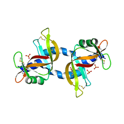 | | The X-ray crystal structure of a dimeric variant of human pancreatic ribonuclease | | 分子名称: | CHLORIDE ION, Ribonuclease pancreatic, SODIUM ION, ... | | 著者 | Pica, A, Merlino, A, Mazzarella, L, Sica, F. | | 登録日 | 2013-05-26 | | 公開日 | 2013-10-02 | | 最終更新日 | 2017-11-15 | | 実験手法 | X-RAY DIFFRACTION (2.7 Å) | | 主引用文献 | Three-dimensional domain swapping and supramolecular protein assembly: insights from the X-ray structure of a dimeric swapped variant of human pancreatic RNase.
Acta Crystallogr.,Sect.D, 69, 2013
|
|
4Y23
 | |
5O7K
 | |
5O7L
 | |
5O7S
 | |
5O7R
 | |
5O7Q
 | |
5LC7
 | |
5LC6
 | |
6HJT
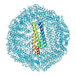 | | The X-ray structure of the horse spleen ferritin nanocage containing Pt, obtained upon encapsulation of a Pt(II) terpyridine compound within the protein cage | | 分子名称: | CADMIUM ION, CHLORIDE ION, DIMETHYL SULFOXIDE, ... | | 著者 | Pica, A, Ferraro, G, Merlino, A. | | 登録日 | 2018-09-04 | | 公開日 | 2018-12-19 | | 最終更新日 | 2024-05-15 | | 実験手法 | X-RAY DIFFRACTION (1.33 Å) | | 主引用文献 | Preparation, structure, cytotoxicity and mechanism of action of ferritin-Pt(II) terpyridine compound nanocomposites.
Nanomedicine (Lond), 13, 2018
|
|
6HJU
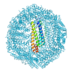 | | The X-ray structure of the horse spleen ferritin nanocage containing Pt, obtained upon encapsulation of a Pt(II) terpyridine compound within the protein cage | | 分子名称: | CADMIUM ION, CHLORIDE ION, DIMETHYL SULFOXIDE, ... | | 著者 | Pica, A, Ferraro, G, Merlino, A. | | 登録日 | 2018-09-04 | | 公開日 | 2018-12-19 | | 最終更新日 | 2024-05-15 | | 実験手法 | X-RAY DIFFRACTION (1.58 Å) | | 主引用文献 | Preparation, structure, cytotoxicity and mechanism of action of ferritin-Pt(II) terpyridine compound nanocomposites.
Nanomedicine (Lond), 13, 2018
|
|
6GXJ
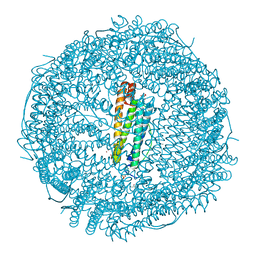 | | X-ray structure of DiRu-1-encapsulated Apoferritin | | 分子名称: | CADMIUM ION, CHLORIDE ION, Ferritin light chain, ... | | 著者 | Pica, A, Ferraro, G, Merlino, A. | | 登録日 | 2018-06-27 | | 公開日 | 2019-02-06 | | 最終更新日 | 2024-01-17 | | 実験手法 | X-RAY DIFFRACTION (1.43 Å) | | 主引用文献 | Encapsulation of the Dinuclear Trithiolato-Bridged Arene Ruthenium Complex Diruthenium-1 in an Apoferritin Nanocage: Structure and Cytotoxicity.
ChemMedChem, 14, 2019
|
|
4I7Y
 | | Crystal Structure of Human Alpha Thrombin in Complex with a 27-mer Aptamer Bound to Exosite II | | 分子名称: | 2-acetamido-2-deoxy-beta-D-glucopyranose, D-phenylalanyl-N-[(2S,3S)-6-{[amino(iminio)methyl]amino}-1-chloro-2-hydroxyhexan-3-yl]-L-prolinamide, DNA (27-MER), ... | | 著者 | Pica, A, Russo Krauss, I, Merlino, A, Mazzarella, L, Sica, F. | | 登録日 | 2012-12-01 | | 公開日 | 2013-10-16 | | 最終更新日 | 2024-10-09 | | 実験手法 | X-RAY DIFFRACTION (2.4 Å) | | 主引用文献 | Duplex-quadruplex motifs in a peculiar structural organization cooperatively contribute to thrombin binding of a DNA aptamer.
Acta Crystallogr.,Sect.D, 69, 2013
|
|
4N4C
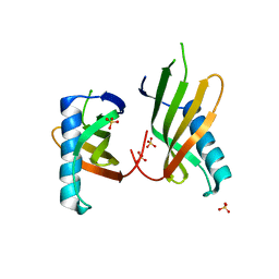
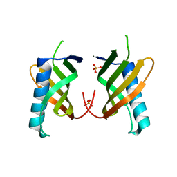
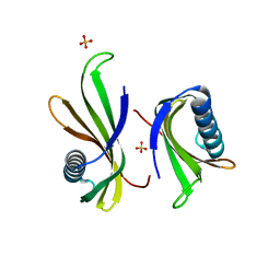
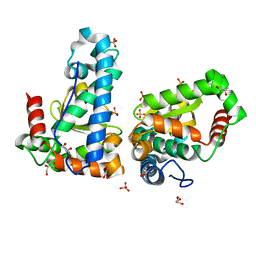
 | | Crystal structure of the C-terminal swapped dimer of a Bovine seminal ribonuclease mutant | | 分子名称: | PHOSPHATE ION, Seminal ribonuclease | | 著者 | Pica, A, Russo Krauss, I, Merlino, A, Sica, F. | | 登録日 | 2013-10-08 | | 公開日 | 2013-11-06 | | 最終更新日 | 2023-09-20 | | 実験手法 | X-RAY DIFFRACTION (2.48 Å) | | 主引用文献 | The multiple forms of bovine seminal ribonuclease: Structure and stability of a C-terminal swapped dimer.
Febs Lett., 587, 2013
|
|
6ZJF
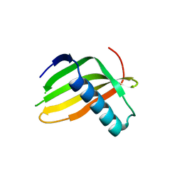
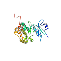 | | Crystal structure of STK17B (DRAK2) in complex with AP-229 | | 分子名称: | 1,2-ETHANEDIOL, 2-[6-(4-cyclopropylphenyl)thieno[3,2-d]pyrimidin-4-yl]sulfanylethanoic acid, Serine/threonine-protein kinase 17B | | 著者 | Chaikuad, A, Picado, A, Willson, T, Knapp, S, Structural Genomics Consortium (SGC) | | 登録日 | 2020-06-28 | | 公開日 | 2020-07-29 | | 最終更新日 | 2024-01-31 | | 実験手法 | X-RAY DIFFRACTION (1.75 Å) | | 主引用文献 | A Chemical Probe for Dark Kinase STK17B Derives Its Potency and High Selectivity through a Unique P-Loop Conformation.
J.Med.Chem., 63, 2020
|
|
4G51
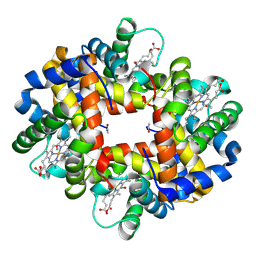 | | Crystallographic analysis of the interaction of nitric oxide with hemoglobin from Trematomus bernacchii in the T quaternary structure (fully ligated state). | | 分子名称: | Hemoglobin subunit alpha, Hemoglobin subunit beta, NITRIC OXIDE, ... | | 著者 | Merlino, A, Balsamo, A, Pica, A, Mazzarella, L, Vergara, A. | | 登録日 | 2012-07-17 | | 公開日 | 2013-01-16 | | 最終更新日 | 2019-02-20 | | 実験手法 | X-RAY DIFFRACTION (2.5 Å) | | 主引用文献 | Selective X-ray-induced NO photodissociation in haemoglobin crystals: evidence from a Raman-assisted crystallographic study.
Acta Crystallogr.,Sect.D, 69, 2013
|
|
4YIP
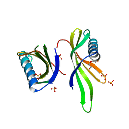
 | | X-ray structure of the iron/manganese cambialistic superoxide dismutase from Streptococcus mutans | | 分子名称: | FE (III) ION, Superoxide dismutase [Mn/Fe] | | 著者 | Russo Krauss, I, Merlino, A, Pica, A, Sica, F. | | 登録日 | 2015-03-02 | | 公開日 | 2016-01-13 | | 最終更新日 | 2024-01-10 | | 実験手法 | X-RAY DIFFRACTION (2.15 Å) | | 主引用文献 | Fine tuning of metal-specific activity in the Mn-like group of cambialistic superoxide dismutases
Rsc Adv, 5, 2015
|
|
4YIO
 | | X-ray structure of the iron/manganese cambialistic superoxide dismutase from Streptococcus thermophilus | | 分子名称: | FE (III) ION, GLYCEROL, SULFATE ION, ... | | 著者 | Russo Krauss, I, Merlino, A, Pica, A, Sica, F. | | 登録日 | 2015-03-02 | | 公開日 | 2016-01-13 | | 最終更新日 | 2024-01-10 | | 実験手法 | X-RAY DIFFRACTION (1.6 Å) | | 主引用文献 | Fine tuning of metal-specific activity in the Mn-like group of cambialistic superoxide dismutases
Rsc Adv, 5, 2015
|
|
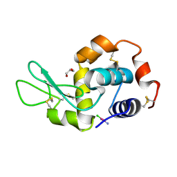
5FCP
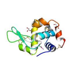 | | X-RAY STRUCTURE OF THE ADDUCT BETWEEN HEN EGG WHITE LYSOZYME AND CISPLATIN AT LONG INCUBATION TIMES | | 分子名称: | CHLORIDE ION, Cisplatin, GLYCEROL, ... | | 著者 | Russo Krauss, I, Ferraro, G, Pica, A, Merlino, A. | | 登録日 | 2015-12-15 | | 公開日 | 2016-04-13 | | 最終更新日 | 2024-01-10 | | 実験手法 | X-RAY DIFFRACTION (1.55 Å) | | 主引用文献 | Effect of temperature on the interaction of cisplatin with the model protein hen egg white lysozyme.
J.Biol.Inorg.Chem., 21, 2016
|
|
5F9X
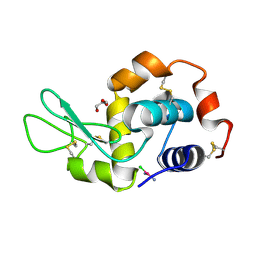 | | X-RAY STRUCTURE OF THE ADDUCT BETWEEN HEN EGG WHITE LYSOZYME AND CISPLATIN UPON 24 HOURS OF INCUBATION AT 55 DEGREES | | 分子名称: | Cisplatin, GLYCEROL, Lysozyme C | | 著者 | Russo Krauss, I, Ferraro, G, Pica, A, Merlino, A. | | 登録日 | 2015-12-10 | | 公開日 | 2016-04-13 | | 最終更新日 | 2024-01-10 | | 実験手法 | X-RAY DIFFRACTION (1.94 Å) | | 主引用文献 | Effect of temperature on the interaction of cisplatin with the model protein hen egg white lysozyme.
J.Biol.Inorg.Chem., 21, 2016
|
|
5F9U
 | | X-RAY STRUCTURE OF THE ADDUCT BETWEEN HEN EGG WHITE LYSOZYME AND CISPLATIN UPON 24 HOURS OF INCUBATION AT 20 DEGREES | | 分子名称: | Cisplatin, GLYCEROL, Lysozyme C | | 著者 | Russo Krauss, I, Ferraro, G, Pica, A, Merlino, A. | | 登録日 | 2015-12-10 | | 公開日 | 2016-04-13 | | 最終更新日 | 2024-01-10 | | 実験手法 | X-RAY DIFFRACTION (1.85 Å) | | 主引用文献 | Effect of temperature on the interaction of cisplatin with the model protein hen egg white lysozyme.
J.Biol.Inorg.Chem., 21, 2016
|
|


