2M57
 
 | |
1TR8
 | |
4V6T
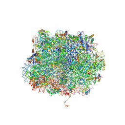
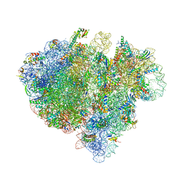 | | Structure of the bacterial ribosome complexed by tmRNA-SmpB and EF-G during translocation and MLD-loading | | 分子名称: | 16S ribosomal RNA, 23S ribosomal RNA, 30S ribosomal protein S10, ... | | 著者 | Ramrath, D.J.F, Yamamoto, H, Rother, K, Wittek, D, Pech, M, Mielke, T, Loerke, J, Scheerer, P, Ivanov, P, Teraoka, Y, Shpanchenko, O, Nierhaus, K.H, Spahn, C.M.T. | | 登録日 | 2012-01-27 | | 公開日 | 2014-07-09 | | 最終更新日 | 2024-02-28 | | 実験手法 | ELECTRON MICROSCOPY (8.3 Å) | | 主引用文献 | The complex of tmRNA-SmpB and EF-G on translocating ribosomes.
Nature, 485, 2012
|
|
5MC6
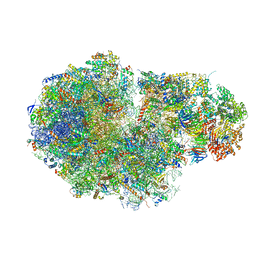 | | Cryo-EM structure of a native ribosome-Ski2-Ski3-Ski8 complex from S. cerevisiae | | 分子名称: | 18S ribosomal RNA, 25S ribosomal RNA, 40S ribosomal protein S0-A, ... | | 著者 | Schmidt, C, Kowalinski, E, Shanmuganathan, V, Defenouillere, Q, Braunger, K, Heuer, A, Pech, M, Namane, A, Berninghausen, O, Fromont-Racine, M, Jacquier, A, Conti, E, Becker, T, Beckmann, R. | | 登録日 | 2016-11-09 | | 公開日 | 2017-01-18 | | 最終更新日 | 2025-07-09 | | 実験手法 | ELECTRON MICROSCOPY (3.8 Å) | | 主引用文献 | The cryo-EM structure of a ribosome-Ski2-Ski3-Ski8 helicase complex.
Science, 354, 2016
|
|
5A6U
 | | Native mammalian ribosome-bound Sec61 protein-conducting channel in the 'non-inserting' state | | 分子名称: | SEC61A, SEC61B, SEC61G | | 著者 | Pfeffer, S, Burbaum, L, Unverdorben, P, Pech, M, Chen, Y, Zimmermann, R, Beckmann, R, Foerster, F. | | 登録日 | 2015-07-01 | | 公開日 | 2015-10-07 | | 最終更新日 | 2024-05-08 | | 実験手法 | ELECTRON MICROSCOPY (9 Å) | | 主引用文献 | Structure of the Native Sec61 Protein-Conducting Channel.
Nat.Commun., 6, 2015
|
|
2YLF
 | |
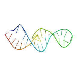
2LU0
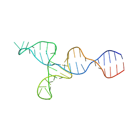 | | NMR solution structure of the kappa-zeta region of S.cerevisiae group II intron ai5(gamma) | | 分子名称: | RNA (49-MER) | | 著者 | Donghi, D, Pechlaner, M, Finazzo, C, Knobloch, B, Sigel, R.K.O. | | 登録日 | 2012-06-05 | | 公開日 | 2013-04-10 | | 最終更新日 | 2024-05-15 | | 実験手法 | SOLUTION NMR | | 主引用文献 | The structural stabilization of the kappa three-way junction by Mg(II) represents the first step in the folding of a group II intron.
Nucleic Acids Res., 41, 2013
|
|
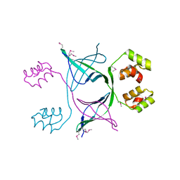
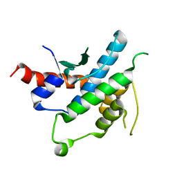
2YLE
 | | Crystal structure of the human Spir-1 KIND FSI domain in complex with the FSI peptide | | 分子名称: | FORMIN-2, PROTEIN SPIRE HOMOLOG 1 | | 著者 | Zeth, K, Pechlivanis, M, Vonrhein, C, Kerkhoff, E. | | 登録日 | 2011-06-01 | | 公開日 | 2011-06-08 | | 最終更新日 | 2024-05-08 | | 実験手法 | X-RAY DIFFRACTION (1.8 Å) | | 主引用文献 | Molecular Basis of Actin Nucleation Factor Cooperativity: Crystal Structure of the Spir-1 Kinase Non-Catalytic C-Lobe Domain (Kind)Formin-2 Formin Spir Interaction Motif (Fsi) Complex.
J.Biol.Chem., 286, 2011
|
|
5GAK
 | | Yeast 60S ribosomal subunit with A-site tRNA, P-site tRNA and eIF-5A | | 分子名称: | 25S rRNA, 4-{(2R)-2-[(1S,3S,5S)-3,5-dimethyl-2-oxocyclohexyl]-2-hydroxyethyl}piperidine-2,6-dione, 5.8S rRNA, ... | | 著者 | Schmidt, C, Becker, T. | | 登録日 | 2015-12-09 | | 公開日 | 2016-02-24 | | 最終更新日 | 2025-07-02 | | 実験手法 | ELECTRON MICROSCOPY (3.88 Å) | | 主引用文献 | Structure of the hypusinylated eukaryotic translation factor eIF-5A bound to the ribosome.
Nucleic Acids Res., 44, 2016
|
|
4FXL
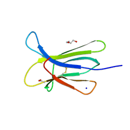 | | Crystal structure of the D76N Beta-2 Microglobulin mutant | | 分子名称: | ACETATE ION, Beta-2-microglobulin, DI(HYDROXYETHYL)ETHER, ... | | 著者 | Ricagno, S, Bellotti, V, Pepys, M.B, Stoppini, M, Bolognesi, M. | | 登録日 | 2012-07-03 | | 公開日 | 2012-08-15 | | 最終更新日 | 2024-11-27 | | 実験手法 | X-RAY DIFFRACTION (1.4 Å) | | 主引用文献 | Hereditary systemic amyloidosis due to Asp76Asn variant beta-2-microglobulin.
N.Engl.J.Med., 366, 2012
|
|
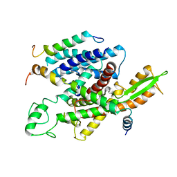
3FRI
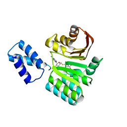 | | Structure of the 16S rRNA methylase RmtB, I222 | | 分子名称: | 16S rRNA methylase, S-ADENOSYL-L-HOMOCYSTEINE | | 著者 | Schmitt, E, Galimand, M, Panvert, M, Dupechez, M, Courvalin, P, Mechulam, Y. | | 登録日 | 2009-01-08 | | 公開日 | 2009-08-11 | | 最終更新日 | 2023-11-01 | | 実験手法 | X-RAY DIFFRACTION (1.8 Å) | | 主引用文献 | Structural bases for 16 S rRNA methylation catalyzed by ArmA and RmtB methyltransferases
J.Mol.Biol., 388, 2009
|
|
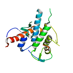
3FRH
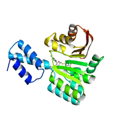 | | Structure of the 16S rRNA methylase RmtB, P21 | | 分子名称: | 16S rRNA methylase, S-ADENOSYL-L-HOMOCYSTEINE | | 著者 | Schmitt, E, Galimand, M, Panvert, M, Dupechez, M, Courvalin, P, Mechulam, Y. | | 登録日 | 2009-01-08 | | 公開日 | 2009-08-11 | | 最終更新日 | 2023-11-01 | | 実験手法 | X-RAY DIFFRACTION (1.2 Å) | | 主引用文献 | Structural bases for 16 S rRNA methylation catalyzed by ArmA and RmtB methyltransferases
J.Mol.Biol., 388, 2009
|
|


