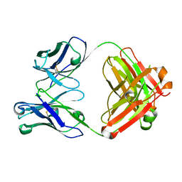
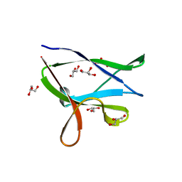
1PRE
 
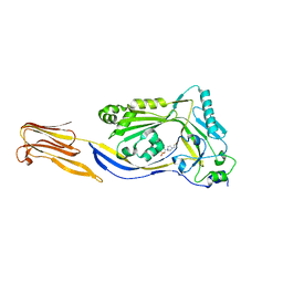
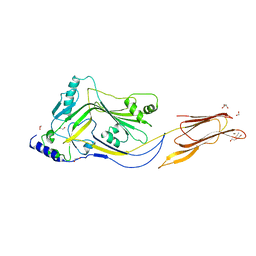
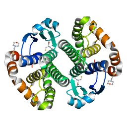
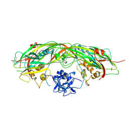 | | PROAEROLYSIN | | 分子名称: | PROAEROLYSIN | | 著者 | Parker, M.W, Buckley, J.T, Postma, J.P.M, Tucker, A.D, Tsernoglou, D. | | 登録日 | 1995-09-15 | | 公開日 | 1996-10-14 | | 最終更新日 | 2011-07-13 | | 実験手法 | X-RAY DIFFRACTION (2.8 Å) | | 主引用文献 | Structure of the Aeromonas toxin proaerolysin in its water-soluble and membrane-channel states.
Nature, 367, 1994
|
|
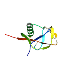
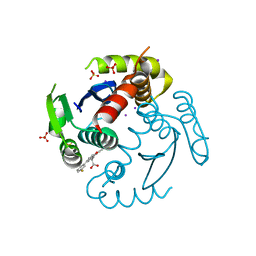
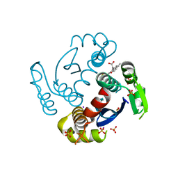
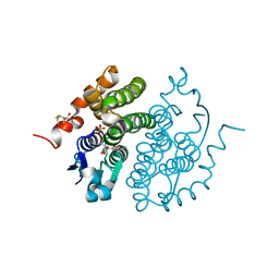
1COL
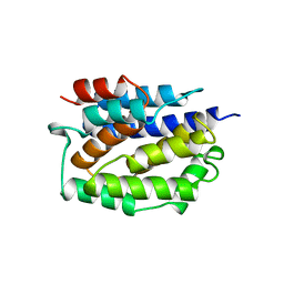 | | REFINED STRUCTURE OF THE PORE-FORMING DOMAIN OF COLICIN A AT 2.4 ANGSTROMS RESOLUTION | | 分子名称: | COLICIN A | | 著者 | Parker, M.W, Postma, J.P.M, Pattus, F, Tucker, A.D, Tsernoglou, D. | | 登録日 | 1991-07-06 | | 公開日 | 1992-07-15 | | 最終更新日 | 2024-02-07 | | 実験手法 | X-RAY DIFFRACTION (2.4 Å) | | 主引用文献 | Refined structure of the pore-forming domain of colicin A at 2.4 A resolution.
J.Mol.Biol., 224, 1992
|
|
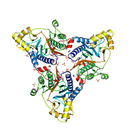
1Z52
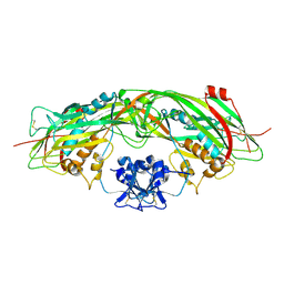 | | Proaerolysin Mutant W373L | | 分子名称: | Aerolysin | | 著者 | Parker, M.W, Feil, S.C, Tang, J.W. | | 登録日 | 2005-03-16 | | 公開日 | 2006-03-07 | | 最終更新日 | 2021-11-10 | | 実験手法 | X-RAY DIFFRACTION (2.38 Å) | | 主引用文献 | Crystal Structure of Proaerolysin at 2.3 A Resolution and Structural Analyses of Single-site Mutants as a Basis for Understanding Membrane Insertion of the Toxin
To be Published
|
|
5DHL
 | |
5DIM
 | |
4QDQ
 | |
4QDR
 | |
4QDS
 | |
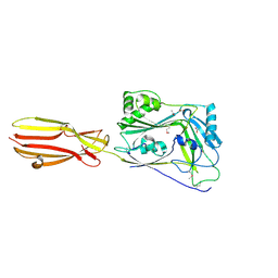
4NKQ
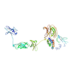 | | Structure of a Cytokine Receptor Complex | | 分子名称: | 2-acetamido-2-deoxy-beta-D-glucopyranose, Cytokine receptor common subunit beta, Granulocyte-macrophage colony-stimulating factor, ... | | 著者 | Parker, M.W, Broughton, S.E. | | 登録日 | 2013-11-13 | | 公開日 | 2015-09-23 | | 最終更新日 | 2020-07-29 | | 実験手法 | X-RAY DIFFRACTION (3.301 Å) | | 主引用文献 | Conformational Changes in the GM-CSF Receptor Suggest a Molecular Mechanism for Affinity Conversion and Receptor Signaling.
Structure, 24, 2016
|
|
4ZGH
 | | Structure of Sugar Binding Protein Pneumolysin | | 分子名称: | 1,2-ETHANEDIOL, DI(HYDROXYETHYL)ETHER, GOLD (I) CYANIDE ION, ... | | 著者 | Parker, M.W, Feil, S.C, Morton, C. | | 登録日 | 2015-04-23 | | 公開日 | 2016-03-09 | | 最終更新日 | 2023-09-27 | | 実験手法 | X-RAY DIFFRACTION (2.9 Å) | | 主引用文献 | Crystal structure of Streptococcus pneumoniae pneumolysin provides key insights into early steps of pore formation.
Sci Rep, 5, 2015
|
|
7SIA
 | |
1FHE
 | |
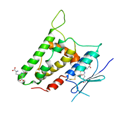
1MD3
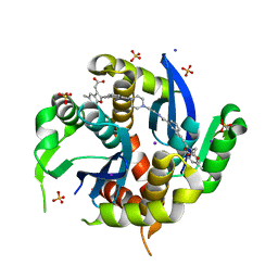
 | | A folding mutant of human class pi glutathione transferase, created by mutating glycine 146 of the wild-type protein to alanine | | 分子名称: | 2-(N-MORPHOLINO)-ETHANESULFONIC ACID, GLUTATHIONE, pi glutathione transferase | | 著者 | Kong, G.K.-W, Dragani, B, Aceto, A, Cocco, R, Mannervik, B, Stenberg, G, McKinstry, W.J, Polekhina, G, Parker, M.W. | | 登録日 | 2002-08-06 | | 公開日 | 2002-08-21 | | 最終更新日 | 2023-10-25 | | 実験手法 | X-RAY DIFFRACTION (2.03 Å) | | 主引用文献 | Contribution of Glycine 146 to a Conserved Folding Module Affecting Stability and Refolding of Human Glutathione Transferase P1-1
J.Biol.Chem., 278, 2003
|
|
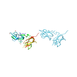
1MWP
 | | N-TERMINAL DOMAIN OF THE AMYLOID PRECURSOR PROTEIN | | 分子名称: | AMYLOID A4 PROTEIN | | 著者 | Rossjohn, J, Cappai, R, Feil, S.C, Henry, A, McKinstry, W.J, Galatis, D, Hesse, L, Multhaup, G, Beyreuther, K, Masters, C.L, Parker, M.W. | | 登録日 | 1999-03-09 | | 公開日 | 2000-03-15 | | 最終更新日 | 2023-12-27 | | 実験手法 | X-RAY DIFFRACTION (1.8 Å) | | 主引用文献 | Crystal structure of the N-terminal, growth factor-like domain of Alzheimer amyloid precursor protein.
Nat.Struct.Biol., 6, 1999
|
|
4FHA
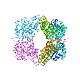 | | Structure of Dihydrodipicolinate Synthase from Streptococcus pneumoniae,bound to pyruvate and lysine | | 分子名称: | Dihydrodipicolinate synthase, LYSINE, SODIUM ION | | 著者 | Perugini, M.A, Dogovski, C, Parker, M.W, Gorman, M.A. | | 登録日 | 2012-06-06 | | 公開日 | 2013-09-18 | | 最終更新日 | 2023-11-29 | | 実験手法 | X-RAY DIFFRACTION (1.88 Å) | | 主引用文献 | Structure, Function, Stability and Knockout Phenotype of Dihydrodipicolinate Synthase from Streptococcus pneumoniae
To be Published
|
|
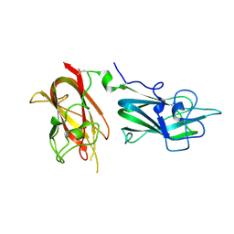
3BKJ
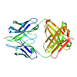 | | Crystal structure of Fab wo2 bound to the n terminal domain of amyloid beta peptide (1-16) | | 分子名称: | Amyloid Beta Peptide, WO2 IgG2a Fab fragment Heavy Chain, WO2 IgG2a Fab fragment Light Chain Kappa | | 著者 | Miles, L.A, Wun, K.S, Crespi, G.A, Fodero-Tavoletti, M, Galatis, D, Bageley, C.J, Beyreuther, K, Masters, C.L, Cappai, R, McKinstry, W.J, Barnham, K.J, Parker, M.W. | | 登録日 | 2007-12-06 | | 公開日 | 2008-04-15 | | 最終更新日 | 2011-07-13 | | 実験手法 | X-RAY DIFFRACTION (1.59 Å) | | 主引用文献 | Amyloid-beta-anti-amyloid-beta complex structure reveals an extended conformation in the immunodominant B-cell epitope.
J.Mol.Biol., 377, 2008
|
|
3BKM
 | | Structure of anti-amyloid-beta Fab WO2 (Form A, P212121) | | 分子名称: | SODIUM ION, WO2 IgG2a Fab fragment Heavy Chain, WO2 IgG2a Fab fragment Light Chain Kappa, ... | | 著者 | Miles, L.A, Wun, K.S, Crespi, G.A, Fodero-Tavoletti, M, Galatis, D, Bageley, C.J, Beyreuther, K, Masters, C.L, Cappai, R, McKinstry, W.J, Barnham, K.J, Parker, M.W. | | 登録日 | 2007-12-07 | | 公開日 | 2008-04-15 | | 最終更新日 | 2023-11-01 | | 実験手法 | X-RAY DIFFRACTION (1.6 Å) | | 主引用文献 | Amyloid-beta-anti-amyloid-beta complex structure reveals an extended conformation in the immunodominant B-cell epitope.
J.Mol.Biol., 377, 2008
|
|
3U0W
 | |
6WC8
 | |
6WE7
 | |
6WM8
 | |
4Y0G
 | | beta2 carbohydrate binding module (CBM) of AMP-activated protein kinase (AMPK) | | 分子名称: | 5'-AMP-activated protein kinase subunit beta-2, GLYCEROL | | 著者 | Mobbs, J, Gorman, M.A, Parker, M.W, Gooley, P.R, Griffin, M. | | 登録日 | 2015-02-06 | | 公開日 | 2015-04-08 | | 最終更新日 | 2024-02-28 | | 実験手法 | X-RAY DIFFRACTION (1.6 Å) | | 主引用文献 | Determinants of oligosaccharide specificity of the carbohydrate-binding modules of AMP-activated protein kinase.
Biochem.J., 468, 2015
|
|
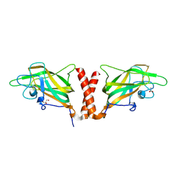
1FW1
 | | Glutathione transferase zeta/maleylacetoacetate isomerase | | 分子名称: | 2,3-DIHYDROXY-1,4-DITHIOBUTANE, GLUTATHIONE, GLUTATHIONE TRANSFERASE ZETA, ... | | 著者 | Polekhina, G, Board, P.G, Blackburn, A.C, Parker, M.W. | | 登録日 | 2000-09-20 | | 公開日 | 2001-09-26 | | 最終更新日 | 2011-07-13 | | 実験手法 | X-RAY DIFFRACTION (1.9 Å) | | 主引用文献 | Crystal structure of maleylacetoacetate isomerase/glutathione transferase zeta reveals the molecular basis for its remarkable catalytic promiscuity.
Biochemistry, 40, 2001
|
|
6XD4
 | | CDC-like protein | | 分子名称: | ACETATE ION, Hemolysin, SODIUM ION | | 著者 | Morton, C.J, Parker, M.W, Lawrence, S.L, Johnstone, B.A, Tweten, R.K. | | 登録日 | 2020-06-09 | | 公開日 | 2021-04-21 | | 実験手法 | X-RAY DIFFRACTION (2.1 Å) | | 主引用文献 | A Key Motif in the Cholesterol-Dependent Cytolysins Reveals a Large Family of Related Proteins.
Mbio, 11, 2020
|
|
2R4V
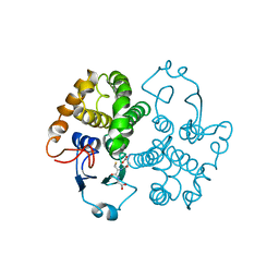
 | | Structure of human CLIC2, crystal form A | | 分子名称: | Chloride intracellular channel protein 2, GLUTATHIONE | | 著者 | Hansen, G, Cromer, B.A, Gorman, M.A, Parker, M.W. | | 登録日 | 2007-09-02 | | 公開日 | 2007-11-13 | | 最終更新日 | 2023-10-25 | | 実験手法 | X-RAY DIFFRACTION (1.85 Å) | | 主引用文献 | Structure of the Janus Protein Human CLIC2
J.Mol.Biol., 374, 2007
|
|


