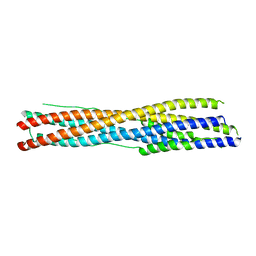
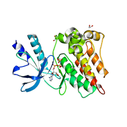
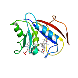
7WH1
 
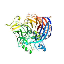 | | structure of C elegans BCMO-2 | | 分子名称: | Beta-Carotene 15,15'-MonoOxygenase, FE (III) ION, GLYCEROL, ... | | 著者 | Pan, W, Liu, L. | | 登録日 | 2021-12-29 | | 公開日 | 2022-02-02 | | 最終更新日 | 2023-11-29 | | 実験手法 | X-RAY DIFFRACTION (1.9 Å) | | 主引用文献 | Structural and Functional Analysis of Nonheme Iron Enzymes BCMO-1 and BCMO-2 from Caenorhabditis elegans .
Front Mol Biosci, 9, 2022
|
|
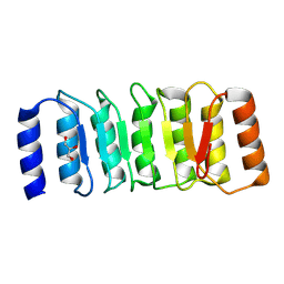
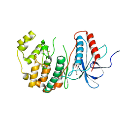
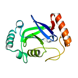
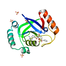
7WH0
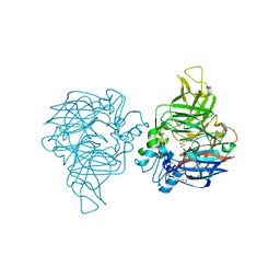 | | structure of C elegans BCMO-1 | | 分子名称: | Beta-Carotene 15,15'-MonoOxygenase, FE (III) ION, GLYCEROL, ... | | 著者 | Pan, W, Liu, L. | | 登録日 | 2021-12-29 | | 公開日 | 2022-02-02 | | 最終更新日 | 2023-11-29 | | 実験手法 | X-RAY DIFFRACTION (1.8 Å) | | 主引用文献 | Structural and Functional Analysis of Nonheme Iron Enzymes BCMO-1 and BCMO-2 from Caenorhabditis elegans .
Front Mol Biosci, 9, 2022
|
|
5YAZ
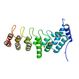 | | Crystal structure of the ANKRD domain of KANK1 | | 分子名称: | ACETATE ION, KN motif and ankyrin repeat domains 1 | | 著者 | Wei, Z, Pan, W. | | 登録日 | 2017-09-02 | | 公開日 | 2017-12-20 | | 最終更新日 | 2023-11-22 | | 実験手法 | X-RAY DIFFRACTION (1.9 Å) | | 主引用文献 | Structural insights into ankyrin repeat-mediated recognition of the kinesin motor protein KIF21A by KANK1, a scaffold protein in focal adhesion.
J. Biol. Chem., 293, 2018
|
|
5YAY
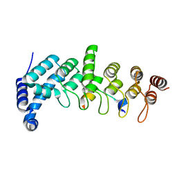 | | Crystal structure of KANK1/KIF21A complex | | 分子名称: | KN motif and ankyrin repeat domains 1, Kinesin-like protein KIF21A | | 著者 | Wei, Z, Pan, W. | | 登録日 | 2017-09-02 | | 公開日 | 2017-12-20 | | 最終更新日 | 2023-11-22 | | 実験手法 | X-RAY DIFFRACTION (1.55 Å) | | 主引用文献 | Structural insights into ankyrin repeat-mediated recognition of the kinesin motor protein KIF21A by KANK1, a scaffold protein in focal adhesion.
J. Biol. Chem., 293, 2018
|
|
7DDX
 | | Crystal structure of KANK1 S1179F mutant in complex wtih eIF4A1 | | 分子名称: | Eukaryotic initiation factor 4A-I, GLYCEROL, KN motif and ankyrin repeat domains 1, ... | | 著者 | Pan, W, Xu, Y, Wei, Z. | | 登録日 | 2020-10-30 | | 公開日 | 2021-09-08 | | 最終更新日 | 2023-11-29 | | 実験手法 | X-RAY DIFFRACTION (2.5 Å) | | 主引用文献 | Nephrotic-syndrome-associated mutation of KANK2 induces pathologic binding competition with physiological interactor KIF21A.
J.Biol.Chem., 297, 2021
|
|
7WXZ
 | | Crystal structure of the recombinant protein HR121 from the S2 protein of SARS-CoV-2 | | 分子名称: | Spike protein S2' | | 著者 | Zheng, Y.T, Ouyang, S, Pang, W, Lu, Y, Zhao, Y.B. | | 登録日 | 2022-02-15 | | 公開日 | 2022-11-23 | | 最終更新日 | 2023-11-29 | | 実験手法 | X-RAY DIFFRACTION (2.41 Å) | | 主引用文献 | A variant-proof SARS-CoV-2 vaccine targeting HR1 domain in S2 subunit of spike protein.
Cell Res., 32, 2022
|
|
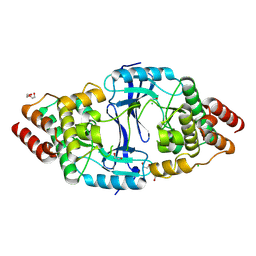
8X5L
 | | The Crystal Structure of PRKACA from Biortus. | | 分子名称: | (2S)-2-(4-chlorophenyl)-2-hydroxy-2-[4-(1H-pyrazol-4-yl)phenyl]ethanaminium, SODIUM ION, cAMP-dependent protein kinase catalytic subunit alpha | | 著者 | Wang, F, Cheng, W, Lv, Z, Lin, D, Pan, W. | | 登録日 | 2023-11-17 | | 公開日 | 2023-12-27 | | 実験手法 | X-RAY DIFFRACTION (2.75 Å) | | 主引用文献 | The Crystal Structure of PRKACA from Biortus.
To Be Published
|
|
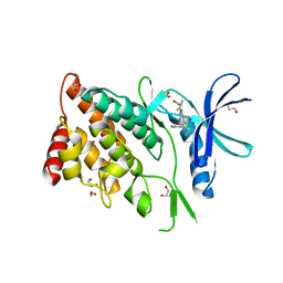
8ZGD
 | | The Crystal Structure of the NLRP1_LRR domain from Biortus. | | 分子名称: | GLYCEROL, NACHT, LRR and PYD domains-containing protein 1, ... | | 著者 | Wang, F, Cheng, W, Yuan, Z, Qi, J, Pan, W. | | 登録日 | 2024-05-09 | | 公開日 | 2024-06-26 | | 実験手法 | X-RAY DIFFRACTION (2.05 Å) | | 主引用文献 | The Crystal Structure of the NLRP1_LRR domain from Biortus.
To Be Published
|
|
8YVV
 | | The Crystal Structure of BTK from Biortus | | 分子名称: | 1,2-ETHANEDIOL, CHLORIDE ION, DI(HYDROXYETHYL)ETHER, ... | | 著者 | Wang, F, Cheng, W, Yuan, Z, Lin, D, Pan, W. | | 登録日 | 2024-03-29 | | 公開日 | 2024-07-03 | | 実験手法 | X-RAY DIFFRACTION (2.25 Å) | | 主引用文献 | The Crystal Structure of BTK from Biortus.
To Be Published
|
|
8WSW
 | | The Crystal Structure of LIMK2a from Biortus | | 分子名称: | 1,2-ETHANEDIOL, LIM domain kinase 2, ~{N}-[5-[2-[2,6-bis(chloranyl)phenyl]-5-[bis(fluoranyl)methyl]pyrazol-3-yl]-1,3-thiazol-2-yl]-2-methyl-propanamide | | 著者 | Wang, F, Cheng, W, Yuan, Z, Lin, D, Pan, W. | | 登録日 | 2023-10-17 | | 公開日 | 2023-11-15 | | 実験手法 | X-RAY DIFFRACTION (2.5 Å) | | 主引用文献 | The Crystal Structure of LIMK2a from Biortus.
To Be Published
|
|
8XFY
 | | The Crystal Structure of RSK2 from Biortus. | | 分子名称: | 2,6-bis(fluoranyl)-4-[4-(4-morpholin-4-ylphenyl)pyridin-3-yl]phenol, Ribosomal protein S6 kinase alpha-3 | | 著者 | Wang, F, Cheng, W, Lv, Z, Lin, D, Pan, W. | | 登録日 | 2023-12-14 | | 公開日 | 2024-03-06 | | 実験手法 | X-RAY DIFFRACTION (3.2 Å) | | 主引用文献 | The Crystal Structure of RSK2 from Biortus.
To Be Published
|
|
8ZKD
 | | The Crystal Structure of the RON from Biortus. | | 分子名称: | 1,2-ETHANEDIOL, MAGNESIUM ION, Macrophage-stimulating protein receptor beta chain, ... | | 著者 | Wang, F, Cheng, W, Yuan, Z, Qi, J, Pan, W. | | 登録日 | 2024-05-16 | | 公開日 | 2024-06-26 | | 実験手法 | X-RAY DIFFRACTION (2.05 Å) | | 主引用文献 | The Crystal Structure of the RON from Biortus.
To Be Published
|
|
8X23
 | | The Crystal Structure of MAPK13 from Biortus. | | 分子名称: | 1,2-ETHANEDIOL, GLYCEROL, Mitogen-activated protein kinase 13 | | 著者 | Wang, F, Cheng, W, Yuan, Z, Lin, D, Pan, W. | | 登録日 | 2023-11-09 | | 公開日 | 2023-12-27 | | 実験手法 | X-RAY DIFFRACTION (1.5 Å) | | 主引用文献 | The Crystal Structure of MAPK13 from Biortus.
To Be Published
|
|
8X2T
 | |
8X2A
 | | The Crystal Structure of BMX from Biortus. | | 分子名称: | 1,2-ETHANEDIOL, 4-[(3S)-3-{[(2E)-but-2-enoyl]amino}piperidin-1-yl]-5-fluoro-2,3-dimethyl-1H-indole-7-carboxamide, CHLORIDE ION, ... | | 著者 | Wang, F, Cheng, W, Yuan, Z, Lin, D, Pan, W. | | 登録日 | 2023-11-09 | | 公開日 | 2023-12-27 | | 実験手法 | X-RAY DIFFRACTION (1.3 Å) | | 主引用文献 | The Crystal Structure of BMX from Biortus.
To Be Published
|
|
5ZTE
 | | Crystal structure of PrxA C119S mutant from Arabidopsis thaliana | | 分子名称: | 2-Cys peroxiredoxin BAS1, chloroplastic | | 著者 | Yang, Y, Cai, W, Wang, J, Pan, W, Liu, L, Wang, M, Zhang, M. | | 登録日 | 2018-05-03 | | 公開日 | 2018-10-10 | | 最終更新日 | 2023-11-22 | | 実験手法 | X-RAY DIFFRACTION (2.6 Å) | | 主引用文献 | Crystal structure of Arabidopsis thaliana peroxiredoxin A C119S mutant.
Acta Crystallogr F Struct Biol Commun, 74, 2018
|
|
2NAE
 | |
4K48
 | | Structure of the Streptococcus pneumoniae leucyl-tRNA synthetase editing domain | | 分子名称: | Leucine--tRNA ligase | | 著者 | Hu, Q.H, Liu, R.J, Fang, Z.P, Zhang, J, Ding, Y.Y, Tan, M, Wang, M, Pan, W, Zhou, H.C, Wang, E.D. | | 登録日 | 2013-04-12 | | 公開日 | 2013-09-04 | | 最終更新日 | 2023-11-08 | | 実験手法 | X-RAY DIFFRACTION (2.49 Å) | | 主引用文献 | Discovery of a potent benzoxaborole-based anti-pneumococcal agent targeting leucyl-tRNA synthetase
Sci Rep, 3, 2013
|
|
4K47
 | | Structure of the Streptococcus pneumoniae leucyl-tRNA synthetase editing domain bound to a benzoxaborole-AMP adduct | | 分子名称: | Leucine--tRNA ligase, SULFATE ION, [(1R,5R,6R,8S)-6-(6-aminopurin-9-yl)-3'-[(R)-oxidanyl(phenyl)methyl]spiro[2,4,7-trioxa-3-boranuidabicyclo[3.3.0]octane-3,9'-8-oxa-9-boranuidabicyclo[4.3.0]nona-1,3,5-triene]-8-yl]methyl dihydrogen phosphate | | 著者 | Hu, Q.H, Liu, R.J, Fang, Z.P, Zhang, J, Ding, Y.Y, Tan, M, Wang, M, Pan, W, Zhou, H.C, Wang, E.D. | | 登録日 | 2013-04-12 | | 公開日 | 2013-09-04 | | 最終更新日 | 2024-03-20 | | 実験手法 | X-RAY DIFFRACTION (2.02 Å) | | 主引用文献 | Discovery of a potent benzoxaborole-based anti-pneumococcal agent targeting leucyl-tRNA synthetase
Sci Rep, 3, 2013
|
|
1CD2
 | | LIGAND INDUCED CONFORMATIONAL CHANGES IN THE CRYSTAL STRUCTURES OF PNEUMOCYSTIS CARINII DIHYDROFOLATE REDUCTASE COMPLEXES WITH FOLATE AND NADP+ | | 分子名称: | DIHYDROFOLATE REDUCTASE, FOLIC ACID, NADP NICOTINAMIDE-ADENINE-DINUCLEOTIDE PHOSPHATE | | 著者 | Cody, V, Galitsky, N, Luft, J.R, Pangborn, W, Blakley, R.L, Gangjee, A. | | 登録日 | 1999-03-05 | | 公開日 | 2000-03-06 | | 最終更新日 | 2023-08-09 | | 実験手法 | X-RAY DIFFRACTION (2.2 Å) | | 主引用文献 | Ligand-induced conformational changes in the crystal structures of Pneumocystis carinii dihydrofolate reductase complexes with folate and NADP+.
Biochemistry, 38, 1999
|
|
1BOZ
 | | STRUCTURE-BASED DESIGN AND SYNTHESIS OF LIPOPHILIC 2,4-DIAMINO-6-SUBSTITUTED QUINAZOLINES AND THEIR EVALUATION AS INHIBITORS OF DIHYDROFOLATE REDUCTASE AND POTENTIAL ANTITUMOR AGENTS | | 分子名称: | N6-(2,5-DIMETHOXY-BENZYL)-N6-METHYL-PYRIDO[2,3-D]PYRIMIDINE-2,4,6-TRIAMINE, NADPH DIHYDRO-NICOTINAMIDE-ADENINE-DINUCLEOTIDE PHOSPHATE, PROTEIN (DIHYDROFOLATE REDUCTASE) | | 著者 | Gangjee, A, Vidwans, A.P, Vasudevan, A, Queener, S.F, Kisliuk, R.L, Cody, V, Li, R, Galitsky, N, Luft, J.R, Pangborn, W. | | 登録日 | 1998-08-06 | | 公開日 | 1998-08-12 | | 最終更新日 | 2024-05-22 | | 実験手法 | X-RAY DIFFRACTION (2.1 Å) | | 主引用文献 | Structure-based design and synthesis of lipophilic 2,4-diamino-6-substituted quinazolines and their evaluation as inhibitors of dihydrofolate reductases and potential antitumor agents.
J.Med.Chem., 41, 1998
|
|
1BQP
 | | THE STRUCTURE OF THE PEA LECTIN-D-MANNOPYRANOSE COMPLEX | | 分子名称: | CALCIUM ION, MANGANESE (II) ION, PROTEIN (LECTIN), ... | | 著者 | Ruzeinikov, S.N, Mikhailova, I.Y, Tsygannik, I.N, Pangborn, W, Duax, W, Pletnev, V.Z. | | 登録日 | 1998-08-17 | | 公開日 | 1998-08-26 | | 最終更新日 | 2023-08-09 | | 実験手法 | X-RAY DIFFRACTION (2.1 Å) | | 主引用文献 | The Structure of the Pea Lectin-D-Mannopyranose Complex at a 2.1 A Resolution
RUSS.J.BIOORGANIC CHEM., 24, 1998
|
|
2L4O
 | | Solution structure of the Streptococcus pneumoniae RrgB pilus backbone D1 domain | | 分子名称: | Cell wall surface anchor family protein | | 著者 | Gentile, M.A, Melchiorre, S, Emolo, C, Moschioni, M, Gianfaldoni, C, Pancotto, L, Ferlenghi, I, Scarselli, M, Pansegrau, W, Veggi, D, Merola, M, Cantini, F, Ruggiero, P, Banci, L, Masignani, V. | | 登録日 | 2010-10-11 | | 公開日 | 2011-03-02 | | 最終更新日 | 2024-05-01 | | 実験手法 | SOLUTION NMR | | 主引用文献 | Solution structure of the Streptococcus pneumoniae RrgB pilus backbone D1 domain: in vivo protection and implications for the molecular mechanisms of pilus polymerization
To be Published
|
|
1BS9
 | | ACETYLXYLAN ESTERASE FROM P. PURPUROGENUM REFINED AT 1.10 ANGSTROMS | | 分子名称: | ACETYL XYLAN ESTERASE, SULFATE ION | | 著者 | Ghosh, D, Erman, M, Sawicki, M.W, Lala, P, Weeks, D.R, Li, N, Pangborn, W, Thiel, D.J, Jornvall, H, Eyzaguirre, J. | | 登録日 | 1998-09-01 | | 公開日 | 1999-05-18 | | 最終更新日 | 2011-07-13 | | 実験手法 | X-RAY DIFFRACTION (1.1 Å) | | 主引用文献 | Determination of a protein structure by iodination: the structure of iodinated acetylxylan esterase.
Acta Crystallogr.,Sect.D, 55, 1999
|
|
2BQP
 | | THE STRUCTURE OF THE PEA LECTIN-D-GLUCOPYRANOSE COMPLEX | | 分子名称: | CALCIUM ION, MANGANESE (II) ION, PROTEIN (PEA LECTIN), ... | | 著者 | Ruzeinikov, S.N, Mikhailova Yu, I, Tsygannik, I.N, Pangborn, W, Duax, W, Pletnev, V.Z. | | 登録日 | 1998-12-08 | | 公開日 | 1998-12-16 | | 最終更新日 | 2023-08-23 | | 実験手法 | X-RAY DIFFRACTION (1.9 Å) | | 主引用文献 | The Structure of Pea Lectin-D-Glucopyranose Complex at a 1.9 A Resolution
RUSS.J.BIOORGANIC CHEM., 23, 1997
|
|


