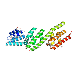
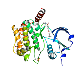
7OTS
 
 | | Crystal structure of human Monoacylglycerol Lipase ABHD6 in complex with oleic acid and octyl glucoside | | 分子名称: | GLYCEROL, Monoacylglycerol lipase ABHD6, OLEIC ACID, ... | | 著者 | Nawrotek, A, Talagas, A, Vuillard, L, Miallau, L. | | 登録日 | 2021-06-10 | | 公開日 | 2021-06-23 | | 最終更新日 | 2024-01-31 | | 実験手法 | X-RAY DIFFRACTION (1.792 Å) | | 主引用文献 | Crystal structure of human Monoacylglycerol Lipase ABHD6 in complex with oleic acid and octyl glucoside
To Be Published
|
|
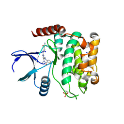
6FNE
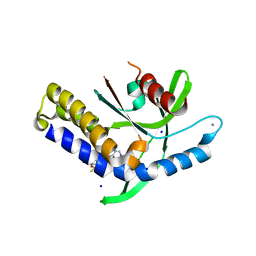
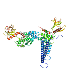
 | | Structure of human Brag2 (Sec7-PH domains) with the inhibitor Bragsin bound to the PH domain | | 分子名称: | (2~{S})-6-methyl-5-nitro-2-(trifluoromethyl)-2,3-dihydrochromen-4-one, IQ motif and SEC7 domain-containing protein 1, NONAETHYLENE GLYCOL | | 著者 | Nawrotek, A, Zeghouf, M, Cherfils, J. | | 登録日 | 2018-02-03 | | 公開日 | 2019-03-13 | | 最終更新日 | 2024-01-17 | | 実験手法 | X-RAY DIFFRACTION (2.501 Å) | | 主引用文献 | PH-domain-binding inhibitors of nucleotide exchange factor BRAG2 disrupt Arf GTPase signaling.
Nat.Chem.Biol., 15, 2019
|
|
8QEL
 | |
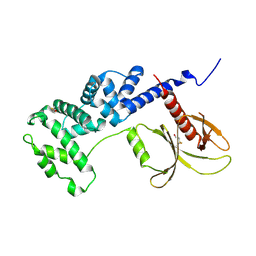
4P40
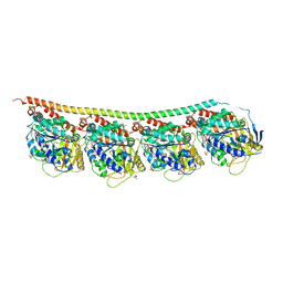
 | | Chlamydia pneumoniae CopN | | 分子名称: | CHLORIDE ION, CopN, DIMETHYLAMINE, ... | | 著者 | Nawrotek, A, Guimaraes, B.G, Knossow, M, Gigant, B. | | 登録日 | 2014-03-10 | | 公開日 | 2014-07-30 | | 最終更新日 | 2023-12-27 | | 実験手法 | X-RAY DIFFRACTION (1.2 Å) | | 主引用文献 | Biochemical and Structural Insights into Microtubule Perturbation by CopN from Chlamydia pneumoniae.
J.Biol.Chem., 289, 2014
|
|
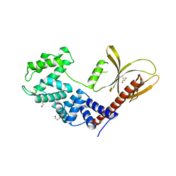
4P3Z
 | | Chlamydia pneumoniae CopN (D29 construct) | | 分子名称: | CopN, GLYCEROL, SULFATE ION | | 著者 | Nawrotek, A, Guimaraes, B.G, Knossow, M, Gigant, B. | | 登録日 | 2014-03-10 | | 公開日 | 2014-07-30 | | 最終更新日 | 2023-12-27 | | 実験手法 | X-RAY DIFFRACTION (1.77 Å) | | 主引用文献 | Biochemical and Structural Insights into Microtubule Perturbation by CopN from Chlamydia pneumoniae.
J.Biol.Chem., 289, 2014
|
|
8AXZ
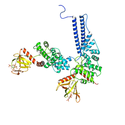
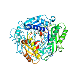 | | Crystal structure of human methionine adenosyltransferase 2A (MAT2A) in complex with S-adenosylmethionine, adenosin and diphosphono-aminophosphonic acid. | | 分子名称: | (DIPHOSPHONO)AMINOPHOSPHONIC ACID, ADENOSINE, DI(HYDROXYETHYL)ETHER, ... | | 著者 | Nawrotek, A, Vuillard, L, Miallau, L. | | 登録日 | 2022-09-01 | | 公開日 | 2022-10-05 | | 最終更新日 | 2024-01-31 | | 実験手法 | X-RAY DIFFRACTION (1.154 Å) | | 主引用文献 | Crystal structure of human methionine adenosyltransferase 2A (MAT2A) in complex with S-adenosylmethionine, adenosin and diphosphono-aminophosphonic acid.
To Be Published
|
|
8B2J
 | | Crystal structure of human STING in complex with ADU-S100 | | 分子名称: | (1~{R},3~{S},6~{R},8~{R},9~{R},10~{S},12~{S},15~{R},17~{R},18~{R})-8,17-bis(6-aminopurin-9-yl)-3,12-bis(oxidanylidene)-3,12-bis(sulfanyl)-2,4,7,11,13,16-hexaoxa-3$l^{5},12$l^{5}-diphosphatricyclo[13.2.1.0^{6,10}]octadecane-9,18-diol, CALCIUM ION, SODIUM ION, ... | | 著者 | Nawrotek, A, Vuillard, L, Miallau, L. | | 登録日 | 2022-09-14 | | 公開日 | 2022-11-30 | | 最終更新日 | 2024-05-01 | | 実験手法 | X-RAY DIFFRACTION (2.174 Å) | | 主引用文献 | Crystal structure of human STING in complex with ADU-S100
To Be Published
|
|
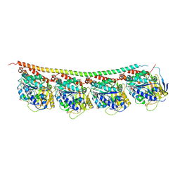
3RYF
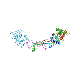
 | | GTP-Tubulin: RB3 Stathmin-like domain complex | | 分子名称: | GUANOSINE-5'-TRIPHOSPHATE, MAGNESIUM ION, SULFATE ION, ... | | 著者 | Nawrotek, A, Knossow, M, Gigant, B. | | 登録日 | 2011-05-11 | | 公開日 | 2011-10-05 | | 最終更新日 | 2023-09-13 | | 実験手法 | X-RAY DIFFRACTION (2.52 Å) | | 主引用文献 | The Determinants That Govern Microtubule Assembly from the Atomic Structure of GTP-Tubulin.
J.Mol.Biol., 412, 2011
|
|
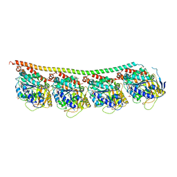
3RYH
 | | GMPCPP-Tubulin: RB3 Stathmin-like domain complex | | 分子名称: | GUANOSINE-5'-TRIPHOSPHATE, MAGNESIUM ION, PHOSPHOMETHYLPHOSPHONIC ACID GUANYLATE ESTER, ... | | 著者 | Nawrotek, A, Knossow, M, Gigant, B. | | 登録日 | 2011-05-11 | | 公開日 | 2011-10-05 | | 最終更新日 | 2023-09-13 | | 実験手法 | X-RAY DIFFRACTION (2.8 Å) | | 主引用文献 | The Determinants That Govern Microtubule Assembly from the Atomic Structure of GTP-Tubulin.
J.Mol.Biol., 412, 2011
|
|
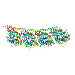
3RYI
 | | GDP-Tubulin: rb3 stathmin-like domain complex | | 分子名称: | GUANOSINE-5'-DIPHOSPHATE, GUANOSINE-5'-TRIPHOSPHATE, MAGNESIUM ION, ... | | 著者 | Nawrotek, A, Knossow, M, Gigant, B. | | 登録日 | 2011-05-11 | | 公開日 | 2011-10-05 | | 最終更新日 | 2023-09-13 | | 実験手法 | X-RAY DIFFRACTION (2.4 Å) | | 主引用文献 | The Determinants That Govern Microtubule Assembly from the Atomic Structure of GTP-Tubulin.
J.Mol.Biol., 412, 2011
|
|
3RYC
 | | Tubulin: RB3 stathmin-like domain complex | | 分子名称: | GUANOSINE-5'-DIPHOSPHATE, GUANOSINE-5'-TRIPHOSPHATE, MAGNESIUM ION, ... | | 著者 | Nawrotek, A, Knossow, M, Gigant, B. | | 登録日 | 2011-05-11 | | 公開日 | 2011-10-05 | | 最終更新日 | 2023-09-13 | | 実験手法 | X-RAY DIFFRACTION (2.1 Å) | | 主引用文献 | The Determinants That Govern Microtubule Assembly from the Atomic Structure of GTP-Tubulin.
J.Mol.Biol., 412, 2011
|
|
7AYM
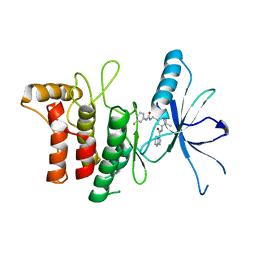 | | Structure of DDR2 Kinase domain in complex with IBZ3 | | 分子名称: | Discoidin domain-containing receptor 2,Epithelial discoidin domain-containing receptor 1, N-[5-({[(3-fluorophenyl)carbamoyl]amino}methyl)-2-methylphenyl]imidazo[1,2-a]pyridine-3-carboxamide | | 著者 | Nawrotek, A, Talagas, A, Vuillard, L.M, Miallau, L. | | 登録日 | 2020-11-12 | | 公開日 | 2020-12-16 | | 最終更新日 | 2024-01-31 | | 実験手法 | X-RAY DIFFRACTION (2.12 Å) | | 主引用文献 | Structure of DDR2 Kinase domain in complex with IBZ3
To Be Published
|
|
7Z4V
 | | Structure of Serine-Threonine kinase STK25 in complex with compound | | 分子名称: | 1,2-ETHANEDIOL, Serine/threonine-protein kinase 25, ~{N}-(5-~{tert}-butyl-1~{H}-pyrazol-3-yl)-4-pyrrolidin-1-ylsulfonyl-benzamide | | 著者 | Nawrotek, A, Vuillard, L, Miallau, L. | | 登録日 | 2022-03-04 | | 公開日 | 2022-04-06 | | 最終更新日 | 2024-01-31 | | 実験手法 | X-RAY DIFFRACTION (1.644 Å) | | 主引用文献 | Targeting non-alcoholic fatty liver disease: Design, X-ray co-crystal structure and synthesis of 'first-in-kind' inhibitors of serine/threonine kinase25.
Bioorg.Med.Chem.Lett., 75, 2022
|
|
8A66
 | | Crystal structure of MST2 in complex with XMU-MP-1 | | 分子名称: | 4-[(5,10-dimethyl-6-oxo-6,10-dihydro-5H-pyrimido[5,4-b]thieno[3,2-e][1,4]diazepin-2-yl)amino]benzenesulfonamide, SODIUM ION, Serine/threonine-protein kinase 3 36kDa subunit | | 著者 | Nawrotek, A, Vuillard, L, Miallau, L, Weber, C. | | 登録日 | 2022-06-16 | | 公開日 | 2022-07-20 | | 最終更新日 | 2024-05-01 | | 実験手法 | X-RAY DIFFRACTION (1.901 Å) | | 主引用文献 | Crystal structure of the Kelch domain of human Keap1in complex with ligand S217879
To Be Published
|
|
5NLV
 | | Brag2 Sec7-PH (390-763) | | 分子名称: | IQ motif and SEC7 domain-containing protein 1 | | 著者 | Nawrotek, A, Cherfils, J. | | 登録日 | 2017-04-05 | | 公開日 | 2017-09-27 | | 最終更新日 | 2024-01-17 | | 実験手法 | X-RAY DIFFRACTION (2.4 Å) | | 主引用文献 | Multiple interactions between an Arf/GEF complex and charged lipids determine activation kinetics on the membrane.
Proc. Natl. Acad. Sci. U.S.A., 114, 2017
|
|
5NLY
 | | Brag2 Sec7-PH (390-763), P212121 | | 分子名称: | IQ motif and SEC7 domain-containing protein 1, PHOSPHATE ION | | 著者 | Nawrotek, A, Cherfils, J. | | 登録日 | 2017-04-05 | | 公開日 | 2017-09-27 | | 最終更新日 | 2024-01-17 | | 実験手法 | X-RAY DIFFRACTION (2 Å) | | 主引用文献 | Multiple interactions between an Arf/GEF complex and charged lipids determine activation kinetics on the membrane.
Proc. Natl. Acad. Sci. U.S.A., 114, 2017
|
|
4UX8
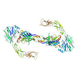 | | RET recognition of GDNF-GFRalpha1 ligand by a composite binding site promotes membrane-proximal self-association | | 分子名称: | 2-acetamido-2-deoxy-beta-D-glucopyranose-(1-4)-2-acetamido-2-deoxy-beta-D-glucopyranose, CALCIUM ION, GDNF FAMILY RECEPTOR ALPHA-1, ... | | 著者 | Goodman, K, Kjaer, S, Beuron, F, Knowles, P, Nawrotek, A, Burns, E, Purkiss, A, George, R, Santoro, M, Morris, E.P, McDonald, N.Q. | | 登録日 | 2014-08-19 | | 公開日 | 2014-10-01 | | 最終更新日 | 2020-07-29 | | 実験手法 | ELECTRON MICROSCOPY (24 Å) | | 主引用文献 | Ret Recognition of Gdnf-Gfralpha1 Ligand by a Composite Binding Site Promotes Membrane-Proximal Self-Association.
Cell Rep., 8, 2014
|
|
7AZB
 | | Structure of DDR2 DS domain in complex with VHH | | 分子名称: | Discoidin domain-containing receptor 2, VHH | | 著者 | Talagas, A, Nawrotek, A, Arrial, A, Vuillard, L.M, Miallau, L. | | 登録日 | 2020-11-16 | | 公開日 | 2020-11-25 | | 最終更新日 | 2024-01-31 | | 実験手法 | X-RAY DIFFRACTION (2.62 Å) | | 主引用文献 | Structure of DDR2 DS domain in complex with VHH
To Be Published
|
|
8A5J
 | | Crystal structure of Human STE20-like kinase 1, MST1 in complex with compound XMU-MP-1 | | 分子名称: | 4-[(5,10-dimethyl-6-oxo-6,10-dihydro-5H-pyrimido[5,4-b]thieno[3,2-e][1,4]diazepin-2-yl)amino]benzenesulfonamide, Serine/threonine-protein kinase 4 37kDa subunit | | 著者 | Nawrotek, A, Vuillard, L, Miallau, L. | | 登録日 | 2022-06-15 | | 公開日 | 2022-07-13 | | 最終更新日 | 2024-05-01 | | 実験手法 | X-RAY DIFFRACTION (2.123 Å) | | 主引用文献 | Crystal structure of the Kelch domain of human Keap1in complex with ligand S217879
To Be Published
|
|
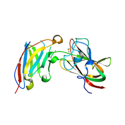
7AMK
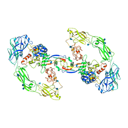
 | | Zebrafish RET Cadherin Like Domains 1 to 4. | | 分子名称: | 2-(N-MORPHOLINO)-ETHANESULFONIC ACID, 2-acetamido-2-deoxy-beta-D-glucopyranose, 2-acetamido-2-deoxy-beta-D-glucopyranose-(1-4)-2-acetamido-2-deoxy-beta-D-glucopyranose, ... | | 著者 | Purkiss, A.G, McDonald, N.Q, Goodman, K.M, Narowtek, A, Knowles, P.P. | | 登録日 | 2020-10-09 | | 公開日 | 2021-02-03 | | 最終更新日 | 2024-01-31 | | 実験手法 | X-RAY DIFFRACTION (2.2 Å) | | 主引用文献 | A two-site flexible clamp mechanism for RET-GDNF-GFR alpha 1 assembly reveals both conformational adaptation and strict geometric spacing.
Structure, 29, 2021
|
|
6U3E
 | |
6U3G
 | |
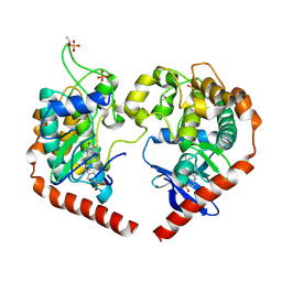
7AB8
 | | Crystal structure of a GDNF-GFRalpha1 complex | | 分子名称: | 1,2-ETHANEDIOL, 2-acetamido-2-deoxy-beta-D-glucopyranose-(1-4)-2-acetamido-2-deoxy-beta-D-glucopyranose, DI(HYDROXYETHYL)ETHER, ... | | 著者 | Adams, S.E, Earl, C.P, Purkiss, A.G, McDonald, N.Q. | | 登録日 | 2020-09-07 | | 公開日 | 2021-01-13 | | 最終更新日 | 2024-01-31 | | 実験手法 | X-RAY DIFFRACTION (2.2 Å) | | 主引用文献 | A two-site flexible clamp mechanism for RET-GDNF-GFR alpha 1 assembly reveals both conformational adaptation and strict geometric spacing.
Structure, 29, 2021
|
|
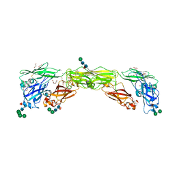
7AML
 | | RET/GDNF/GFRa1 extracellular complex Cryo-EM structure | | 分子名称: | 2-acetamido-2-deoxy-beta-D-glucopyranose, CALCIUM ION, GDNF family receptor alpha, ... | | 著者 | Adams, S.E, Earl, C.P, Purkiss, A.G, McDonald, N.Q. | | 登録日 | 2020-10-09 | | 公開日 | 2021-01-13 | | 最終更新日 | 2021-07-14 | | 実験手法 | ELECTRON MICROSCOPY (3.5 Å) | | 主引用文献 | A two-site flexible clamp mechanism for RET-GDNF-GFR alpha 1 assembly reveals both conformational adaptation and strict geometric spacing.
Structure, 29, 2021
|
|


