1SR9
 
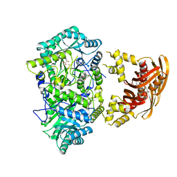
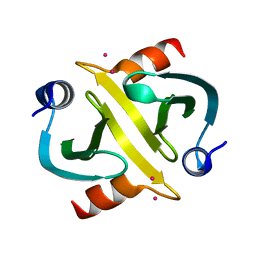
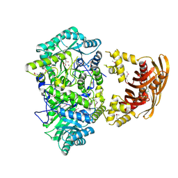 | | Crystal Structure of LeuA from Mycobacterium tuberculosis | | 分子名称: | 2-isopropylmalate synthase, 3-METHYL-2-OXOBUTANOIC ACID, CHLORIDE ION, ... | | 著者 | Koon, N, Squire, C.J, Baker, E.N. | | 登録日 | 2004-03-22 | | 公開日 | 2004-05-18 | | 最終更新日 | 2017-10-11 | | 実験手法 | X-RAY DIFFRACTION (2 Å) | | 主引用文献 | Crystal structure of LeuA from Mycobacterium tuberculosis, a key enzyme in leucine biosynthesis
Proc.Natl.Acad.Sci.USA, 101, 2004
|
|
3FIG
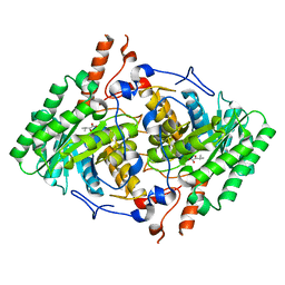
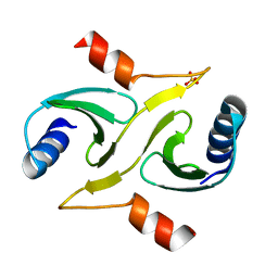
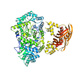 | | Crystal Structure of Leucine-bound LeuA from Mycobacterium tuberculosis | | 分子名称: | 2-isopropylmalate synthase, GLYCEROL, LEUCINE, ... | | 著者 | Koon, N, Squire, C.J, Baker, E.N. | | 登録日 | 2008-12-11 | | 公開日 | 2008-12-23 | | 最終更新日 | 2023-11-01 | | 実験手法 | X-RAY DIFFRACTION (2.3 Å) | | 主引用文献 | Crystal structure of LeuA from Mycobacterium tuberculosis, a key enzyme in leucine biosynthesis.
Proc.Natl.Acad.Sci.USA, 101, 2004
|
|
3HQ1
 | |
3HPZ
 | |
3HPS
 | |
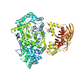
3U6W
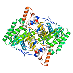
 | | Truncated M. tuberculosis LeuA (1-425) complexed with KIV | | 分子名称: | 2-isopropylmalate synthase, 3-METHYL-2-OXOBUTANOIC ACID, GLYCEROL, ... | | 著者 | Koon, N, Baker, E.N, Squire, C.J. | | 登録日 | 2011-10-13 | | 公開日 | 2012-03-14 | | 最終更新日 | 2023-09-13 | | 実験手法 | X-RAY DIFFRACTION (2.21 Å) | | 主引用文献 | Removal of the C-terminal regulatory domain of alpha-isopropylmalate synthase disrupts functional substrate binding.
Biochemistry, 51, 2012
|
|
3HPX
 | |
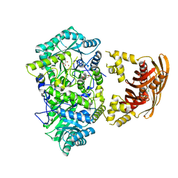
3RMJ
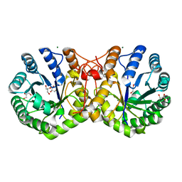 | | Crystal structure of truncated alpha-Isopropylmalate Synthase from Neisseria meningitidis | | 分子名称: | 2-isopropylmalate synthase, GLYCEROL, MAGNESIUM ION, ... | | 著者 | Huisman, F.H.A, Baker, H.M, Koon, N, Baker, E.N, Parker, E.J. | | 登録日 | 2011-04-20 | | 公開日 | 2012-03-14 | | 最終更新日 | 2023-11-01 | | 実験手法 | X-RAY DIFFRACTION (1.95 Å) | | 主引用文献 | Removal of the C-terminal regulatory domain of alpha-isopropylmalate synthase disrupts functional substrate binding
Biochemistry, 51, 2012
|
|
3IR8
 | | Red fluorescent protein mKeima at pH 7.0 | | 分子名称: | Large stokes shift fluorescent protein | | 著者 | Henderson, J.N, Osborn, M.F, Koon, N, Gepshtein, R, Huppert, D, Remington, S.J. | | 登録日 | 2009-08-21 | | 公開日 | 2009-09-08 | | 最終更新日 | 2023-11-15 | | 実験手法 | X-RAY DIFFRACTION (1.63 Å) | | 主引用文献 | Excited state proton transfer in the red fluorescent protein mKeima.
J.Am.Chem.Soc., 131, 2009
|
|
2IY2
 | |
2IYJ
 | |


