8P4K
 
 | | Vaccinia Virus palisade layer A10 trimer | | 分子名称: | Core protein OPG136 | | 著者 | Datler, J, Hansen, J.M, Thader, A, Schloegl, A, Hodirnau, V.V, Schur, F.K.M. | | 登録日 | 2023-05-22 | | 公開日 | 2024-01-17 | | 最終更新日 | 2024-02-21 | | 実験手法 | ELECTRON MICROSCOPY (3.8 Å) | | 主引用文献 | Multi-modal cryo-EM reveals trimers of protein A10 to form the palisade layer in poxvirus cores.
Nat.Struct.Mol.Biol., 2024
|
|
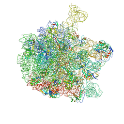
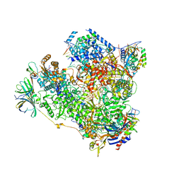
6QDW
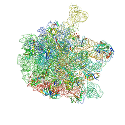 | | Cryo-EM structure of the 50S ribosomal subunit at 2.83 Angstroms with modeled GBC SecM peptide | | 分子名称: | 23S rRNA, 50S ribosomal protein L13, 50S ribosomal protein L14, ... | | 著者 | Schulte, L, Reitz, J, Hodirnau, V.V, Kudlinzki, D, Mao, J, Glaubitz, C, Frangakis, A, Schwalbe, H. | | 登録日 | 2019-01-03 | | 公開日 | 2020-01-15 | | 最終更新日 | 2020-12-02 | | 実験手法 | ELECTRON MICROSCOPY (2.83 Å) | | 主引用文献 | Cysteine oxidation and disulfide formation in the ribosomal exit tunnel.
Nat Commun, 11, 2020
|
|
7AQK
 | | Model of the actin filament Arp2/3 complex branch junction in cells | | 分子名称: | Actin, alpha skeletal muscle, ACTA1, ... | | 著者 | Faessler, F, Dimchev, G, Hodirnau, V.V, Wan, W, Schur, F.K.M. | | 登録日 | 2020-10-22 | | 公開日 | 2020-12-02 | | 最終更新日 | 2024-05-01 | | 実験手法 | ELECTRON MICROSCOPY (9 Å) | | 主引用文献 | Cryo-electron tomography structure of Arp2/3 complex in cells reveals new insights into the branch junction.
Nat Commun, 11, 2020
|
|
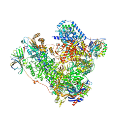
6YS3
 | | Cryo-EM structure of the 50S ribosomal subunit at 2.58 Angstroms with modeled GBC SecM peptide | | 分子名称: | 23S rRNA, 50S ribosomal protein L13, 50S ribosomal protein L14, ... | | 著者 | Schulte, L, Reitz, J, Kudlinzki, D, Hodirnau, V.V, Frangakis, A, Schwalbe, H. | | 登録日 | 2020-04-20 | | 公開日 | 2020-09-30 | | 実験手法 | ELECTRON MICROSCOPY (2.58 Å) | | 主引用文献 | Cryo-EM structure of the 50S ribosomal subunit at 2.58 Angstroms with modeled GBC SecM peptide
Nat Commun, 2020
|
|
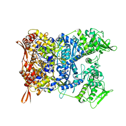
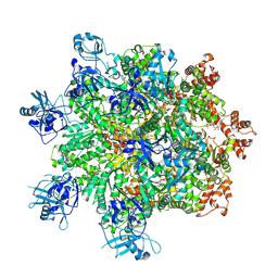
5M3F
 | | Yeast RNA polymerase I elongation complex at 3.8A | | 分子名称: | DNA-directed RNA polymerase I subunit RPA12, DNA-directed RNA polymerase I subunit RPA135, DNA-directed RNA polymerase I subunit RPA14, ... | | 著者 | Neyer, S, Kunz, M, Geiss, C, Hantsche, M, Hodirnau, V.-V, Seybert, A, Engel, C, Scheffer, M.P, Cramer, P, Frangakis, A.S. | | 登録日 | 2016-10-14 | | 公開日 | 2016-11-23 | | 最終更新日 | 2019-12-11 | | 実験手法 | ELECTRON MICROSCOPY (3.8 Å) | | 主引用文献 | Structure of RNA polymerase I transcribing ribosomal DNA genes.
Nature, 540, 2016
|
|
5M3M
 | | Free monomeric RNA polymerase I at 4.0A resolution | | 分子名称: | DNA-directed RNA polymerase I subunit RPA12, DNA-directed RNA polymerase I subunit RPA135, DNA-directed RNA polymerase I subunit RPA14, ... | | 著者 | Neyer, S, Kunz, M, Geiss, C, Hantsche, M, Hodirnau, V.-V, Seybert, A, Engel, C, Scheffer, M.P, Cramer, P, Frangakis, A.S. | | 登録日 | 2016-10-15 | | 公開日 | 2016-11-23 | | 最終更新日 | 2024-05-15 | | 実験手法 | ELECTRON MICROSCOPY (4 Å) | | 主引用文献 | Structure of RNA polymerase I transcribing ribosomal DNA genes.
Nature, 540, 2016
|
|
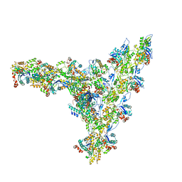
7Z34
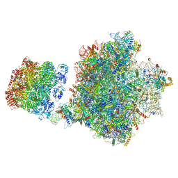 | | Structure of pre-60S particle bound to DRG1(AFG2). | | 分子名称: | 35S pre-ribosomal RNA, 5.8S rRNA, 5S rRNA, ... | | 著者 | Prattes, M, Grishkovskaya, I, Bergler, H, Haselbach, D. | | 登録日 | 2022-03-01 | | 公開日 | 2022-09-21 | | 最終更新日 | 2023-07-19 | | 実験手法 | ELECTRON MICROSCOPY (3.8 Å) | | 主引用文献 | Visualizing maturation factor extraction from the nascent ribosome by the AAA-ATPase Drg1.
Nat.Struct.Mol.Biol., 29, 2022
|
|
7Z11
 | | Structure of substrate bound DRG1 (AFG2) | | 分子名称: | ATPase family gene 2 protein, PHOSPHOTHIOPHOSPHORIC ACID-ADENYLATE ESTER, peptide substrate | | 著者 | Prattes, M, Grishkovskaya, I, Bergler, H, Haselbach, D. | | 登録日 | 2022-02-24 | | 公開日 | 2022-09-21 | | 最終更新日 | 2024-07-17 | | 実験手法 | ELECTRON MICROSCOPY (3.2 Å) | | 主引用文献 | Visualizing maturation factor extraction from the nascent ribosome by the AAA-ATPase Drg1.
Nat.Struct.Mol.Biol., 29, 2022
|
|
7NKU
 | | diazaborine bound Drg1(AFG2) | | 分子名称: | 6-METHYL-2(PROPANE-1-SULFONYL)-2H-THIENO[3,2-D][1,2,3]DIAZABORININ-1-OL, ATPase family gene 2 protein, PHOSPHOTHIOPHOSPHORIC ACID-ADENYLATE ESTER | | 著者 | Prattes, M, Bergler, H, Haselbach, D. | | 登録日 | 2021-02-19 | | 公開日 | 2021-06-23 | | 実験手法 | ELECTRON MICROSCOPY (3.4 Å) | | 主引用文献 | Structural basis for inhibition of the AAA-ATPase Drg1 by diazaborine.
Nat Commun, 12, 2021
|
|


