Loading
PDBj
メニューPDBj@FacebookPDBj@TwitterPDBj@YouTubewwPDB FoundationwwPDB
RCSB PDBPDBeBMRBAdv. SearchSearch help
Search by PDB author
3TG0
DownloadVisualize
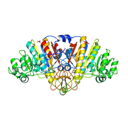
BU of 3tg0 by Molmil
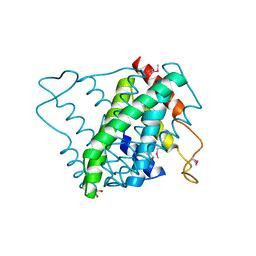
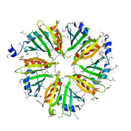
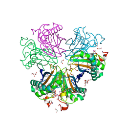
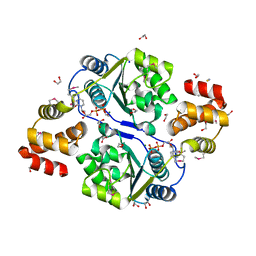
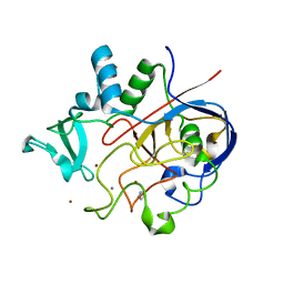
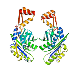
E. coli alkaline phosphatase with bound inorganic phosphate
分子名称: Alkaline phosphatase, MAGNESIUM ION, PHOSPHATE ION, ...
著者Bobyr, E, Lassila, J.K, Wiersma-Koch, H.I, Fenn, T.D, Lee, J.J, Nikolic-Hughes, I, Hodgson, K.O, Rees, D.C, Hedman, B, Herschlag, D.
登録日2011-08-16
公開日2011-11-09
最終更新日2023-09-13
実験手法X-RAY DIFFRACTION (1.2 Å)
主引用文献High-resolution analysis of Zn(2+) coordination in the alkaline phosphatase superfamily by EXAFS and x-ray crystallography.
J.Mol.Biol., 415, 2012
3HBZ
DownloadVisualize
BU of 3hbz by Molmil
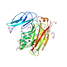
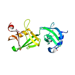
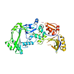
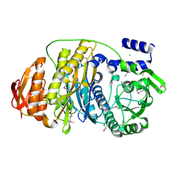
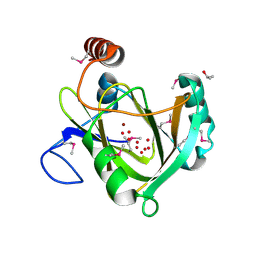
Crystal structure of a putative glycoside hydrolase (bt_2081) from bacteroides thetaiotaomicron vpi-5482 at 2.05 A resolution
分子名称: ACETATE ION, CACODYLATE ION, CALCIUM ION, ...
著者Joint Center for Structural Genomics (JCSG)
登録日2009-05-05
公開日2009-05-26
最終更新日2023-02-01
実験手法X-RAY DIFFRACTION (2.05 Å)
主引用文献Structure of Bacteroides thetaiotaomicron BT2081 at 2.05 A resolution: the first structural representative of a new protein family that may play a role in carbohydrate metabolism.
Acta Crystallogr.,Sect.F, 66, 2010
2FNO
DownloadVisualize
BU of 2fno by Molmil
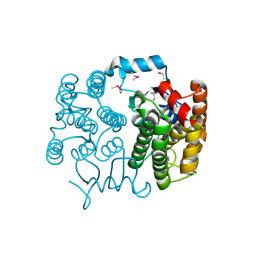
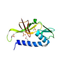
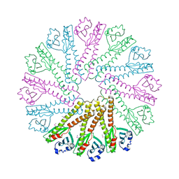
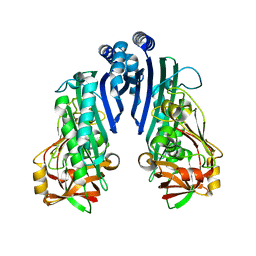
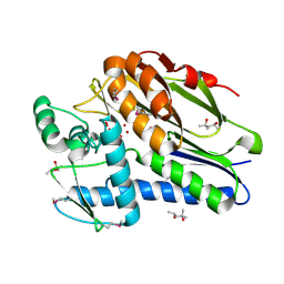
Crystal structure of a glutathione s-transferase (atu5508) from agrobacterium tumefaciens str. c58 at 2.00 A resolution
分子名称: AGR_pAT_752p, THIOCYANATE ION
著者Joint Center for Structural Genomics (JCSG)
登録日2006-01-11
公開日2006-02-14
最終更新日2023-01-25
実験手法X-RAY DIFFRACTION (2 Å)
主引用文献Comparative structural analysis of a novel glutathioneS-transferase (ATU5508) from Agrobacterium tumefaciens at 2.0 A resolution.
Proteins, 65, 2006
3NL9
DownloadVisualize
BU of 3nl9 by Molmil
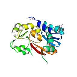
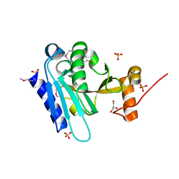
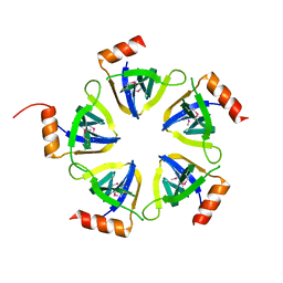
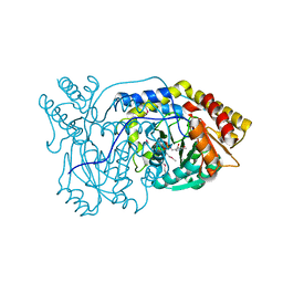
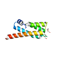
Crystal structure of a putative NTP pyrophosphohydrolase (Exig_1061) from EXIGUOBACTERIUM SP. 255-15 at 1.78 A resolution
分子名称: 1,2-ETHANEDIOL, putative NTP pyrophosphohydrolase
著者Joint Center for Structural Genomics (JCSG)
登録日2010-06-21
公開日2010-07-21
最終更新日2023-02-01
実験手法X-RAY DIFFRACTION (1.78 Å)
主引用文献Structure of a putative NTP pyrophosphohydrolase: YP_001813558.1 from Exiguobacterium sibiricum 255-15.
Acta Crystallogr.,Sect.F, 66, 2010
3L5O
DownloadVisualize
BU of 3l5o by Molmil
Crystal structure of protein with unknown function from DUF364 family (ZP_00559375.1) from Desulfitobacterium hafniense DCB-2 at 2.01 A resolution
分子名称: 1,2-ETHANEDIOL, CHLORIDE ION, IMIDAZOLE, ...
著者Joint Center for Structural Genomics, Joint Center for Structural Genomics (JCSG)
登録日2009-12-22
公開日2010-02-02
最終更新日2023-02-01
実験手法X-RAY DIFFRACTION (2.01 Å)
主引用文献Structure of the first representative of Pfam family PF04016 (DUF364) reveals enolase and Rossmann-like folds that combine to form a unique active site with a possible role in heavy-metal chelation.
Acta Crystallogr.,Sect.F, 66, 2010
3HSA
DownloadVisualize
BU of 3hsa by Molmil
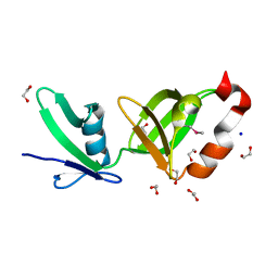
Crystal structure of pleckstrin homology domain (YP_926556.1) from SHEWANELLA AMAZONENSIS SB2B at 1.99 A resolution
分子名称: DI(HYDROXYETHYL)ETHER, GLYCEROL, pleckstrin homology domain
著者Joint Center for Structural Genomics (JCSG)
登録日2009-06-10
公開日2009-06-23
最終更新日2023-02-01
実験手法X-RAY DIFFRACTION (1.99 Å)
主引用文献Bacterial pleckstrin homology domains: a prokaryotic origin for the PH domain.
J.Mol.Biol., 396, 2010
2RE3
DownloadVisualize
BU of 2re3 by Molmil
CRYSTAL STRUCTURE OF a DUF1285 family protein (SPO_0140) FROM SILICIBACTER POMEROYI DSS-3 AT 2.50 A RESOLUTION
分子名称: GLYCEROL, Uncharacterized protein
著者Joint Center for Structural Genomics (JCSG)
登録日2007-09-25
公開日2007-10-09
最終更新日2023-01-25
実験手法X-RAY DIFFRACTION (2.5 Å)
主引用文献Structures of the first representatives of Pfam family PF06938 (DUF1285) reveal a new fold with repeated structural motifs and possible involvement in signal transduction.
Acta Crystallogr.,Sect.F, 66, 2010
3IRB
DownloadVisualize
BU of 3irb by Molmil
Crystal structure of protein with unknown function from DUF35 family (13815350) from SULFOLOBUS SOLFATARICUS at 1.80 A resolution
分子名称: ACETIC ACID, SULFATE ION, ZINC ION, ...
著者Joint Center for Structural Genomics (JCSG)
登録日2009-08-21
公開日2009-09-01
最終更新日2023-02-01
実験手法X-RAY DIFFRACTION (1.8 Å)
主引用文献The structure of SSO2064, the first representative of Pfam family PF01796, reveals a novel two-domain zinc-ribbon OB-fold architecture with a potential acyl-CoA-binding role.
Acta Crystallogr.,Sect.F, 66, 2010
3K5J
DownloadVisualize
BU of 3k5j by Molmil
Crystal structure of Putative SUFU (suppressor of fused protein) homolog (YP_208451.1) from Neisseria gonorrhoeae FA 1090 at 1.40 A resolution
分子名称: GLYCEROL, SULFATE ION, Suppressor of fused family protein
著者Joint Center for Structural Genomics (JCSG)
登録日2009-10-07
公開日2010-01-26
最終更新日2019-07-17
実験手法X-RAY DIFFRACTION (1.4 Å)
主引用文献The crystal structure of a bacterial Sufu-like protein defines a novel group of bacterial proteins that are similar to the N-terminal domain of human Sufu.
Protein Sci., 19, 2010
3BYQ
DownloadVisualize
BU of 3byq by Molmil
Crystal structure of a duf1185 family protein (bb2672) from bordetella bronchiseptica rb50 at 1.70 A resolution
分子名称: 1,2-ETHANEDIOL, CHLORIDE ION, TETRAETHYLENE GLYCOL, ...
著者Joint Center for Structural Genomics (JCSG)
登録日2008-01-16
公開日2008-01-29
最終更新日2023-02-01
実験手法X-RAY DIFFRACTION (1.7 Å)
主引用文献Structures of the first representatives of Pfam family PF06684 (DUF1185) reveal a novel variant of the Bacillus chorismate mutase fold and suggest a role in amino-acid metabolism.
Acta Crystallogr.,Sect.F, 66, 2010
3KK7
DownloadVisualize
BU of 3kk7 by Molmil
Crystal structure of Putative cell invasion protein with MAC/Perforin domain (NP_812351.1) from BACTERIODES THETAIOTAOMICRON VPI-5482 at 2.46 A resolution
分子名称: (4S)-2-METHYL-2,4-PENTANEDIOL, 1,2-ETHANEDIOL, CHLORIDE ION, ...
著者Joint Center for Structural Genomics (JCSG)
登録日2009-11-04
公開日2009-11-24
最終更新日2023-02-01
実験手法X-RAY DIFFRACTION (2.46 Å)
主引用文献Structure of a membrane-attack complex/perforin (MACPF) family protein from the human gut symbiont Bacteroides thetaiotaomicron.
Acta Crystallogr.,Sect.F, 66, 2010
3B77
DownloadVisualize
BU of 3b77 by Molmil
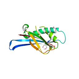
Crystal structure of a ph domain containing bacterial protein (exig_2160) from exiguobacterium sibiricum 255-15 at 2.42 A resolution
分子名称: Uncharacterized protein
著者Joint Center for Structural Genomics (JCSG)
登録日2007-10-30
公開日2007-11-13
最終更新日2023-01-25
実験手法X-RAY DIFFRACTION (2.42 Å)
主引用文献Bacterial pleckstrin homology domains: a prokaryotic origin for the PH domain.
J.Mol.Biol., 396, 2010
3BY7
DownloadVisualize
BU of 3by7 by Molmil
CRYSTAL STRUCTURE OF A PROTEIN STRUCTURALLY SIMILAR TO SM/LSM-LIKE RNA-BINDING PROTEINS (JCVI_PEP_1096686650277) FROM UNCULTURED MARINE ORGANISM AT 2.60 A RESOLUTION
分子名称: uncharacterized protein
著者Joint Center for Structural Genomics (JCSG)
登録日2008-01-15
公開日2008-01-29
最終更新日2023-02-01
実験手法X-RAY DIFFRACTION (2.6 Å)
主引用文献Crystal structure of a novel Sm-like protein of putative cyanophage origin at 2.60 A resolution.
Proteins, 75, 2009
3BOS
DownloadVisualize
BU of 3bos by Molmil
Crystal structure of a putative dna replication regulator HDA (SAMA_1916) from Shewanella amazonensis sb2b at 1.75 A resolution
分子名称: 1,2-ETHANEDIOL, CHLORIDE ION, CYTIDINE-5'-DIPHOSPHATE, ...
著者Joint Center for Structural Genomics (JCSG)
登録日2007-12-17
公開日2008-01-15
最終更新日2023-01-25
実験手法X-RAY DIFFRACTION (1.75 Å)
主引用文献A structural basis for the regulatory inactivation of DnaA.
J.Mol.Biol., 385, 2009
2RA9
DownloadVisualize
BU of 2ra9 by Molmil
Crystal structure of a duf1285 family protein (sbal_2486) from shewanella baltica os155 at 1.40 A resolution
分子名称: 1,2-ETHANEDIOL, SODIUM ION, Uncharacterized protein DUF1285
著者Joint Center for Structural Genomics (JCSG)
登録日2007-09-14
公開日2007-10-16
最終更新日2023-01-25
実験手法X-RAY DIFFRACTION (1.4 Å)
主引用文献Structures of the first representatives of Pfam family PF06938 (DUF1285) reveal a new fold with repeated structural motifs and possible involvement in signal transduction.
Acta Crystallogr.,Sect.F, 66, 2010
2QTP
DownloadVisualize
BU of 2qtp by Molmil
Crystal structure of a duf1185 family protein (spo0826) from silicibacter pomeroyi dss-3 at 2.10 A resolution
分子名称: Uncharacterized protein
著者Joint Center for Structural Genomics (JCSG)
登録日2007-08-02
公開日2007-08-21
最終更新日2023-01-25
実験手法X-RAY DIFFRACTION (2.1 Å)
主引用文献Structures of the first representatives of Pfam family PF06684 (DUF1185) reveal a novel variant of the Bacillus chorismate mutase fold and suggest a role in amino-acid metabolism.
Acta Crystallogr.,Sect.F, 66, 2010
4PNJ
DownloadVisualize
BU of 4pnj by Molmil
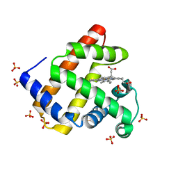
Recombinant Sperm Whale P6 Myoglobin Solved with Single Pulse Free Electron Laser Data
分子名称: Myoglobin, PROTOPORPHYRIN IX CONTAINING FE, SULFATE ION
著者Cohen, A, Gonzalez, A, Lam, W, Lyubimov, A, Sauter, N, Tsai, Y, Uervirojnangkoorn, M, Brunger, A, Soltis, M.
登録日2014-05-23
公開日2014-11-05
最終更新日2023-09-27
実験手法X-RAY DIFFRACTION (1.36 Å)
主引用文献Goniometer-based femtosecond crystallography with X-ray free electron lasers.
Proc.Natl.Acad.Sci.USA, 111, 2014
6MUJ
DownloadVisualize
BU of 6muj by Molmil
Formylglycine generating enzyme bound to copper
分子名称: 2,3-DIHYDROXY-1,4-DITHIOBUTANE, CALCIUM ION, COPPER (II) ION, ...
著者Lafrance-Vanasse, J, Appel, M.J, Tsai, C.-L, Bertozzi, C, Tainer, J.A.
登録日2018-10-23
公開日2019-02-27
最終更新日2023-10-11
実験手法X-RAY DIFFRACTION (2.249 Å)
主引用文献Formylglycine-generating enzyme binds substrate directly at a mononuclear Cu(I) center to initiate O2activation.
Proc. Natl. Acad. Sci. U.S.A., 116, 2019
1VK3
DownloadVisualize
BU of 1vk3 by Molmil
Crystal structure of Phosphoribosylformylglycinamidine synthase II (TM1246) from Thermotoga maritima at 2.15 A resolution
分子名称: CHLORIDE ION, Phosphoribosylformylglycinamidine synthase II
著者Joint Center for Structural Genomics (JCSG)
登録日2004-04-23
公開日2004-05-11
最終更新日2023-01-25
実験手法X-RAY DIFFRACTION (2.15 Å)
主引用文献Crystal structure of phosphoribosylformylglycinamidine synthase II (smPurL) from Thermotoga maritima at 2.15 A resolution.
Proteins, 63, 2006
1VL4
DownloadVisualize
BU of 1vl4 by Molmil
CRYSTAL STRUCTURE OF A PUTATIVE MODULATOR OF A DNA GYRASE (TM0727) FROM THERMOTOGA MARITIMA MSB8 AT 1.95 A RESOLUTION
分子名称: pmbA-related protein
著者Joint Center for Structural Genomics (JCSG)
登録日2004-07-09
公開日2004-09-21
最終更新日2024-05-22
実験手法X-RAY DIFFRACTION (1.95 Å)
主引用文献Crystal structure of a putative modulator of DNA gyrase (pmbA) from Thermotoga maritima at 1.95 A resolution reveals a new fold.
Proteins, 61, 2005
1VJO
DownloadVisualize
BU of 1vjo by Molmil
Crystal structure of Alanine--glyoxylate aminotransferase (ALR1004) from Nostoc sp. at 1.70 A resolution
分子名称: PYRIDOXAL-5'-PHOSPHATE, alanine--glyoxylate aminotransferase
著者Joint Center for Structural Genomics (JCSG)
登録日2004-03-16
公開日2004-03-23
最終更新日2023-01-25
実験手法X-RAY DIFFRACTION (1.7 Å)
主引用文献Crystal structure of an alanine-glyoxylate aminotransferase from Anabaena sp. at 1.70 A resolution reveals a noncovalently linked PLP cofactor
Proteins, 58, 2005
1VQ0
DownloadVisualize
BU of 1vq0 by Molmil
Crystal structure of 33 kDa chaperonin (Heat shock protein 33 homolog) (HSP33) (TM1394) from Thermotoga maritima at 2.20 A resolution
分子名称: 1,2-ETHANEDIOL, 33 kDa chaperonin, CHLORIDE ION, ...
著者Joint Center for Structural Genomics (JCSG)
登録日2004-11-30
公開日2004-12-14
最終更新日2023-09-20
実験手法X-RAY DIFFRACTION (2.2 Å)
主引用文献Crystal structure of Hsp33 chaperone (TM1394) from Thermotoga maritima at 2.20 A resolution.
Proteins, 61, 2005
1VR3
DownloadVisualize
BU of 1vr3 by Molmil
Crystal structure of Acireductone dioxygenase (13543033) from Mus musculus at 2.06 A resolution
分子名称: Acireductone dioxygenase, ISOPROPYL ALCOHOL, NICKEL (II) ION, ...
著者Joint Center for Structural Genomics (JCSG)
登録日2005-01-28
公開日2005-02-15
最終更新日2023-01-25
実験手法X-RAY DIFFRACTION (2.06 Å)
主引用文献Crystal structure of acireductone dioxygenase (ARD) from Mus musculus at 2.06 angstrom resolution.
Proteins, 64, 2006
1VRM
DownloadVisualize
BU of 1vrm by Molmil
Crystal structure of the apbe protein (tm1553) from thermotoga maritima msb8 at 1.58 A resolution
分子名称: (4R)-2-METHYLPENTANE-2,4-DIOL, (4S)-2-METHYL-2,4-PENTANEDIOL, UNKNOWN LIGAND, ...
著者Joint Center for Structural Genomics (JCSG)
登録日2005-03-10
公開日2005-03-22
最終更新日2023-01-25
実験手法X-RAY DIFFRACTION (1.58 Å)
主引用文献Crystal structure of the ApbE protein (TM1553) from Thermotoga maritima at 1.58 A resolution.
Proteins, 64, 2006
2OOC
DownloadVisualize
BU of 2ooc by Molmil
Crystal structure of Histidine Phosphotransferase ShpA (NP_419930.1) from Caulobacter crescentus at 1.52 A resolution
分子名称: GLYCEROL, Histidine phosphotransferase, TETRAETHYLENE GLYCOL
著者Joint Center for Structural Genomics (JCSG)
登録日2007-01-25
公開日2007-02-06
最終更新日2023-01-25
実験手法X-RAY DIFFRACTION (1.52 Å)
主引用文献Crystal structure of histidine phosphotransfer protein ShpA, an essential regulator of stalk biogenesis in Caulobacter crescentus.
J.Mol.Biol., 390, 2009

222415

件を2024-07-10に公開中

PDB statisticsPDBj update infoContact PDBjnumon