5NXS
 
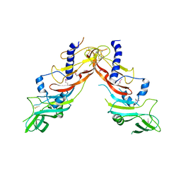 | |
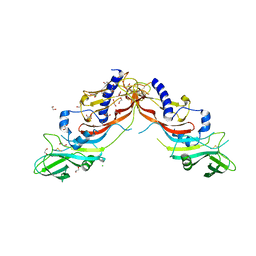
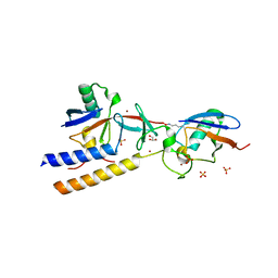
5NTU
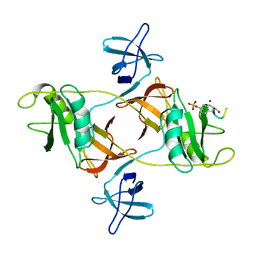
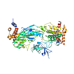
 | | Crystal Structure of human Pro-myostatin Precursor at 2.6 A Resolution | | 分子名称: | 1,2-ETHANEDIOL, CHLORIDE ION, Growth/differentiation factor 8 | | 著者 | Cotton, T.R, Fischer, G, Hyvonen, M. | | 登録日 | 2017-04-28 | | 公開日 | 2018-01-17 | | 最終更新日 | 2018-02-21 | | 実験手法 | X-RAY DIFFRACTION (2.58 Å) | | 主引用文献 | Structure of the human myostatin precursor and determinants of growth factor latency.
EMBO J., 37, 2018
|
|
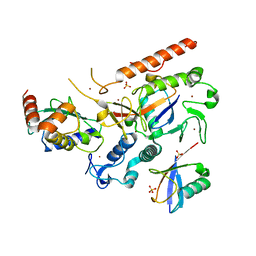
7M4O
 | |
7M4M
 | |
7M4N
 | |
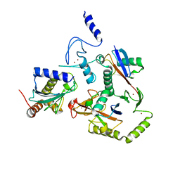
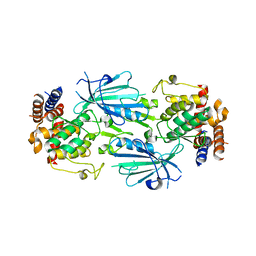
8EB0
 | | RNF216/E2-Ub/Ub transthiolation complex | | 分子名称: | E3 ubiquitin-protein ligase RNF216, SULFATE ION, Ubiquitin, ... | | 著者 | Cotton, T.R, Wang, X.S, Lechtenberg, B.C. | | 登録日 | 2022-08-30 | | 公開日 | 2023-01-18 | | 最終更新日 | 2023-10-25 | | 実験手法 | X-RAY DIFFRACTION (3.03 Å) | | 主引用文献 | The unifying catalytic mechanism of the RING-between-RING E3 ubiquitin ligase family.
Nat Commun, 14, 2023
|
|
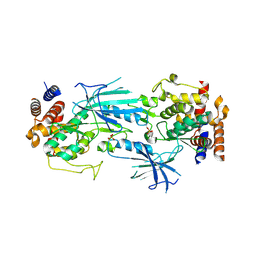
8EAZ
 | | HOIL-1/E2-Ub/Ub transthiolation complex | | 分子名称: | RanBP-type and C3HC4-type zinc finger-containing protein 1, Ubiquitin, Ubiquitin-conjugating enzyme E2 L3, ... | | 著者 | Wang, X.S, Cotton, T.R, Lechtenberg, B.C. | | 登録日 | 2022-08-30 | | 公開日 | 2023-01-18 | | 最終更新日 | 2023-10-25 | | 実験手法 | X-RAY DIFFRACTION (3.08 Å) | | 主引用文献 | The unifying catalytic mechanism of the RING-between-RING E3 ubiquitin ligase family.
Nat Commun, 14, 2023
|
|
8DGN
 | |
8DGO
 | |
8DGM
 | | 14-3-3 epsilon bound to phosphorylated PEAK1 (pT1165) peptide | | 分子名称: | 1,2-ETHANEDIOL, 14-3-3 protein epsilon, Inactive tyrosine-protein kinase PEAK1 | | 著者 | Roy, M.J, Hardy, J.M, Lucet, I.S. | | 登録日 | 2022-06-24 | | 公開日 | 2023-06-07 | | 最終更新日 | 2023-10-25 | | 実験手法 | X-RAY DIFFRACTION (3.2 Å) | | 主引用文献 | Structural mapping of PEAK pseudokinase interactions identifies 14-3-3 as a molecular switch for PEAK3 signaling.
Nat Commun, 14, 2023
|
|
8DGP
 | | 14-3-3 epsilon bound to phosphorylated PEAK3 (pS69) peptide | | 分子名称: | 1,2-ETHANEDIOL, 14-3-3 protein epsilon, Phosphorylated PEAK3 (pS69) peptide, ... | | 著者 | Roy, M.J, Hardy, J.M, Lucet, I.S. | | 登録日 | 2022-06-24 | | 公開日 | 2023-06-07 | | 最終更新日 | 2023-10-25 | | 実験手法 | X-RAY DIFFRACTION (2.7 Å) | | 主引用文献 | Structural mapping of PEAK pseudokinase interactions identifies 14-3-3 as a molecular switch for PEAK3 signaling.
Nat Commun, 14, 2023
|
|
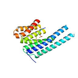
7T3X
 | | Structure of unphosphorylated Pediculus humanus (Ph) PINK1 D334A mutant | | 分子名称: | Serine/threonine-protein kinase PINK1 | | 著者 | Gan, Z.Y, Leis, A, Dewson, G, Glukhova, A, Komander, D. | | 登録日 | 2021-12-09 | | 公開日 | 2021-12-22 | | 最終更新日 | 2023-10-18 | | 実験手法 | X-RAY DIFFRACTION (3.53 Å) | | 主引用文献 | Activation mechanism of PINK1.
Nature, 602, 2022
|
|
7T4K
 | | Structure of dimeric phosphorylated Pediculus humanus (Ph) PINK1 with kinked alpha-C helix in chain B | | 分子名称: | Serine/threonine-protein kinase PINK1, putative | | 著者 | Gan, Z.Y, Leis, A, Dewson, G, Glukhova, A, Komander, D. | | 登録日 | 2021-12-10 | | 公開日 | 2022-01-12 | | 最終更新日 | 2022-02-23 | | 実験手法 | ELECTRON MICROSCOPY (3.25 Å) | | 主引用文献 | Activation mechanism of PINK1.
Nature, 602, 2022
|
|
7T4N
 | | Structure of dimeric unphosphorylated Pediculus humanus (Ph) PINK1 D357A mutant | | 分子名称: | Serine/threonine-protein kinase PINK1, putative | | 著者 | Gan, Z.Y, Leis, A, Dewson, G, Glukhova, A, Komander, D. | | 登録日 | 2021-12-10 | | 公開日 | 2022-01-12 | | 最終更新日 | 2024-02-28 | | 実験手法 | ELECTRON MICROSCOPY (2.35 Å) | | 主引用文献 | Activation mechanism of PINK1.
Nature, 602, 2022
|
|
7T4L
 | | Structure of dimeric phosphorylated Pediculus humanus (Ph) PINK1 with extended alpha-C helix in chain B | | 分子名称: | Serine/threonine-protein kinase PINK1, putative | | 著者 | Gan, Z.Y, Leis, A, Dewson, G, Glukhova, A, Komander, D. | | 登録日 | 2021-12-10 | | 公開日 | 2022-01-12 | | 最終更新日 | 2022-02-23 | | 実験手法 | ELECTRON MICROSCOPY (3.28 Å) | | 主引用文献 | Activation mechanism of PINK1.
Nature, 602, 2022
|
|
7T4M
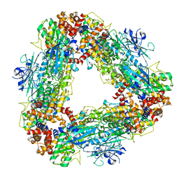 | | Structure of dodecameric unphosphorylated Pediculus humanus (Ph) PINK1 D357A mutant | | 分子名称: | Serine/threonine-protein kinase PINK1, putative | | 著者 | Gan, Z.Y, Leis, A, Dewson, G, Glukhova, A, Komander, D. | | 登録日 | 2021-12-10 | | 公開日 | 2022-01-12 | | 最終更新日 | 2024-02-28 | | 実験手法 | ELECTRON MICROSCOPY (2.48 Å) | | 主引用文献 | Activation mechanism of PINK1.
Nature, 602, 2022
|
|


