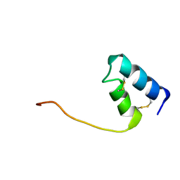
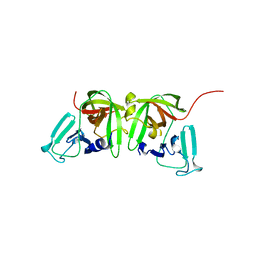
5E5Q
 
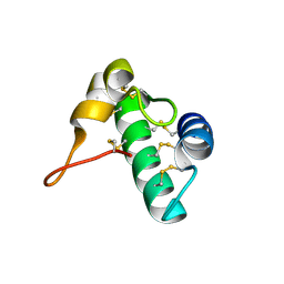 | | Racemic snakin-1 in P21/c | | 分子名称: | Snakin-1 | | 著者 | Yeung, H, Squire, C.J, Yosaatmadja, Y, Panjikar, S, Baker, E.N, Harris, P.W.R, Brimble, M.A. | | 登録日 | 2015-10-09 | | 公開日 | 2016-05-18 | | 最終更新日 | 2016-07-20 | | 実験手法 | X-RAY DIFFRACTION (1.6 Å) | | 主引用文献 | Radiation Damage and Racemic Protein Crystallography Reveal the Unique Structure of the GASA/Snakin Protein Superfamily.
Angew.Chem.Int.Ed.Engl., 55, 2016
|
|
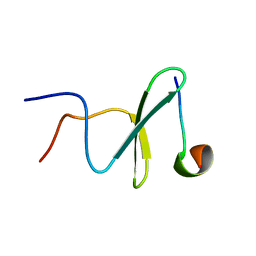
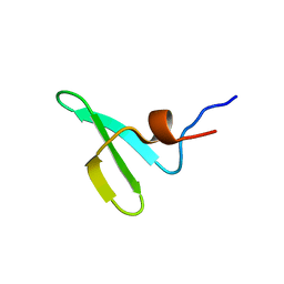
5E5Y
 | | Quasi-racemic snakin-1 in P1 before radiation damage | | 分子名称: | 1,2-ETHANEDIOL, D- snakin-1, FORMIC ACID, ... | | 著者 | Yeung, H, Squire, C.J, Yosaatmadja, Y, Panjikar, S, Baker, E.N, Harris, P.W.R, Brimble, M.A. | | 登録日 | 2015-10-09 | | 公開日 | 2016-05-18 | | 最終更新日 | 2016-07-20 | | 実験手法 | X-RAY DIFFRACTION (1.506 Å) | | 主引用文献 | Radiation Damage and Racemic Protein Crystallography Reveal the Unique Structure of the GASA/Snakin Protein Superfamily.
Angew.Chem.Int.Ed.Engl., 55, 2016
|
|
2M3O
 | | Structure and dynamics of a human Nedd4 WW domain-ENaC complex | | 分子名称: | Amiloride-sensitive sodium channel subunit alpha, E3 ubiquitin-protein ligase NEDD4 | | 著者 | Bobby, R, Medini, K, Neudecker, P, Lee, V, MacDonald, F.J, Brimble, M.A, Lott, J, Dingley, A.J. | | 登録日 | 2013-01-23 | | 公開日 | 2013-08-28 | | 最終更新日 | 2024-05-01 | | 実験手法 | SOLUTION NMR | | 主引用文献 | Structure and dynamics of human Nedd4-1 WW3 in complex with the alpha ENaC PY motif.
Biochim.Biophys.Acta, 1834, 2013
|
|
5E5T
 | | Quasi-racemic snakin-1 in P1 after radiation damage | | 分子名称: | 1,2-ETHANEDIOL, D- snakin-1, FORMIC ACID, ... | | 著者 | Yeung, H, Squire, C.J, Yosaatmadja, Y, Panjikar, S, Baker, E.N, Harris, P.W.R, Brimble, M.A. | | 登録日 | 2015-10-09 | | 公開日 | 2016-05-18 | | 最終更新日 | 2016-07-20 | | 実験手法 | X-RAY DIFFRACTION (1.572 Å) | | 主引用文献 | Radiation Damage and Racemic Protein Crystallography Reveal the Unique Structure of the GASA/Snakin Protein Superfamily.
Angew.Chem.Int.Ed.Engl., 55, 2016
|
|
6MW1
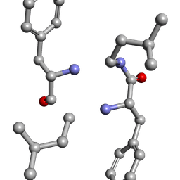 | |
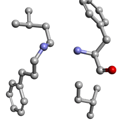
6MW0
 | | Mle-Phe-Mle-D-Phe. Linear tetrapeptide related to pseudoxylallemycin A. | | 分子名称: | METHANOL, Mle-Phe-Mle-D-Phe Linear tetrapeptide related to pseudoxylallemycin A | | 著者 | Cameron, A.J, Harris, P.W.R, Brimble, M.A, Squire, C.J. | | 登録日 | 2018-10-29 | | 公開日 | 2019-09-11 | | 実験手法 | X-RAY DIFFRACTION (0.78 Å) | | 主引用文献 | Investigations of the key macrolactamisation step in the synthesis of cyclic tetrapeptide pseudoxylallemycin A.
Org.Biomol.Chem., 17, 2019
|
|
6MW2
 | |
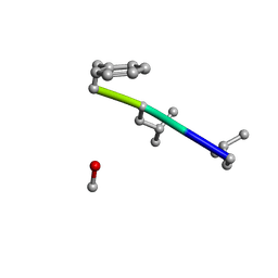
6MVZ
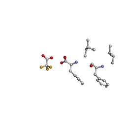 | | Mle-Phe-Mle-Phe. Linear precursor of pseudoxylallemycin A. | | 分子名称: | Linear precursor of pseudoxylallemycin A, trifluoroacetic acid | | 著者 | Cameron, A.J, Harris, P.W.R, Brimble, M.A, Squire, C.J. | | 登録日 | 2018-10-29 | | 公開日 | 2019-09-11 | | 実験手法 | X-RAY DIFFRACTION (0.83 Å) | | 主引用文献 | Investigations of the key macrolactamisation step in the synthesis of cyclic tetrapeptide pseudoxylallemycin A.
Org.Biomol.Chem., 17, 2019
|
|
4RWG
 | |
4RWF
 | |
8DFZ
 | |
4N31
 | |
5VCJ
 | | Structure of alpha-galactosylphytosphingosine bound by CD1d and in complex with the Va14Vb8.2 TCR | | 分子名称: | (2S,3S,4R)-2-amino-3,4-dihydroxyoctadecyl alpha-D-galactopyranoside, 2-acetamido-2-deoxy-beta-D-glucopyranose, Antigen-presenting glycoprotein CD1d1, ... | | 著者 | Wang, J, Zajonc, D.M. | | 登録日 | 2017-03-31 | | 公開日 | 2018-04-04 | | 最終更新日 | 2023-10-04 | | 実験手法 | X-RAY DIFFRACTION (3.16 Å) | | 主引用文献 | Enhancing T cell responses and tumour immunity by vaccination with peptides conjugated to a weak NKT cell agonist.
Org. Biomol. Chem., 17, 2019
|
|
8U1W
 | | Structure of Norovirus (Hu/GII.4/Sydney/NSW0514/2012/AU) protease bound to inhibitor NV-004 | | 分子名称: | ACETATE ION, GLYCEROL, Peptidase C37, ... | | 著者 | Eruera, A.R, Campbell, A.C, Krause, K.L. | | 登録日 | 2023-09-03 | | 公開日 | 2023-12-20 | | 実験手法 | X-RAY DIFFRACTION (1.84 Å) | | 主引用文献 | Crystal Structure of Inhibitor-Bound GII.4 Sydney 2012 Norovirus 3C-Like Protease.
Viruses, 15, 2023
|
|
8U1V
 | |
5AHT
 | |
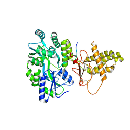
4WVH
 | | Crystal structure of the Type-I signal peptidase from Staphylococcus aureus (SpsB) in complex with a substrate peptide (pep1). | | 分子名称: | Maltose-binding periplasmic protein,Signal peptidase IB, alpha-D-glucopyranose-(1-4)-alpha-D-glucopyranose, substrate peptide (pep1) | | 著者 | Young, P.G, Ting, Y.T, Baker, E.N. | | 登録日 | 2014-11-05 | | 公開日 | 2015-09-23 | | 最終更新日 | 2023-09-27 | | 実験手法 | X-RAY DIFFRACTION (2.1 Å) | | 主引用文献 | Peptide binding to a bacterial signal peptidase visualized by peptide tethering and carrier-driven crystallization.
IUCrJ, 3, 2016
|
|
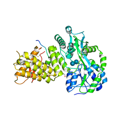
4WVJ
 | | Crystal structure of the Type-I signal peptidase from Staphylococcus aureus (SpsB) in complex with an inhibitor peptide (pep3). | | 分子名称: | Maltose-binding periplasmic protein,Signal peptidase IB, alpha-D-glucopyranose-(1-4)-alpha-D-glucopyranose, inhibitor peptide (PEP3) | | 著者 | Young, P.G, Ting, Y.T, Baker, E.N. | | 登録日 | 2014-11-05 | | 公開日 | 2015-09-23 | | 最終更新日 | 2023-09-27 | | 実験手法 | X-RAY DIFFRACTION (1.95 Å) | | 主引用文献 | Peptide binding to a bacterial signal peptidase visualized by peptide tethering and carrier-driven crystallization.
IUCrJ, 3, 2016
|
|
4WVG
 | |
4WVI
 | | Crystal structure of the Type-I signal peptidase from Staphylococcus aureus (SpsB) in complex with a substrate peptide (pep2). | | 分子名称: | Maltose-binding periplasmic protein,Signal peptidase IB, alpha-D-glucopyranose-(1-4)-alpha-D-glucopyranose, substrate peptide (pep2) | | 著者 | Young, P.G, Ting, Y.T, Baker, E.N. | | 登録日 | 2014-11-05 | | 公開日 | 2015-09-23 | | 最終更新日 | 2023-09-27 | | 実験手法 | X-RAY DIFFRACTION (1.9 Å) | | 主引用文献 | Peptide binding to a bacterial signal peptidase visualized by peptide tethering and carrier-driven crystallization.
IUCrJ, 3, 2016
|
|


