5JQS
 
 | |
5JKN
 | |
6Y6R
 | |
6YJG
 | |
6Z49
 | |
6Z7V
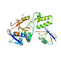
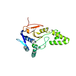
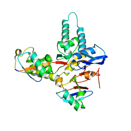
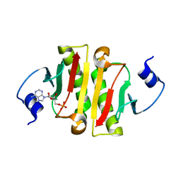
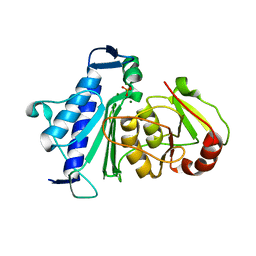
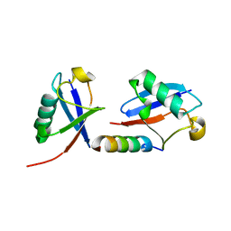
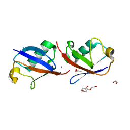
 | | Crystal structure of Mindy2 (C266A) in complex with Lys48 linked di-ubiquitin (K48-Ub2) | | 分子名称: | POTASSIUM ION, Polyubiquitin-C, TETRAETHYLENE GLYCOL, ... | | 著者 | Abdul Rehman, S.A, Lange, S.M, Kulathu, Y. | | 登録日 | 2020-06-01 | | 公開日 | 2021-06-09 | | 最終更新日 | 2024-01-24 | | 実験手法 | X-RAY DIFFRACTION (2.65 Å) | | 主引用文献 | Mechanism of activation and regulation of deubiquitinase activity in MINDY1 and MINDY2.
Mol.Cell, 81, 2021
|
|
6Z90
 | |
4EHS
 | |
6TUV
 | |
6TXB
 | |
4IM9
 | |
5XW3
 | |
4ZGL
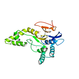
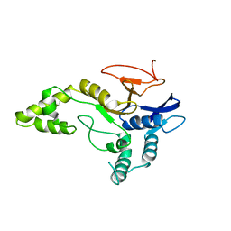
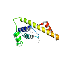
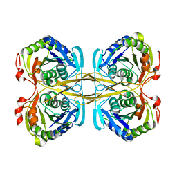
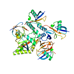
 | | Hit Like Protein | | 分子名称: | ADENOSINE MONOPHOSPHATE, Uncharacterized HIT-like protein HP_0404 | | 著者 | Tarique, K.F, Devi, S, Abdul Rehman, S.A, Gourinath, S. | | 登録日 | 2015-04-23 | | 公開日 | 2015-05-27 | | 最終更新日 | 2023-11-08 | | 実験手法 | X-RAY DIFFRACTION (2.95 Å) | | 主引用文献 | Crystal structure of HINT from Helicobacter pylori.
Acta Crystallogr.,Sect.F, 72, 2016
|
|
4ZG5
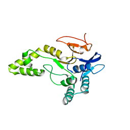
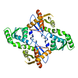
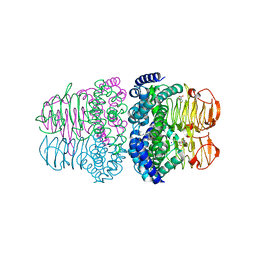
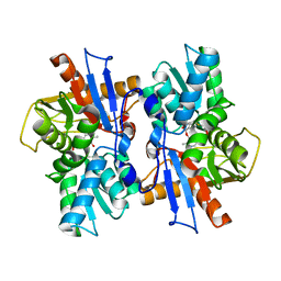
 | | Structural and functional insights into Survival endonuclease, an important virulence factor of Brucella abortus | | 分子名称: | 5'-nucleotidase SurE, MAGNESIUM ION | | 著者 | Tarique, K.F, Abdul Rehman, S.A, Devi, S, Gourinath, S. | | 登録日 | 2015-04-22 | | 公開日 | 2015-05-06 | | 最終更新日 | 2023-11-08 | | 実験手法 | X-RAY DIFFRACTION (1.9 Å) | | 主引用文献 | Structural and functional insights into the stationary-phase survival protein SurE, an important virulence factor of Brucella abortus
Acta Crystallogr.,Sect.F, 72, 2016
|
|
4H7O
 | |
4QEZ
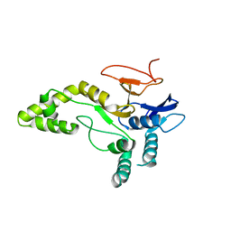
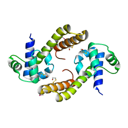
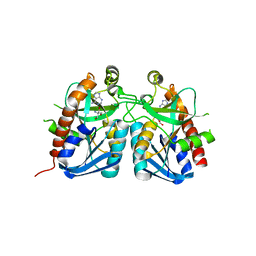 | | Crystal structure of 5'-methylthioadenosine/S-adenosylhomocysteine nucleosidase from Bacillus anthracis | | 分子名称: | 2-AMINO-2-HYDROXYMETHYL-PROPANE-1,3-DIOL, 5'-methylthioadenosine/S-adenosylhomocysteine nucleosidase, ADENINE | | 著者 | Tarique, K.F, Devi, S, Abdul Rehman, S.A, Gourinath, S. | | 登録日 | 2014-05-19 | | 公開日 | 2014-06-18 | | 最終更新日 | 2023-11-08 | | 実験手法 | X-RAY DIFFRACTION (2.7 Å) | | 主引用文献 | Crystal structure of 5'-methylthioadenosine/S-adenosylhomocysteine nucleosidase from Bacillus anthracis
To be Published
|
|
4QXD
 | | Crystal structure of Inositol Polyphosphate 1-Phosphatase from Entamoeba histolytica | | 分子名称: | 3'(2'),5'-bisphosphate nucleotidase, putative, MAGNESIUM ION, ... | | 著者 | Tarique, K.F, Abdul Rehman, S.A, Betzel, C, Gourinath, S. | | 登録日 | 2014-07-19 | | 公開日 | 2014-08-06 | | 最終更新日 | 2024-04-03 | | 実験手法 | X-RAY DIFFRACTION (2.55 Å) | | 主引用文献 | Structure-based identification of inositol polyphosphate 1-phosphatase from Entamoeba histolytica
Acta Crystallogr.,Sect.D, 70, 2014
|
|
4S1Z
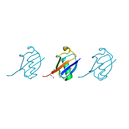
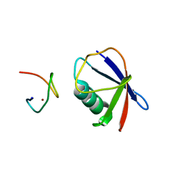 | | Crystal structure of TRABID NZF1 in complex with K29 linked di-Ubiquitin | | 分子名称: | Ubiquitin, Ubiquitin thioesterase ZRANB1, ZINC ION | | 著者 | Kristariyanto, Y.A, Abdul Rehman, S.A, Campbell, D.G, Morrice, N.A, Johnson, C, Toth, R, Kulathu, Y. | | 登録日 | 2015-01-16 | | 公開日 | 2015-04-08 | | 最終更新日 | 2023-09-20 | | 実験手法 | X-RAY DIFFRACTION (3.03 Å) | | 主引用文献 | K29-selective ubiquitin binding domain reveals structural basis of specificity and heterotypic nature of k29 polyubiquitin.
Mol.Cell, 58, 2015
|
|
4S22
 | | Crystal structure of K29 linked di-Ubiquitin | | 分子名称: | 1,2-ETHANEDIOL, GLYCEROL, IODIDE ION, ... | | 著者 | Kristariyanto, Y.A, Abdul Rehman, S.A, Campbell, D.G, Morrice, N.A, Johnson, C, Toth, R, Kulathu, Y. | | 登録日 | 2015-01-17 | | 公開日 | 2015-04-08 | | 最終更新日 | 2023-09-20 | | 実験手法 | X-RAY DIFFRACTION (2.3 Å) | | 主引用文献 | K29-selective ubiquitin binding domain reveals structural basis of specificity and heterotypic nature of k29 polyubiquitin.
Mol.Cell, 58, 2015
|
|
6FGE
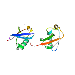
 | | Crystal structure of human ZUFSP/ZUP1 in complex with ubiquitin | | 分子名称: | ACETATE ION, AMMONIUM ION, DI(HYDROXYETHYL)ETHER, ... | | 著者 | Kwasna, D, Abdul Rehman, S.A, Kulathu, Y. | | 登録日 | 2018-01-10 | | 公開日 | 2018-04-04 | | 最終更新日 | 2018-04-18 | | 実験手法 | X-RAY DIFFRACTION (1.74 Å) | | 主引用文献 | Discovery and Characterization of ZUFSP/ZUP1, a Distinct Deubiquitinase Class Important for Genome Stability.
Mol. Cell, 70, 2018
|
|
5MN9
 | |
7NPI
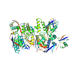
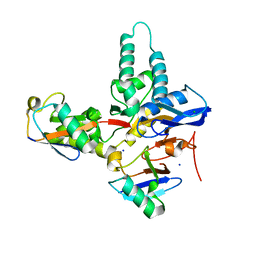
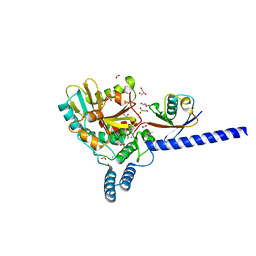
 | | Crystal structure of Mindy2 (C266A) in complex with Lys48-linked penta-ubiquitin (K48-Ub5) | | 分子名称: | CHLORIDE ION, Polyubiquitin-C, SODIUM ION, ... | | 著者 | Lange, S.M, Armstrong, L.A, Kulathu, Y. | | 登録日 | 2021-02-26 | | 公開日 | 2021-09-15 | | 最終更新日 | 2024-01-31 | | 実験手法 | X-RAY DIFFRACTION (2.81 Å) | | 主引用文献 | Mechanism of activation and regulation of deubiquitinase activity in MINDY1 and MINDY2.
Mol.Cell, 81, 2021
|
|
6AHI
 | |
4Y1H
 | | Crystal structure of K33 linked tri-Ubiquitin | | 分子名称: | 1,2-ETHANEDIOL, Ubiquitin-40S ribosomal protein S27a | | 著者 | Kristariyanto, Y.A, Abdul Rehman, S.A, Choi, S.Y, Ritorto, S, Campbell, D.G, Morrice, N.A, Toth, R, Kulathu, Y. | | 登録日 | 2015-02-07 | | 公開日 | 2015-03-18 | | 最終更新日 | 2024-01-10 | | 実験手法 | X-RAY DIFFRACTION (1.4 Å) | | 主引用文献 | Assembly and structure of Lys33-linked polyubiquitin reveals distinct conformations.
Biochem.J., 467, 2015
|
|
4XYZ
 | | Crystal structure of K33 linked di-Ubiquitin | | 分子名称: | 1,2-ETHANEDIOL, ACETATE ION, IODIDE ION, ... | | 著者 | Kristariyanto, Y.A, Abdul Rehman, S.A, Choi, S.Y, Ritorto, S, Campbell, D.G, Morrice, N.A, Toth, R, Kulathu, Y. | | 登録日 | 2015-02-03 | | 公開日 | 2015-03-18 | | 最終更新日 | 2024-01-10 | | 実験手法 | X-RAY DIFFRACTION (1.65 Å) | | 主引用文献 | Assembly and structure of Lys33-linked polyubiquitin reveals distinct conformations.
Biochem.J., 467, 2015
|
|


