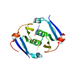
3TEK
 
 | | ThermoDBP: a non-canonical single-stranded DNA binding protein with a novel structure and mechanism | | 分子名称: | ThermoDBP-single stranded DNA binding protein | | 著者 | White, M.F, Paytubi, S, Liu, H, Graham, S, McMahon, S.A, Naismith, J.H. | | 登録日 | 2011-08-15 | | 公開日 | 2011-11-23 | | 最終更新日 | 2024-02-28 | | 実験手法 | X-RAY DIFFRACTION (2 Å) | | 主引用文献 | Displacement of the canonical single-stranded DNA-binding protein in the Thermoproteales.
Proc.Natl.Acad.Sci.USA, 109, 2012
|
|
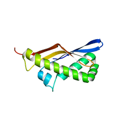
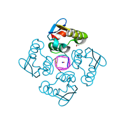
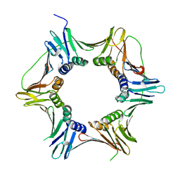
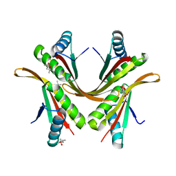
1H0Y
 | | Structure of Alba: an archaeal chromatin protein modulated by acetylation | | 分子名称: | DNA BINDING PROTEIN SSO10B, SULFATE ION | | 著者 | Wardleworth, B.N, Russell, R.J.M, Bell, S.D, Taylor, G.L, White, M.F. | | 登録日 | 2002-07-01 | | 公開日 | 2002-09-05 | | 最終更新日 | 2023-12-13 | | 実験手法 | X-RAY DIFFRACTION (2.8 Å) | | 主引用文献 | Structure of Alba: An Archaeal Chromatin Protein Modulated by Acetylation
Embo J., 21, 2002
|
|
2X5T
 | | Crystal structure of ORF131 from Sulfolobus islandicus rudivirus 1 | | 分子名称: | MALONATE ION, ORF 131 | | 著者 | Oke, M, Carter, L.G, Johnson, K.A, Liu, H, Mcmahon, S.A, Naismith, J.H, White, M.F. | | 登録日 | 2010-02-10 | | 公開日 | 2010-07-28 | | 最終更新日 | 2023-12-20 | | 実験手法 | X-RAY DIFFRACTION (2.2 Å) | | 主引用文献 | The Scottish Structural Proteomics Facility: Targets, Methods and Outputs.
J.Struct.Funct.Genomics, 11, 2010
|
|
2X7B
 | | Crystal structure of the N-terminal acetylase Ard1 from Sulfolobus solfataricus P2 | | 分子名称: | CHLORIDE ION, COENZYME A, N-ACETYLTRANSFERASE SSO0209 | | 著者 | Oke, M, Carter, L.G, Johnson, K.A, Liu, H, Mcmahon, S.A, Mackay, D, White, M.F, Taylor, G.L, Naismith, J.H. | | 登録日 | 2010-02-25 | | 公開日 | 2010-07-21 | | 最終更新日 | 2023-12-20 | | 実験手法 | X-RAY DIFFRACTION (1.95 Å) | | 主引用文献 | The Scottish Structural Proteomics Facility: Targets, Methods and Outputs.
J.Struct.Funct.Genomics, 11, 2010
|
|
2X5R
 | | Crystal Structure of the hypothetical protein ORF126 from Pyrobaculum spherical virus | | 分子名称: | HYPOTHETICAL PROTEIN ORF126, ZINC ION | | 著者 | Oke, M, Carter, L.G, Johnson, K.A, Liu, H, Mcmahon, S.A, White, M.F, Naismith, J.H. | | 登録日 | 2010-02-10 | | 公開日 | 2010-07-28 | | 最終更新日 | 2024-05-08 | | 実験手法 | X-RAY DIFFRACTION (2 Å) | | 主引用文献 | The Scottish Structural Proteomics Facility: Targets, Methods and Outputs.
J.Struct.Funct.Genomics, 11, 2010
|
|
2X4K
 | | Crystal structure of SAR1376, a putative 4-oxalocrotonate tautomerase from the methicillin-resistant Staphylococcus aureus (MRSA) | | 分子名称: | 4-OXALOCROTONATE TAUTOMERASE, ACETATE ION, PHOSPHATE ION, ... | | 著者 | Oke, M, Carter, L.G, Johnson, K.A, Liu, H, Mcmahon, S.A, White, M.F, Naismith, J.H. | | 登録日 | 2010-02-01 | | 公開日 | 2010-07-21 | | 最終更新日 | 2024-05-08 | | 実験手法 | X-RAY DIFFRACTION (1.1 Å) | | 主引用文献 | The Scottish Structural Proteomics Facility: Targets, Methods and Outputs.
J.Struct.Funct.Genomics, 11, 2010
|
|
2X3N
 | | Crystal structure of pqsL, a probable FAD-dependent monooxygenase from Pseudomonas aeruginosa | | 分子名称: | FLAVIN-ADENINE DINUCLEOTIDE, PROBABLE FAD-DEPENDENT MONOOXYGENASE | | 著者 | Oke, M, Carter, L.G, Johnson, K.A, Liu, H, Mcmahon, S.A, White, M.F, Naismith, J.H. | | 登録日 | 2010-01-25 | | 公開日 | 2010-07-21 | | 最終更新日 | 2024-05-08 | | 実験手法 | X-RAY DIFFRACTION (1.75 Å) | | 主引用文献 | The Scottish Structural Proteomics Facility: Targets, Methods and Outputs.
J.Struct.Funct.Genomics, 11, 2010
|
|
2X4H
 | | Crystal Structure of the hypothetical protein SSo2273 from Sulfolobus solfataricus | | 分子名称: | HYPOTHETICAL PROTEIN SSO2273, ZINC ION | | 著者 | Oke, M, Carter, L.G, Johnson, K.A, Liu, H, Mcmahon, S.A, White, M.F, Naismith, J.H. | | 登録日 | 2010-01-31 | | 公開日 | 2010-07-21 | | 最終更新日 | 2024-05-08 | | 実験手法 | X-RAY DIFFRACTION (2.3 Å) | | 主引用文献 | The Scottish Structural Proteomics Facility: Targets, Methods and Outputs.
J.Struct.Funct.Genomics, 11, 2010
|
|
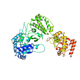
1HH1
 | | THE STRUCTURE OF HJC, A HOLLIDAY JUNCTION RESOLVING ENZYME FROM SULFOLOBUS SOLFATARICUS | | 分子名称: | HOLLIDAY JUNCTION RESOLVING ENZYME HJC | | 著者 | Bond, C.S, Kvaratskhelia, M, Richard, D, White, M.F, Hunter, W.N. | | 登録日 | 2000-12-18 | | 公開日 | 2001-04-06 | | 最終更新日 | 2024-05-08 | | 実験手法 | X-RAY DIFFRACTION (2.15 Å) | | 主引用文献 | Structure of Hjc, a Holliday Junction Resolvase, from Sulfolobus Solfataricus
Proc.Natl.Acad.Sci.USA, 98, 2001
|
|
7BDV
 | | Structure of Can2 from Sulfobacillus thermosulfidooxidans in complex with cyclic tetra-adenylate (cA4) | | 分子名称: | Can2, Cyclic tetraadenosine monophosphate (cA4) | | 著者 | McQuarrie, S, McMahon, S.A, Gloster, T.M, White, M.F, Graham, S, Zhu, W, Gruschow, S. | | 登録日 | 2020-12-22 | | 公開日 | 2021-03-03 | | 最終更新日 | 2023-12-13 | | 実験手法 | X-RAY DIFFRACTION (2.02 Å) | | 主引用文献 | The CRISPR ancillary effector Can2 is a dual-specificity nuclease potentiating type III CRISPR defence.
Nucleic Acids Res., 49, 2021
|
|
6YUD
 | | Structure of Csx3/Crn3 from Archaeoglobus fulgidus in complex with cyclic tetra-adenylate (cA4) | | 分子名称: | Cyclic tetraadenosine monophosphate (cA4), Uncharacterized protein AF_1864 | | 著者 | McQuarrie, S, Gloster, T.M, White, M.F, Graham, S, Athukoralage, J.S, Gruschow, S. | | 登録日 | 2020-04-27 | | 公開日 | 2020-08-19 | | 最終更新日 | 2024-01-24 | | 実験手法 | X-RAY DIFFRACTION (1.84 Å) | | 主引用文献 | Tetramerisation of the CRISPR ring nuclease Crn3/Csx3 facilitates cyclic oligoadenylate cleavage.
Elife, 9, 2020
|
|
8QJK
 | | Structure of the cytoplasmic domain of csx23 from Vibrio cholera in complex with cyclic tetra-adenylate (cA4) | | 分子名称: | ACETYL GROUP, Cyclic tetraadenosine monophosphate (cA4), SODIUM ION, ... | | 著者 | McMahon, S.A, McQuarrie, S, Gloster, T.M, Gruschow, S, White, M.F. | | 登録日 | 2023-09-13 | | 公開日 | 2024-08-07 | | 実験手法 | X-RAY DIFFRACTION (1.761 Å) | | 主引用文献 | A cyclic-nucleotide binding membrane protein provides CRISPR-mediated antiphage defence in Vibrio cholera
To Be Published
|
|
8PCW
 | | Structure of Csm6' from Streptococcus thermophilus | | 分子名称: | CRISPR system endoribonuclease Csm6' | | 著者 | McQuarrie, S.J, Athukoralage, J.S, McMahon, S.A, Graham, S, Ackerman, K, Bode, B.E, White, M.F, Gloster, T.M. | | 登録日 | 2023-06-11 | | 公開日 | 2023-10-04 | | 最終更新日 | 2023-11-08 | | 実験手法 | X-RAY DIFFRACTION (3.54 Å) | | 主引用文献 | Activation of Csm6 ribonuclease by cyclic nucleotide binding: in an emergency, twist to open.
Nucleic Acids Res., 51, 2023
|
|
8PE3
 | | Structure of Csm6' from Streptococcus thermophilus in complex with cyclic hexa-adenylate (cA6) | | 分子名称: | CRISPR system endoribonuclease Csm6', Cyclic hexaadenosine monophosphate (cA6), RNA | | 著者 | McQuarrie, S.J, Athukoralage, J.S, McMahon, S.A, Graham, S, Ackerman, K, Bode, B.E, White, M.F, Gloster, T.M. | | 登録日 | 2023-06-13 | | 公開日 | 2023-10-04 | | 最終更新日 | 2023-11-08 | | 実験手法 | X-RAY DIFFRACTION (1.96 Å) | | 主引用文献 | Activation of Csm6 ribonuclease by cyclic nucleotide binding: in an emergency, twist to open.
Nucleic Acids Res., 51, 2023
|
|
7QQK
 | | TIR-SAVED effector bound to cA3 | | 分子名称: | RNA (5'-R(P*AP*AP*A)-3'), TIR_SAVED fusion protein | | 著者 | Spagnolo, L, White, M.F, Hogrel, G, Guild, A. | | 登録日 | 2022-01-09 | | 公開日 | 2022-06-15 | | 最終更新日 | 2024-07-17 | | 実験手法 | ELECTRON MICROSCOPY (3.8 Å) | | 主引用文献 | Cyclic nucleotide-induced helical structure activates a TIR immune effector.
Nature, 608, 2022
|
|
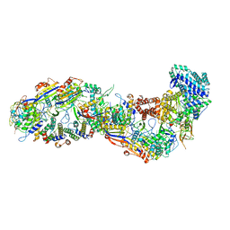
8ANE
 | | Structure of the type I-G CRISPR effector | | 分子名称: | Cas7, RNA (66-MER) | | 著者 | Shangguan, Q, Graham, S, Sundaramoorthy, R, White, M.F. | | 登録日 | 2022-08-05 | | 公開日 | 2022-11-09 | | 最終更新日 | 2024-07-24 | | 実験手法 | ELECTRON MICROSCOPY (3.2 Å) | | 主引用文献 | Structure and mechanism of the type I-G CRISPR effector.
Nucleic Acids Res., 50, 2022
|
|
8B2X
 | |
8BMW
 | | SsoCsm | | 分子名称: | CRISPR-associated Cas7 paralog (Type III-D), CRISPR-associated protein Cas10 (Type III-D), CRISPR-associated protein Cas5 (Type III-D), ... | | 著者 | Spagnolo, L, White, M.F. | | 登録日 | 2022-11-11 | | 公開日 | 2023-03-01 | | 最終更新日 | 2023-03-22 | | 実験手法 | ELECTRON MICROSCOPY (3.5 Å) | | 主引用文献 | Structure of the Saccharolobus solfataricus type III-D CRISPR effector.
Curr Res Struct Biol, 5, 2023
|
|
3FFE
 | | Structure of Achromobactin Synthetase Protein D, (AcsD) | | 分子名称: | AcsD | | 著者 | McMahon, S.A, Liu, H, Carter, L, Oke, M, Johnson, K.A, Schmelz, S, Challis, G.L, White, M.F, Naismith, J.H, Scottish Structural Proteomics Facility (SSPF) | | 登録日 | 2008-12-03 | | 公開日 | 2009-02-03 | | 最終更新日 | 2023-12-27 | | 実験手法 | X-RAY DIFFRACTION (2.25 Å) | | 主引用文献 | AcsD catalyzes enantioselective citrate desymmetrization in siderophore biosynthesis
Nat.Chem.Biol., 5, 2009
|
|
2IVY
 | | Crystal structure of hypothetical protein sso1404 from Sulfolobus solfataricus P2 | | 分子名称: | HYPOTHETICAL PROTEIN SSO1404 | | 著者 | Yan, X, Carter, L.G, Dorward, M, Liu, H, McMahon, S.A, Oke, M, Powers, H, White, M.F, Naismith, J.H. | | 登録日 | 2006-06-22 | | 公開日 | 2006-06-28 | | 最終更新日 | 2023-12-13 | | 実験手法 | X-RAY DIFFRACTION (1.4 Å) | | 主引用文献 | The Scottish Structural Proteomics Facility: Targets, Methods and Outputs.
J.Struct.Funct.Genomics, 11, 2010
|
|
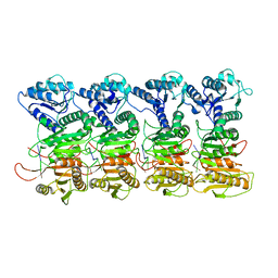
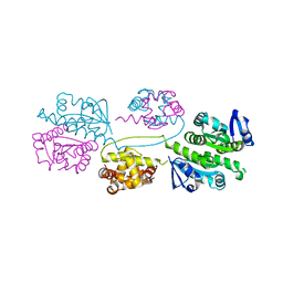
2IX2
 | | Crystal structure of the heterotrimeric PCNA from Sulfolobus solfataricus | | 分子名称: | DNA POLYMERASE SLIDING CLAMP A, DNA POLYMERASE SLIDING CLAMP B, DNA POLYMERASE SLIDING CLAMP C | | 著者 | Williams, G.J, Johnson, K, McMahon, S.A, Carter, L, Oke, M, Liu, H, Taylor, G.L, White, M.F, Naismith, J.H. | | 登録日 | 2006-07-05 | | 公開日 | 2006-10-04 | | 最終更新日 | 2023-12-13 | | 実験手法 | X-RAY DIFFRACTION (2.2 Å) | | 主引用文献 | Structure of the Heterotrimeric PCNA from Sulfolobus Solfataricus.
Acta Crystallogr.,Sect.F, 62, 2006
|
|
2BGW
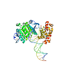 | | XPF from Aeropyrum pernix, complex with DNA | | 分子名称: | 5'-D(*GP*AP*TP*CP*AP*CP*AP*GP*AP*TP *GP*CP*TP*GP*A)-3', 5'-D(*TP*CP*AP*GP*CP*AP*TP*CP*TP*GP *TP*GP*AP*TP*C)-3', MAGNESIUM ION, ... | | 著者 | Newman, M, Murray-Rust, J, Lally, J, Rudolf, J, Fadden, A, Knowles, P.P, White, M.F, McDonald, N.Q. | | 登録日 | 2005-01-06 | | 公開日 | 2005-02-23 | | 最終更新日 | 2023-12-13 | | 実験手法 | X-RAY DIFFRACTION (2.8 Å) | | 主引用文献 | Structure of an XPF endonuclease with and without DNA suggests a model for substrate recognition.
EMBO J., 24, 2005
|
|
2BKE
 | | Conformational Flexibility Revealed by the Crystal Structure of a Crenarchaeal RadA | | 分子名称: | CHLORIDE ION, DNA REPAIR AND RECOMBINATION PROTEIN RADA | | 著者 | Ariza, A, Richard, D.L, White, M.F, Bond, C.S. | | 登録日 | 2005-02-15 | | 公開日 | 2005-03-16 | | 最終更新日 | 2023-12-13 | | 実験手法 | X-RAY DIFFRACTION (3.2 Å) | | 主引用文献 | Conformational Flexibility Revealed by the Crystal Structure of a Crenarchaeal Rada
Nucleic Acids Res., 33, 2005
|
|
2BHN
 | | XPF from Aeropyrum pernix | | 分子名称: | XPF ENDONUCLEASE | | 著者 | Newman, M, Murray-Rust, J, Lally, J, Rudolf, J, Fadden, A, Knowles, P.P, White, M.F, McDonald, N.Q. | | 登録日 | 2005-01-14 | | 公開日 | 2005-02-23 | | 最終更新日 | 2023-12-13 | | 実験手法 | X-RAY DIFFRACTION (3.2 Å) | | 主引用文献 | Structure of an XPF endonuclease with and without DNA suggests a model for substrate recognition.
EMBO J., 24, 2005
|
|
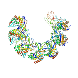
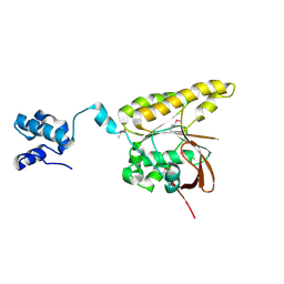
2BKY
 | | Crystal structure of the Alba1:Alba2 heterodimer from sulfolobus solfataricus | | 分子名称: | (4S)-2-METHYL-2,4-PENTANEDIOL, DNA/RNA-BINDING PROTEIN ALBA 1, DNA/RNA-BINDING PROTEIN ALBA 2 | | 著者 | Jelinska, C, Conroy, M.J, Craven, C.J, Bullough, P.A, Waltho, J.P, Taylor, G.L, White, M.F. | | 登録日 | 2005-02-22 | | 公開日 | 2005-07-14 | | 最終更新日 | 2024-05-08 | | 実験手法 | X-RAY DIFFRACTION (1.7 Å) | | 主引用文献 | Obligate Heterodimerization of the Archaeal Alba2 Protein with Alba1 Provides a Mechanism for Control of DNA Packaging.
Structure, 13, 2005
|
|


