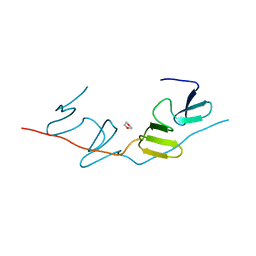
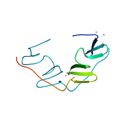
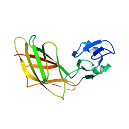
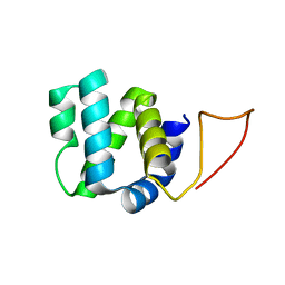
2B1O
 
 | | Solution Structure of Ca2+-bound DdCAD-1 | | 分子名称: | CALCIUM ION, Calcium-dependent cell adhesion molecule-1 | | 著者 | Lin, Z, Sriskanthadevan, S, Huang, H.B, Siu, C.H, Yang, D.W. | | 登録日 | 2005-09-16 | | 公開日 | 2006-09-26 | | 最終更新日 | 2024-05-29 | | 実験手法 | SOLUTION NMR | | 主引用文献 | Solution structures of the adhesion molecule DdCAD-1 reveal new insights into Ca(2+)-dependent cell-cell adhesion
Nat.Struct.Mol.Biol., 13, 2006
|
|
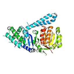
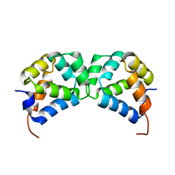
7BQF
 | | Dimerization of SAV1 WW tandem | | 分子名称: | 1,4-DIETHYLENE DIOXIDE, Protein salvador homolog 1 | | 著者 | Lin, Z, Zhang, M. | | 登録日 | 2020-03-24 | | 公開日 | 2020-09-23 | | 最終更新日 | 2023-11-29 | | 実験手法 | X-RAY DIFFRACTION (1.70037615 Å) | | 主引用文献 | A WW Tandem-Mediated Dimerization Mode of SAV1 Essential for Hippo Signaling.
Cell Rep, 32, 2020
|
|
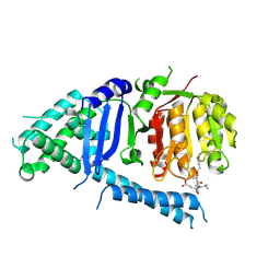
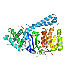
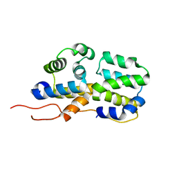
7BQG
 | | Complex structure of SAV1 and Dendrin | | 分子名称: | POTASSIUM ION, Protein salvador homolog 1,Dendrin | | 著者 | Lin, Z, Zhang, M. | | 登録日 | 2020-03-24 | | 公開日 | 2020-09-23 | | 最終更新日 | 2023-11-29 | | 実験手法 | X-RAY DIFFRACTION (1.55010867 Å) | | 主引用文献 | A WW Tandem-Mediated Dimerization Mode of SAV1 Essential for Hippo Signaling.
Cell Rep, 32, 2020
|
|
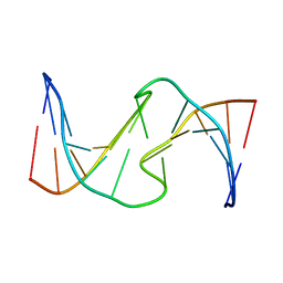
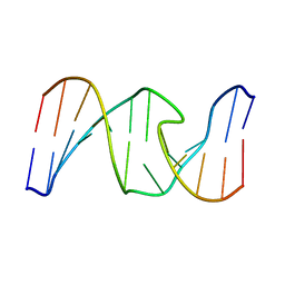
6T59
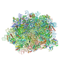 | | Structure of rabbit 80S ribosome translating beta-tubulin in complex with tetratricopeptide protein 5 and nascent chain-associated complex | | 分子名称: | 28S ribosomal RNA, 5.8S ribosomal RNA, 5S ribosomal RNA, ... | | 著者 | Lin, Z, Gasic, I, Chandrasekaran, V, Peters, N, Shao, S, Ramakrishnan, V, Mitchison, T.J, Hegde, R.S. | | 登録日 | 2019-10-15 | | 公開日 | 2019-11-27 | | 最終更新日 | 2020-01-15 | | 実験手法 | ELECTRON MICROSCOPY (3.11 Å) | | 主引用文献 | TTC5 mediates autoregulation of tubulin via mRNA degradation.
Science, 367, 2020
|
|
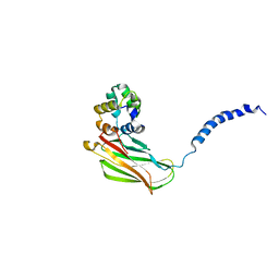
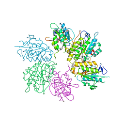
3PB1
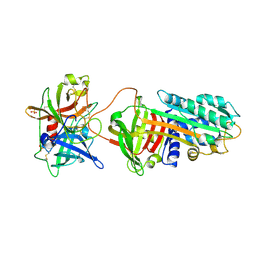 | | Crystal Structure of a Michaelis Complex between Plasminogen Activator Inhibitor-1 and Urokinase-type Plasminogen Activator | | 分子名称: | Plasminogen activator inhibitor 1, Plasminogen activator, urokinase, ... | | 著者 | Lin, Z, Jiang, L, Huang, M, Structure 2 Function Project (S2F) | | 登録日 | 2010-10-20 | | 公開日 | 2010-12-29 | | 最終更新日 | 2023-11-01 | | 実験手法 | X-RAY DIFFRACTION (2.3 Å) | | 主引用文献 | Structural basis for recognition of urokinase-type plasminogen activator by plasminogen activator inhibitor-1.
J.Biol.Chem., 286, 2011
|
|
5Z6R
 | | SPASTIN AAA WITH ATP | | 分子名称: | ADENOSINE-5'-TRIPHOSPHATE, Spastin | | 著者 | Lin, Z, Wang, C, Shen, Y. | | 登録日 | 2018-01-25 | | 公開日 | 2019-01-30 | | 最終更新日 | 2024-03-27 | | 実験手法 | X-RAY DIFFRACTION (3 Å) | | 主引用文献 | The Aaa Family Protein Spastin Severs A Microtubule
To Be Published
|
|
6J68
 | | Structure of KIBRA and LATS1 Complex | | 分子名称: | Peptide from Serine/threonine-protein kinase LATS1, Protein KIBRA | | 著者 | Lin, Z, Yang, Z, Ji, Z, Zhang, M. | | 登録日 | 2019-01-14 | | 公開日 | 2019-09-25 | | 最終更新日 | 2024-03-27 | | 実験手法 | X-RAY DIFFRACTION (2.495 Å) | | 主引用文献 | Decoding WW domain tandem-mediated target recognitions in tissue growth and cell polarity.
Elife, 8, 2019
|
|
6JJY
 | | Crystal Structure of KIBRA and beta-Dystroglycan | | 分子名称: | Peptide from Dystroglycan, Protein KIBRA, SULFATE ION | | 著者 | Lin, Z, Yang, Z, Ji, Z, Zhang, M. | | 登録日 | 2019-02-27 | | 公開日 | 2019-09-25 | | 最終更新日 | 2023-11-22 | | 実験手法 | X-RAY DIFFRACTION (2.298 Å) | | 主引用文献 | Decoding WW domain tandem-mediated target recognitions in tissue growth and cell polarity.
Elife, 8, 2019
|
|
6JK0
 | | Crystal Structure of YAP1 and Dendrin complex | | 分子名称: | CALCIUM ION, Transcriptional coactivator YAP1,Dendrin | | 著者 | Lin, Z, Yang, Z, Ji, Z, Zhang, M. | | 登録日 | 2019-02-27 | | 公開日 | 2019-09-25 | | 最終更新日 | 2023-11-22 | | 実験手法 | X-RAY DIFFRACTION (3.1 Å) | | 主引用文献 | Decoding WW domain tandem-mediated target recognitions in tissue growth and cell polarity.
Elife, 8, 2019
|
|
6JK1
 | | Crystal Structure of YAP1 and Dendrin complex 2 | | 分子名称: | Dendrin,Transcriptional coactivator YAP1 | | 著者 | Lin, Z, Yang, Z, Ji, Z, Zhang, M. | | 登録日 | 2019-02-27 | | 公開日 | 2019-09-25 | | 最終更新日 | 2023-11-22 | | 実験手法 | X-RAY DIFFRACTION (2 Å) | | 主引用文献 | Decoding WW domain tandem-mediated target recognitions in tissue growth and cell polarity.
Elife, 8, 2019
|
|
6JJW
 | | Crystal Structure of KIBRA and PTPN14 complex | | 分子名称: | CHLORIDE ION, FORMIC ACID, GLYCEROL, ... | | 著者 | Lin, Z, Yang, Z, Ji, Z, Zhang, M. | | 登録日 | 2019-02-27 | | 公開日 | 2019-09-25 | | 最終更新日 | 2023-11-22 | | 実験手法 | X-RAY DIFFRACTION (2.4 Å) | | 主引用文献 | Decoding WW domain tandem-mediated target recognitions in tissue growth and cell polarity.
Elife, 8, 2019
|
|
6JJZ
 | | Crystal Structure of MAGI2 and Dendrin complex | | 分子名称: | ACETATE ION, Membrane-associated guanylate kinase, WW and PDZ domain-containing protein 2, ... | | 著者 | Lin, Z, Zhang, H, Yang, Z, Ji, Z, Zhang, M, Zhu, J. | | 登録日 | 2019-02-27 | | 公開日 | 2019-09-25 | | 最終更新日 | 2023-11-22 | | 実験手法 | X-RAY DIFFRACTION (1.65 Å) | | 主引用文献 | Decoding WW domain tandem-mediated target recognitions in tissue growth and cell polarity.
Elife, 8, 2019
|
|
6JJX
 | | Crystal Structure of KIBRA and Angiomotin complex | | 分子名称: | Peptide from Angiomotin, Protein KIBRA | | 著者 | Lin, Z, Yang, Z, Ji, Z, Zhang, M. | | 登録日 | 2019-02-27 | | 公開日 | 2019-09-25 | | 最終更新日 | 2023-11-22 | | 実験手法 | X-RAY DIFFRACTION (2 Å) | | 主引用文献 | Decoding WW domain tandem-mediated target recognitions in tissue growth and cell polarity.
Elife, 8, 2019
|
|
1YHP
 | |
5FBY
 | | Crystal structure of ctSPD | | 分子名称: | cleaved peptide, separase | | 著者 | Lin, Z, Luo, X, Yu, H. | | 登録日 | 2015-12-14 | | 公開日 | 2016-03-30 | | 最終更新日 | 2024-03-06 | | 実験手法 | X-RAY DIFFRACTION (1.898 Å) | | 主引用文献 | Structural basis of cohesin cleavage by separase.
Nature, 532, 2016
|
|
5FC3
 | |
5FC2
 | |
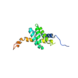
2N7Z
 | | Solution structure of RIP2 CARD | | 分子名称: | Receptor-interacting serine/threonine-protein kinase 2 | | 著者 | Lin, Z, Ibanez, C.F. | | 登録日 | 2015-09-28 | | 公開日 | 2015-12-23 | | 最終更新日 | 2024-05-01 | | 実験手法 | SOLUTION NMR | | 主引用文献 | Structural basis of death domain signaling in the p75 neurotrophin receptor
Elife, 4, 2015
|
|
2N97
 | | DD homodimer | | 分子名称: | Tumor necrosis factor receptor superfamily member 16 | | 著者 | Lin, Z, Ibanez, C.F. | | 登録日 | 2015-11-07 | | 公開日 | 2015-12-23 | | 最終更新日 | 2024-05-15 | | 実験手法 | SOLUTION NMR | | 主引用文献 | Structural basis of death domain signaling in the p75 neurotrophin receptor
Elife, 4, 2015
|
|
2N83
 | | p75NTR DD:RIP2 CARD | | 分子名称: | Receptor-interacting serine/threonine-protein kinase 2, Tumor necrosis factor receptor superfamily member 16 | | 著者 | Lin, Z, Ibanez, C.F. | | 登録日 | 2015-10-02 | | 公開日 | 2015-12-23 | | 最終更新日 | 2024-05-01 | | 実験手法 | SOLUTION NMR | | 主引用文献 | Structural basis of death domain signaling in the p75 neurotrophin receptor
Elife, 4, 2015
|
|
2N80
 | | p75NTR DD:RhoGDI | | 分子名称: | Rho GDP-dissociation inhibitor 1, Tumor necrosis factor receptor superfamily member 16 | | 著者 | Lin, Z, Ibanez, C.F. | | 登録日 | 2015-09-30 | | 公開日 | 2015-12-23 | | 最終更新日 | 2024-05-01 | | 実験手法 | SOLUTION NMR | | 主引用文献 | Structural basis of death domain signaling in the p75 neurotrophin receptor
Elife, 4, 2015
|
|
4EJS
 | | Structure of yeast elongator subcomplex Elp456 | | 分子名称: | Elongator complex protein 4, Elongator complex protein 5, Elongator complex protein 6 | | 著者 | Lin, Z, Zhao, W, Long, J, Shen, Y. | | 登録日 | 2012-04-07 | | 公開日 | 2012-05-02 | | 最終更新日 | 2024-03-20 | | 実験手法 | X-RAY DIFFRACTION (2.606 Å) | | 主引用文献 | Crystal structure of elongator subcomplex Elp4-6
J.Biol.Chem., 287, 2012
|
|
1HT7
 | |
1HT4
 | |
2K3N
 | |


