3L6J
 
 | |
7DW9
 | |
3TVY
 | |
7EO0
 | |
7WGR
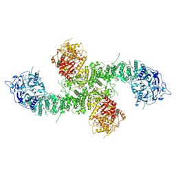
 | | Cryo-electron microscopic structure of the 2-oxoglutarate dehydrogenase (E1) component of the human alpha-ketoglutarate (2-oxoglutarate) dehydrogenase complex | | 分子名称: | 2-oxoglutarate dehydrogenase, mitochondrial, CALCIUM ION, ... | | 著者 | Yu, X, Yang, W, Zhong, Y.H, Ma, X.M, Gao, Y.Z. | | 登録日 | 2021-12-28 | | 公開日 | 2022-06-01 | | 最終更新日 | 2024-06-26 | | 実験手法 | ELECTRON MICROSCOPY (2.92 Å) | | 主引用文献 | Structural basis for the activity and regulation of human alpha-ketoglutarate dehydrogenase revealed by Cryo-EM
Biochem.Biophys.Res.Commun., 602, 2022
|
|
7E4M
 | |
7E4O
 | |
7E4N
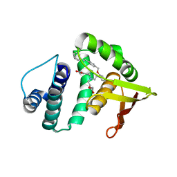
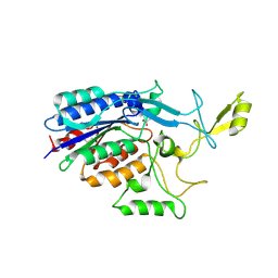
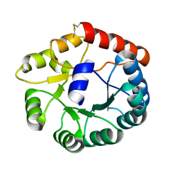
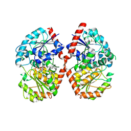
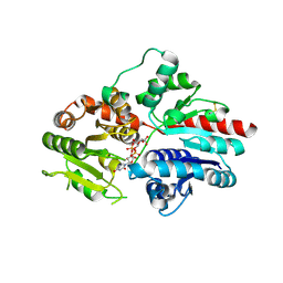
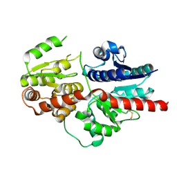
 | | Crystal structure of Sat1646 | | 分子名称: | MAGNESIUM ION, Sat1646 | | 著者 | Yu, J.H, Xing, B.Y, Ma, M. | | 登録日 | 2021-02-14 | | 公開日 | 2021-10-06 | | 最終更新日 | 2024-05-29 | | 実験手法 | X-RAY DIFFRACTION (1.94 Å) | | 主引用文献 | Functional characterization and structural bases of two class I diterpene synthases in pimarane-type diterpene biosynthesis
Commun Chem, 4, 2021
|
|
3TXA
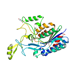
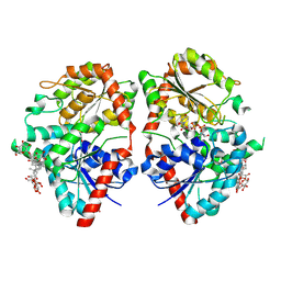
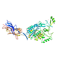 | | Structural Analysis of Adhesive Tip pilin, GBS104 from Group B Streptococcus agalactiae | | 分子名称: | CADMIUM ION, Cell wall surface anchor family protein, LITHIUM ION, ... | | 著者 | Krishnan, V, Narayana, S.V.L. | | 登録日 | 2011-09-23 | | 公開日 | 2013-03-27 | | 最終更新日 | 2023-09-13 | | 実験手法 | X-RAY DIFFRACTION (2.619 Å) | | 主引用文献 | Structure of Streptococcus agalactiae tip pilin GBS104: a model for GBS pili assembly and host interactions.
Acta Crystallogr.,Sect.D, 69, 2013
|
|
3TW0
 | |
8Y0R
 | |
8Y0Q
 | |
8D4X
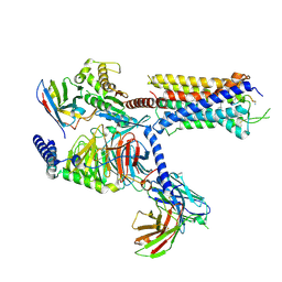
 | | Structure of the human UBR5 HECT-type E3 ubiquitin ligase in a dimeric form | | 分子名称: | E3 ubiquitin-protein ligase UBR5, ZINC ION | | 著者 | Wang, F, He, Q, Lin, G, Li, H. | | 登録日 | 2022-06-02 | | 公開日 | 2023-04-19 | | 最終更新日 | 2024-06-12 | | 実験手法 | ELECTRON MICROSCOPY (2.8 Å) | | 主引用文献 | Structure of the human UBR5 E3 ubiquitin ligase.
Structure, 31, 2023
|
|
8DGN
 | |
8DGM
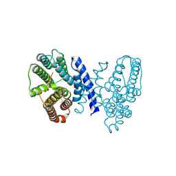
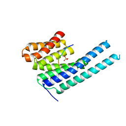
 | | 14-3-3 epsilon bound to phosphorylated PEAK1 (pT1165) peptide | | 分子名称: | 1,2-ETHANEDIOL, 14-3-3 protein epsilon, Inactive tyrosine-protein kinase PEAK1 | | 著者 | Roy, M.J, Hardy, J.M, Lucet, I.S. | | 登録日 | 2022-06-24 | | 公開日 | 2023-06-07 | | 最終更新日 | 2023-10-25 | | 実験手法 | X-RAY DIFFRACTION (3.2 Å) | | 主引用文献 | Structural mapping of PEAK pseudokinase interactions identifies 14-3-3 as a molecular switch for PEAK3 signaling.
Nat Commun, 14, 2023
|
|
8DGO
 | |
8DGP
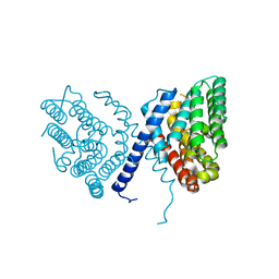
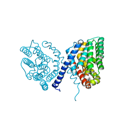
 | | 14-3-3 epsilon bound to phosphorylated PEAK3 (pS69) peptide | | 分子名称: | 1,2-ETHANEDIOL, 14-3-3 protein epsilon, Phosphorylated PEAK3 (pS69) peptide, ... | | 著者 | Roy, M.J, Hardy, J.M, Lucet, I.S. | | 登録日 | 2022-06-24 | | 公開日 | 2023-06-07 | | 最終更新日 | 2023-10-25 | | 実験手法 | X-RAY DIFFRACTION (2.7 Å) | | 主引用文献 | Structural mapping of PEAK pseudokinase interactions identifies 14-3-3 as a molecular switch for PEAK3 signaling.
Nat Commun, 14, 2023
|
|
8JGC
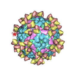
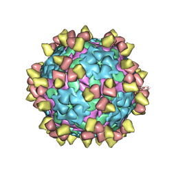
 | | Cryo-EM structure of Mi3 fused with LOV2 | | 分子名称: | LOV domain-containing protein,2-dehydro-3-deoxyphosphogluconate aldolase/4-hydroxy-2-oxoglutarate aldolase | | 著者 | Zhang, H.W, Kang, W, Xue, C. | | 登録日 | 2023-05-20 | | 公開日 | 2024-04-24 | | 実験手法 | ELECTRON MICROSCOPY (3.44 Å) | | 主引用文献 | Dynamic Metabolons Using Stimuli-Responsive Protein Cages.
J.Am.Chem.Soc., 146, 2024
|
|
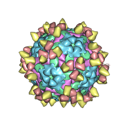
8JGA
 | | Cryo-EM structure of Mi3 fused with FKBP | | 分子名称: | Peptidyl-prolyl cis-trans isomerase FKBP1A,2-dehydro-3-deoxyphosphogluconate aldolase/4-hydroxy-2-oxoglutarate aldolase | | 著者 | Zhang, H.W, Kang, W, Xue, C. | | 登録日 | 2023-05-20 | | 公開日 | 2024-04-24 | | 最終更新日 | 2024-10-16 | | 実験手法 | ELECTRON MICROSCOPY (3.68 Å) | | 主引用文献 | Dynamic Metabolons Using Stimuli-Responsive Protein Cages.
J.Am.Chem.Soc., 146, 2024
|
|
8E0Q
 | | Structure of the human UBR5 HECT-type E3 ubiquitin ligase in a C2 symmetric dimeric form | | 分子名称: | E3 ubiquitin-protein ligase UBR5, ZINC ION | | 著者 | Wang, F, He, Q, Lin, G, Li, H. | | 登録日 | 2022-08-09 | | 公開日 | 2023-04-19 | | 最終更新日 | 2024-06-12 | | 実験手法 | ELECTRON MICROSCOPY (2.66 Å) | | 主引用文献 | Structure of the human UBR5 E3 ubiquitin ligase.
Structure, 31, 2023
|
|
8EWI
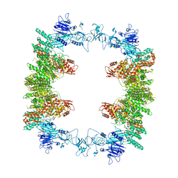 | | Structure of the human UBR5 HECT-type E3 ubiquitin ligase in a tetrameric form | | 分子名称: | E3 ubiquitin-protein ligase UBR5, ZINC ION | | 著者 | Wang, F, He, Q, Lin, G, Li, H. | | 登録日 | 2022-10-23 | | 公開日 | 2023-04-19 | | 最終更新日 | 2024-06-19 | | 実験手法 | ELECTRON MICROSCOPY (3.5 Å) | | 主引用文献 | Structure of the human UBR5 E3 ubiquitin ligase.
Structure, 31, 2023
|
|
8HJF
 | | Crystal structure of glycosyltransferase SgUGT94-289-3 in complex with M5, state 2 | | 分子名称: | (20S)-2,5,8,11,14,17-HEXAMETHYL-3,6,9,12,15,18-HEXAOXAHENICOSANE-1,20-DIOL, (2S,3S,4S,5R,6R)-2-(hydroxymethyl)-6-[[(2R,3S,4S,5R,6R)-6-[[(3S,8S,9R,10R,11R,13R,14S,17R)-17-[(2S,5R)-5-[(2S,3R,4S,5S,6R)-3-[(2R,3R,4S,5S,6S)-6-(hydroxymethyl)-3,4,5-tris(oxidanyl)oxan-2-yl]oxy-6-[[(2R,3R,4S,5S,6S)-6-(hydroxymethyl)-3,4,5-tris(oxidanyl)oxan-2-yl]oxymethyl]-4,5-bis(oxidanyl)oxan-2-yl]oxy-6-methyl-6-oxidanyl-heptan-2-yl]-4,4,9,13,14-pentamethyl-11-oxidanyl-2,3,7,8,10,11,12,15,16,17-decahydro-1H-cyclopenta[a]phenanthren-3-yl]oxy]-3,4,5-tris(oxidanyl)oxan-2-yl]methoxy]oxane-3,4,5-triol, 2-AMINO-2-HYDROXYMETHYL-PROPANE-1,3-DIOL, ... | | 著者 | Li, M, Zhang, S, Cui, S. | | 登録日 | 2022-11-23 | | 公開日 | 2024-05-29 | | 最終更新日 | 2024-08-14 | | 実験手法 | X-RAY DIFFRACTION (2 Å) | | 主引用文献 | Structural insights into the catalytic selectivity of glycosyltransferase SgUGT94-289-3 towards mogrosides.
Nat Commun, 15, 2024
|
|
8HJP
 | | Crystal structure of glycosyltransferase SgUGT94-289-3 in complex with UDP state 1 | | 分子名称: | (20S)-2,5,8,11,14,17-HEXAMETHYL-3,6,9,12,15,18-HEXAOXAHENICOSANE-1,20-DIOL, URIDINE-5'-DIPHOSPHATE, glycosyltranseferease | | 著者 | Li, M, Zhang, S, Cui, S. | | 登録日 | 2022-11-23 | | 公開日 | 2024-05-29 | | 最終更新日 | 2024-08-14 | | 実験手法 | X-RAY DIFFRACTION (2.2 Å) | | 主引用文献 | Structural insights into the catalytic selectivity of glycosyltransferase SgUGT94-289-3 towards mogrosides.
Nat Commun, 15, 2024
|
|
8HJN
 | |
8HJQ
 | |


