8EWG
 
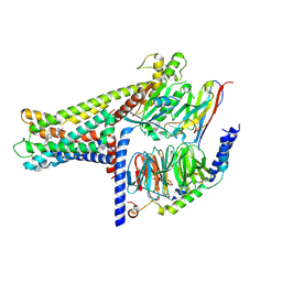
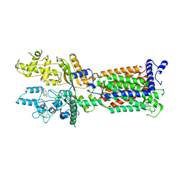
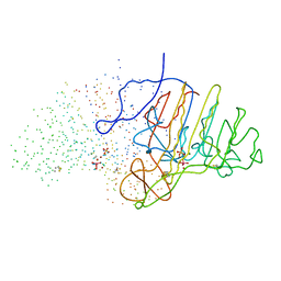
 | | Cryo-EM structure of a riboendonclease | | 分子名称: | CRISPR-associated endonuclease Cas9, RNA (56-MER) | | 著者 | Li, Z, Wang, F. | | 登録日 | 2022-10-23 | | 公開日 | 2023-08-30 | | 最終更新日 | 2024-05-01 | | 実験手法 | ELECTRON MICROSCOPY (2.9 Å) | | 主引用文献 | Structural Basis for the Ribonuclease Activity of a Thermostable CRISPR-Cas13a from Thermoclostridium caenicola.
J.Mol.Biol., 435, 2023
|
|
4L53
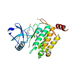 | | Crystal Structure of (1R,4R)-4-{4-[7-amino-2-(1,2,3-benzothiadiazol-7-yl)-3-chlorofuro[2,3-c]pyridin-4-yl]-1H-pyrazol-1-yl}cyclohexan-1-ol bound to TAK1-TAB1 | | 分子名称: | 1,2-ETHANEDIOL, Mitogen-activated protein kinase kinase kinase 7, TGF-beta-activated kinase 1 and MAP3K7-binding protein 1 chimera, ... | | 著者 | Wang, J, Hornberger, K.R, Crew, A.P, Jestel, A, Maskos, K, Moertl, M. | | 登録日 | 2013-06-10 | | 公開日 | 2013-07-03 | | 最終更新日 | 2024-02-28 | | 実験手法 | X-RAY DIFFRACTION (2.55 Å) | | 主引用文献 | Discovery of 7-aminofuro[2,3-c]pyridine inhibitors of TAK1: Optimization of kinase selectivity and pharmacokinetics.
Bioorg.Med.Chem.Lett., 23, 2013
|
|
4OR9
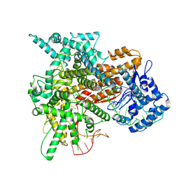
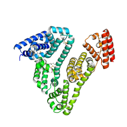
 | | Crystal structure of human calcineurin | | 分子名称: | CALCIUM ION, Calcineurin subunit B type 1, FE (III) ION, ... | | 著者 | Li, S.J, Wang, J, Wu, J.W, Wang, Z.X. | | 登録日 | 2014-02-11 | | 公開日 | 2015-05-20 | | 最終更新日 | 2023-11-08 | | 実験手法 | X-RAY DIFFRACTION (2.23 Å) | | 主引用文献 | Cooperative autoinhibition and multi-level activation mechanisms of calcineurin
To be Published
|
|
4ORA
 | | Crystal structure of a human calcineurin mutant | | 分子名称: | CALCIUM ION, Calcineurin subunit B type 1, FE (III) ION, ... | | 著者 | Li, S.J, Wang, J, Wu, J.W, Wang, Z.X. | | 登録日 | 2014-02-11 | | 公開日 | 2015-05-20 | | 最終更新日 | 2023-11-08 | | 実験手法 | X-RAY DIFFRACTION (2.747 Å) | | 主引用文献 | Cooperative autoinhibition and multi-level activation mechanisms of calcineurin
To be Published
|
|
7X9E
 | | Crystal structure of the 76E1 Fab in complex with a SARS-CoV-2 spike peptide | | 分子名称: | 76E1 Fab Heavy Chain, 76E1 Fab Light Chain, Spike peptide | | 著者 | Chen, X, Zhang, T, Ding, J, Sun, X, Sun, B. | | 登録日 | 2022-03-15 | | 公開日 | 2022-05-11 | | 最終更新日 | 2023-11-29 | | 実験手法 | X-RAY DIFFRACTION (2.6 Å) | | 主引用文献 | Neutralization mechanism of a human antibody with pan-coronavirus reactivity including SARS-CoV-2.
Nat Microbiol, 7, 2022
|
|
7CK8
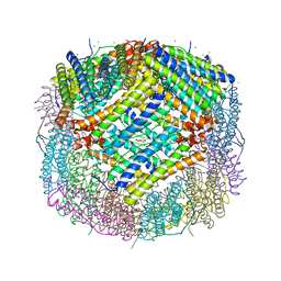 | | Crystal structure of human ferritin heavy chain mutant C90S/C102S/C130S | | 分子名称: | CHLORIDE ION, FE (III) ION, Ferritin heavy chain, ... | | 著者 | Chen, X, Jiang, B, Yan, X, Fan, K. | | 登録日 | 2020-07-16 | | 公開日 | 2021-05-26 | | 最終更新日 | 2024-03-27 | | 実験手法 | X-RAY DIFFRACTION (1.8 Å) | | 主引用文献 | A natural drug entry channel in the ferritin nanocage.
Nano Today, 35, 2020
|
|
7XV9
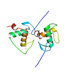 | | Crystal structure of the Human TR4 DNA-Binding Domain | | 分子名称: | Nuclear receptor subfamily 2 group C member 2, ZINC ION | | 著者 | Liu, Y, Chen, Z. | | 登録日 | 2022-05-21 | | 公開日 | 2022-12-28 | | 最終更新日 | 2023-11-29 | | 実験手法 | X-RAY DIFFRACTION (1.599 Å) | | 主引用文献 | Structures of human TR4LBD-JAZF1 and TR4DBD-DNA complexes reveal the molecular basis of transcriptional regulation.
Nucleic Acids Res., 51, 2023
|
|
7XV8
 | |
7XV6
 | |
7XVA
 | |
7WCM
 | | Cryo-EM structure of GPR119-Gs Complex with small molecule agonist MBX-2982 | | 分子名称: | 2-[1-(5-ethylpyrimidin-2-yl)piperidin-4-yl]-4-[[4-(1,2,3,4-tetrazol-1-yl)phenoxy]methyl]-1,3-thiazole, Glucose-dependent insulinotropic receptor, Guanine nucleotide-binding protein G(I)/G(S)/G(O) subunit gamma-2, ... | | 著者 | Qiao, A.N, Wu, S, Ye, S. | | 登録日 | 2021-12-20 | | 公開日 | 2022-12-21 | | 最終更新日 | 2022-12-28 | | 実験手法 | ELECTRON MICROSCOPY (2.33 Å) | | 主引用文献 | Activation and signaling mechanism revealed by GPR119-G s complex structures.
Nat Commun, 13, 2022
|
|
7WCN
 | | Cryo-EM structure of GPR119-Gs Complex with small molecule agonist AR231453 | | 分子名称: | Glucose-dependent insulinotropic receptor, Guanine nucleotide-binding protein G(I)/G(S)/G(O) subunit gamma-2, Guanine nucleotide-binding protein G(I)/G(S)/G(T) subunit beta-1, ... | | 著者 | Qiao, A.N, Wu, S, Ye, S. | | 登録日 | 2021-12-20 | | 公開日 | 2022-12-21 | | 最終更新日 | 2022-12-28 | | 実験手法 | ELECTRON MICROSCOPY (2.87 Å) | | 主引用文献 | Activation and signaling mechanism revealed by GPR119-G s complex structures.
Nat Commun, 13, 2022
|
|
8H4U
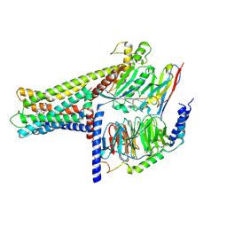
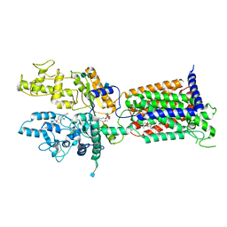
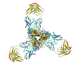
 | | Cryo-EM structure of a riboendonuclease | | 分子名称: | CRISPR-associated endonuclease Cas9 | | 著者 | Li, Z, Wang, F. | | 登録日 | 2022-10-11 | | 公開日 | 2023-08-30 | | 最終更新日 | 2024-03-13 | | 実験手法 | ELECTRON MICROSCOPY (3.5 Å) | | 主引用文献 | Structural Basis for the Ribonuclease Activity of a Thermostable CRISPR-Cas13a from Thermoclostridium caenicola.
J.Mol.Biol., 435, 2023
|
|
6V6T
 | |
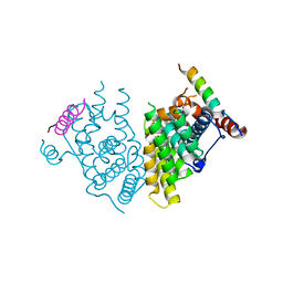
3UIV
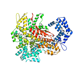
 | | Human serum albumin-myristate-amantadine hydrochloride complex | | 分子名称: | (3S,5S,7S)-tricyclo[3.3.1.1~3,7~]decan-1-amine, MYRISTIC ACID, Serum albumin | | 著者 | Yang, F, Ma, Z, Ma, L, Yang, G. | | 登録日 | 2011-11-06 | | 公開日 | 2012-11-07 | | 最終更新日 | 2023-11-01 | | 実験手法 | X-RAY DIFFRACTION (2.2 Å) | | 主引用文献 | Structural basis of the drug-binding specificity of human serum albumin
TO BE PUBLISHED
|
|
2JN7
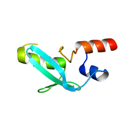
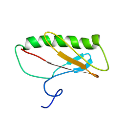 | | Northeast Structural Genomics Consortium Target ER411 | | 分子名称: | protein yfjZ | | 著者 | Tian, F, Zhao, L, Jiang, M, Cunningham, K, Ma, L, Xiao, R, Liu, J, Baran, M, Swapna, G.V.T, Acton, T.B, Rost, B, Montelione, G.T, Prestegard, J.H, Northeast Structural Genomics Consortium (NESG) | | 登録日 | 2006-12-28 | | 公開日 | 2007-01-09 | | 最終更新日 | 2024-05-08 | | 実験手法 | SOLUTION NMR | | 主引用文献 | NMR solution structure of E.Coli hypothetical protein YFJZ
To be Published
|
|
7T69
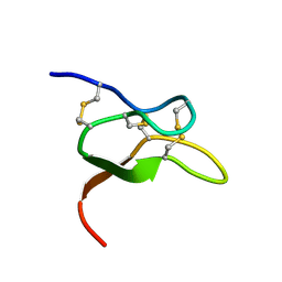
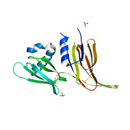 | | Crystal structure of Avr3 (SIX1) from Fusarium oxysporum f. sp. lycopersici | | 分子名称: | Avr3 (SIX1), Secreted in xylem 1, SULFATE ION | | 著者 | Yu, D.S, Outram, M.A, Ericsson, D.J, Jones, D.A, Williams, S.J. | | 登録日 | 2021-12-13 | | 公開日 | 2023-01-11 | | 最終更新日 | 2023-08-16 | | 実験手法 | X-RAY DIFFRACTION (1.68 Å) | | 主引用文献 | The structural repertoire of Fusarium oxysporum f. sp. lycopersici effectors revealed by experimental and computational studies
Elife, 2023
|
|
7T6A
 | | Crystal structure of Avr1 (SIX4) from Fusarium oxysporum f. sp. lycopersici | | 分子名称: | Avr1 (FolSIX4), Avirulence protein 1, Avr1 (SIX4), ... | | 著者 | Yu, D.S, Outram, M.A, Ericsson, D.J, Jones, D.A, Williams, S.J. | | 登録日 | 2021-12-13 | | 公開日 | 2023-01-11 | | 最終更新日 | 2023-08-16 | | 実験手法 | X-RAY DIFFRACTION (1.65 Å) | | 主引用文献 | The structural repertoire of Fusarium oxysporum f. sp. lycopersici effectors revealed by experimental and computational studies
Elife, 2023
|
|
7V6Z
 | | Cryo-EM structure of Patched1 (V1084A mutant) in lipid nanodisc, 3.64 angstrom (reprocessed with the dataset of 7dzp) | | 分子名称: | 2-acetamido-2-deoxy-beta-D-glucopyranose, 2-acetamido-2-deoxy-beta-D-glucopyranose-(1-4)-2-acetamido-2-deoxy-beta-D-glucopyranose, CHOLESTEROL, ... | | 著者 | Luo, Y, Zhao, Y, Qu, Q, Li, D. | | 登録日 | 2021-08-20 | | 公開日 | 2021-09-22 | | 最終更新日 | 2022-02-16 | | 実験手法 | ELECTRON MICROSCOPY (3.64 Å) | | 主引用文献 | Cryo-EM study of patched in lipid nanodisc suggests a structural basis for its clustering in caveolae.
Structure, 29, 2021
|
|
7V6Y
 | | Cryo-EM structure of Patched in lipid nanodisc - the wildtype, 3.5 angstrom (re-processed with dataset of 7dzq) | | 分子名称: | (2S)-2-azanyl-3-[[(2S)-3-butanoyloxy-2-dec-9-enoyloxy-propoxy]-oxidanyl-phosphoryl]oxy-propanoic acid, 2-acetamido-2-deoxy-beta-D-glucopyranose, 2-acetamido-2-deoxy-beta-D-glucopyranose-(1-4)-2-acetamido-2-deoxy-beta-D-glucopyranose, ... | | 著者 | Luo, Y, Zhao, Y, Qu, Q, Li, D. | | 登録日 | 2021-08-20 | | 公開日 | 2021-09-22 | | 最終更新日 | 2022-02-16 | | 実験手法 | ELECTRON MICROSCOPY (3.5 Å) | | 主引用文献 | Cryo-EM study of patched in lipid nanodisc suggests a structural basis for its clustering in caveolae.
Structure, 29, 2021
|
|
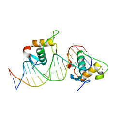
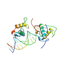
7YDW
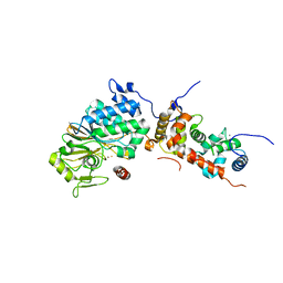
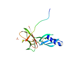
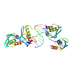 | | Crystal structure of the MPND-DNA complex | | 分子名称: | DNA (5'-D(P*AP*AP*AP*AP*AP*AP*AP*AP*AP*A)-3'), DNA (5'-D(P*TP*TP*TP*TP*TP*TP*TP*TP*TP*T)-3'), MPN domain-containing protein | | 著者 | Yang, M, Chen, Z. | | 登録日 | 2022-07-04 | | 公開日 | 2023-02-15 | | 最終更新日 | 2024-05-29 | | 実験手法 | X-RAY DIFFRACTION (2.47 Å) | | 主引用文献 | Structures of MPND Reveal the Molecular Recognition of Nucleosomes.
Int J Mol Sci, 24, 2023
|
|
7YDT
 | | Crystal structure of mouse MPND | | 分子名称: | MPN domain containing protein | | 著者 | Yang, M, Chen, Z. | | 登録日 | 2022-07-04 | | 公開日 | 2023-02-15 | | 最終更新日 | 2024-05-29 | | 実験手法 | X-RAY DIFFRACTION (2.055 Å) | | 主引用文献 | Structures of MPND Reveal the Molecular Recognition of Nucleosomes.
Int J Mol Sci, 24, 2023
|
|
1B8J
 | | ALKALINE PHOSPHATASE COMPLEXED WITH VANADATE | | 分子名称: | MAGNESIUM ION, PROTEIN (ALKALINE PHOSPHATASE), SULFATE ION, ... | | 著者 | Holtz, K.M, Stec, B, Kantrowitz, E.R. | | 登録日 | 1999-02-01 | | 公開日 | 1999-02-18 | | 最終更新日 | 2023-08-09 | | 実験手法 | X-RAY DIFFRACTION (1.9 Å) | | 主引用文献 | A model of the transition state in the alkaline phosphatase reaction.
J.Biol.Chem., 274, 1999
|
|
7X6L
 | |
7X6O
 | |


