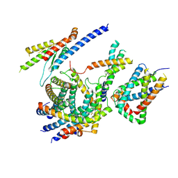
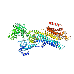
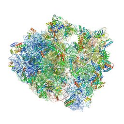
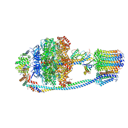
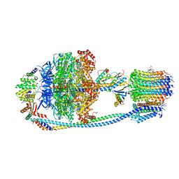
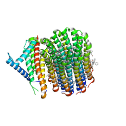
7K65
 
 | | Hedgehog receptor Patched (PTCH1) in complex with conformation selective nanobody TI23 | | 分子名称: | 2-acetamido-2-deoxy-beta-D-glucopyranose, 2-acetamido-2-deoxy-beta-D-glucopyranose-(1-4)-2-acetamido-2-deoxy-beta-D-glucopyranose, 2-{[(4-O-alpha-D-glucopyranosyl-alpha-D-glucopyranosyl)oxy]methyl}-4-{[(3beta,9beta,14beta,17beta,25R)-spirost-5-en-3-yl]oxy}butyl 4-O-alpha-D-glucopyranosyl-alpha-D-glucopyranoside, ... | | 著者 | Zhang, Y, Bulkley, D.P, Liang, J, Manglik, A, Cheng, Y, Beachy, P.A. | | 登録日 | 2020-09-18 | | 公開日 | 2021-03-17 | | 実験手法 | ELECTRON MICROSCOPY (3.4 Å) | | 主引用文献 | Hedgehog pathway activation through nanobody-mediated conformational blockade of the Patched sterol conduit.
Proc.Natl.Acad.Sci.USA, 117, 2020
|
|
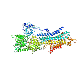
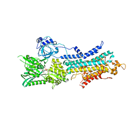
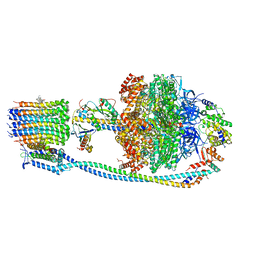
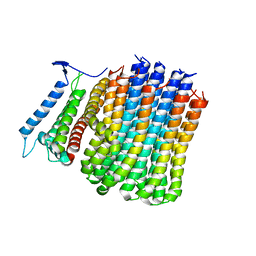
6LO8
 | | Cryo-EM structure of the TIM22 complex from yeast | | 分子名称: | Mitochondrial import inner membrane translocase subunit TIM10, Mitochondrial import inner membrane translocase subunit TIM12, Mitochondrial import inner membrane translocase subunit TIM18, ... | | 著者 | Zhang, Y, Zhou, X, Wu, X, Li, L. | | 登録日 | 2020-01-04 | | 公開日 | 2020-09-30 | | 最終更新日 | 2021-03-17 | | 実験手法 | ELECTRON MICROSCOPY (3.83 Å) | | 主引用文献 | Structure of the mitochondrial TIM22 complex from yeast.
Cell Res., 31, 2021
|
|
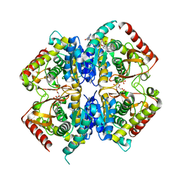
6LN6
 | | CryoEM structure of SERCA2b T1032stop in E1-2Ca2+-AMPPCP (class2) | | 分子名称: | CALCIUM ION, MAGNESIUM ION, PHOSPHOMETHYLPHOSPHONIC ACID ADENYLATE ESTER, ... | | 著者 | Zhang, Y, Tsutsumi, A, Watanabe, S, Inaba, K. | | 登録日 | 2019-12-28 | | 公開日 | 2020-08-26 | | 最終更新日 | 2020-09-16 | | 実験手法 | ELECTRON MICROSCOPY (2.9 Å) | | 主引用文献 | Cryo-EM structures of SERCA2b reveal the mechanism of regulation by the luminal extension tail.
Sci Adv, 6, 2020
|
|
3UUX
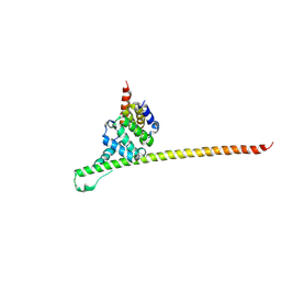 | | Crystal structure of yeast Fis1 in complex with Mdv1 fragment containing N-terminal extension and coiled coil domains | | 分子名称: | Mitochondria fission 1 protein, Mitochondrial division protein 1 | | 著者 | Zhang, Y, Chan, N.C, Gristick, H, Chan, D.C. | | 登録日 | 2011-11-28 | | 公開日 | 2012-02-08 | | 最終更新日 | 2012-04-25 | | 実験手法 | X-RAY DIFFRACTION (3.9 Å) | | 主引用文献 | Crystal structure of mitochondrial fission complex reveals scaffolding function for mitochondrial division 1 (mdv1) coiled coil.
J.Biol.Chem., 287, 2012
|
|
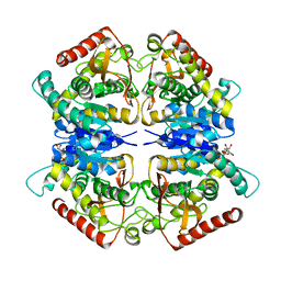
6LLE
 | | CryoEM structure of SERCA2b WT in E1-2Ca2+-AMPPCP state. | | 分子名称: | CALCIUM ION, MAGNESIUM ION, PHOSPHOMETHYLPHOSPHONIC ACID ADENYLATE ESTER, ... | | 著者 | Zhang, Y, Tsutsumi, A, Watanabe, S, Inaba, K. | | 登録日 | 2019-12-23 | | 公開日 | 2020-08-26 | | 最終更新日 | 2024-03-27 | | 実験手法 | ELECTRON MICROSCOPY (2.9 Å) | | 主引用文献 | Cryo-EM structures of SERCA2b reveal the mechanism of regulation by the luminal extension tail.
Sci Adv, 6, 2020
|
|
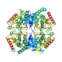
6LLY
 | | CryoEM structure of SERCA2b WT in E2-BeF3- state | | 分子名称: | BERYLLIUM TRIFLUORIDE ION, MAGNESIUM ION, Sarcoplasmic/endoplasmic reticulum calcium ATPase 2 | | 著者 | Zhang, Y, Tsutsumi, A, Watanabe, S, Inaba, K. | | 登録日 | 2019-12-24 | | 公開日 | 2020-08-26 | | 最終更新日 | 2024-03-27 | | 実験手法 | ELECTRON MICROSCOPY (2.8 Å) | | 主引用文献 | Cryo-EM structures of SERCA2b reveal the mechanism of regulation by the luminal extension tail.
Sci Adv, 6, 2020
|
|
5UQ7
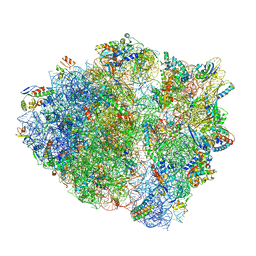 | | 70S ribosome complex with dnaX mRNA stemloop and E-site tRNA ("in" conformation) | | 分子名称: | 16S rRNA, 23S rRNA, 30S ribosomal protein S10, ... | | 著者 | Zhang, Y, Hong, S, Skiniotis, G, Dunham, C.M. | | 登録日 | 2017-02-07 | | 公開日 | 2018-03-07 | | 最終更新日 | 2018-07-18 | | 実験手法 | ELECTRON MICROSCOPY (3.5 Å) | | 主引用文献 | Alternative Mode of E-Site tRNA Binding in the Presence of a Downstream mRNA Stem Loop at the Entrance Channel.
Structure, 26, 2018
|
|
5V22
 | |
5V21
 | |
5UQ8
 | | 70S ribosome complex with dnaX mRNA stem-loop and E-site tRNA ("out" conformation) | | 分子名称: | 16S rRNA, 23S rRNA, 30S ribosomal protein S10, ... | | 著者 | Zhang, Y, Hong, S, Skiniotis, G, Dunham, C.M. | | 登録日 | 2017-02-07 | | 公開日 | 2018-03-07 | | 最終更新日 | 2018-03-21 | | 実験手法 | ELECTRON MICROSCOPY (3.2 Å) | | 主引用文献 | Alternative Mode of E-Site tRNA Binding in the Presence of a Downstream mRNA Stem Loop at the Entrance Channel.
Structure, 26, 2018
|
|
5VAI
 | | Cryo-EM structure of the activated Glucagon-like peptide-1 receptor in complex with G protein | | 分子名称: | Glucagon-like peptide 1, Guanine nucleotide-binding protein G(I)/G(S)/G(O) subunit gamma-2, Guanine nucleotide-binding protein G(I)/G(S)/G(T) subunit beta-1, ... | | 著者 | Zhang, Y, Sun, B, Feng, D, Hu, H, Chu, M, Qu, Q, Tarrasch, J.T, Li, S, Kobilka, T.S, Kobilka, B.K, Skiniotis, G. | | 登録日 | 2017-03-27 | | 公開日 | 2017-05-24 | | 最終更新日 | 2019-12-18 | | 実験手法 | ELECTRON MICROSCOPY (4.1 Å) | | 主引用文献 | Cryo-EM structure of the activated GLP-1 receptor in complex with a G protein.
Nature, 546, 2017
|
|
3PQF
 | |
3PQE
 | |
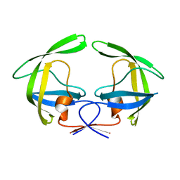
3PME
 | | Crystal structure of the receptor binding domain of botulinum neurotoxin C/D mosaic serotype | | 分子名称: | GLYCEROL, SULFATE ION, Type C neurotoxin | | 著者 | Zhang, Y, Buchko, G.W, Qin, L, Robinson, H, Varnum, S.M, Seattle Structural Genomics Center for Infectious Disease (SSGCID) | | 登録日 | 2010-11-16 | | 公開日 | 2010-12-15 | | 最終更新日 | 2011-07-13 | | 実験手法 | X-RAY DIFFRACTION (1.56 Å) | | 主引用文献 | Crystal structure of the receptor binding domain of the botulinum C-D mosaic neurotoxin reveals potential roles of lysines 1118 and 1136 in membrane interactions.
Biochem.Biophys.Res.Commun., 404, 2011
|
|
3PQD
 | |
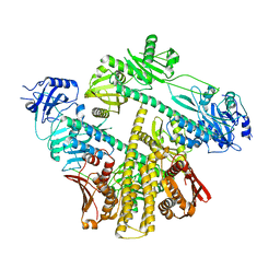
8J0T
 | | Cryo-EM structure of Mycobacterium tuberculosis ATP synthase in the apo-form | | 分子名称: | ADENOSINE-5'-DIPHOSPHATE, ADENOSINE-5'-TRIPHOSPHATE, ATP synthase epsilon chain, ... | | 著者 | Zhang, Y, Lai, Y, Liu, F, Rao, Z, Gong, H. | | 登録日 | 2023-04-11 | | 公開日 | 2024-05-22 | | 実験手法 | ELECTRON MICROSCOPY (2.8 Å) | | 主引用文献 | Structure of Mycobacterium tuberculosis ATP synthase
To Be Published
|
|
8J0S
 | | Cryo-EM structure of Mycobacterium tuberculosis ATP synthase in complex with bedaquiline(BDQ) | | 分子名称: | ADENOSINE-5'-DIPHOSPHATE, ADENOSINE-5'-TRIPHOSPHATE, ATP synthase epsilon chain, ... | | 著者 | Zhang, Y, Lai, Y, Liu, F, Rao, Z, Gong, H. | | 登録日 | 2023-04-11 | | 公開日 | 2024-05-22 | | 実験手法 | ELECTRON MICROSCOPY (2.58 Å) | | 主引用文献 | Structure of Mycobacterium tuberculosis ATP synthase
To Be Published
|
|
8JR0
 | | Cryo-EM structure of Mycobacterium tuberculosis ATP synthase in complex with TBAJ-587 | | 分子名称: | (1~{S},2~{S})-1-(6-bromanyl-2-methoxy-quinolin-3-yl)-2-(2,6-dimethoxypyridin-4-yl)-4-(dimethylamino)-1-(2-fluoranyl-3-methoxy-phenyl)butan-2-ol, ADENOSINE-5'-DIPHOSPHATE, ADENOSINE-5'-TRIPHOSPHATE, ... | | 著者 | Zhang, Y, Lai, Y, Liu, F, Rao, Z, Gong, H. | | 登録日 | 2023-06-15 | | 公開日 | 2024-05-01 | | 実験手法 | ELECTRON MICROSCOPY (2.8 Å) | | 主引用文献 | Structure of Mycobacterium tuberculosis ATP synthase
To Be Published
|
|
8JR1
 | | Cryo-EM structure of Mycobacterium tuberculosis ATP synthase Fo in complex with TBAJ-587 | | 分子名称: | (1~{S},2~{S})-1-(6-bromanyl-2-methoxy-quinolin-3-yl)-2-(2,6-dimethoxypyridin-4-yl)-4-(dimethylamino)-1-(2-fluoranyl-3-methoxy-phenyl)butan-2-ol, ATP synthase subunit a, ATP synthase subunit c | | 著者 | Zhang, Y, Lai, Y, Liu, F, Rao, Z, Gong, H. | | 登録日 | 2023-06-15 | | 公開日 | 2024-05-01 | | 実験手法 | ELECTRON MICROSCOPY (3.17 Å) | | 主引用文献 | Structure of Mycobacterium tuberculosis ATP synthase
To Be Published
|
|
8J57
 | | Cryo-EM structure of Mycobacterium tuberculosis ATP synthase Fo in complex with bedaquiline(BDQ) | | 分子名称: | ATP synthase subunit a, ATP synthase subunit c, Bedaquiline | | 著者 | Zhang, Y, Lai, Y, Liu, F, Rao, Z, Gong, H. | | 登録日 | 2023-04-21 | | 公開日 | 2024-05-01 | | 実験手法 | ELECTRON MICROSCOPY (2.85 Å) | | 主引用文献 | Structure of Mycobacterium tuberculosis ATP synthase
To Be Published
|
|
8J58
 | | Cryo-EM structure of Mycobacterium tuberculosis ATP synthase Fo in the apo-form | | 分子名称: | ATP synthase subunit a, ATP synthase subunit c | | 著者 | Zhang, Y, Lai, Y, Liu, F, Rao, Z, Gong, H. | | 登録日 | 2023-04-21 | | 公開日 | 2024-05-01 | | 実験手法 | ELECTRON MICROSCOPY (3.15 Å) | | 主引用文献 | Structure of Mycobacterium tuberculosis ATP synthase
To Be Published
|
|
8JY0
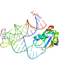 | | Crystal structure of RhoBAST complexed with TMR-DN | | 分子名称: | 2,4-dinitroaniline, 5-aminocarbonyl-2-[3-(dimethylamino)-6-dimethylazaniumylidene-xanthen-9-yl]benzoate, GUANOSINE-5'-DIPHOSPHATE, ... | | 著者 | Zhang, Y, Xiao, Y, Xu, Z, Fang, X. | | 登録日 | 2023-07-02 | | 公開日 | 2024-05-29 | | 実験手法 | X-RAY DIFFRACTION (2.75 Å) | | 主引用文献 | Structural mechanisms for binding and activation of a contact-quenched fluorophore by RhoBAST.
Nat Commun, 15, 2024
|
|
4OXW
 | |
4NPU
 | |
8ISI
 | | Photochromobilin-free form of Arabidopsis thaliana phytochrome A - apo-AtphyA | | 分子名称: | Phytochrome A | | 著者 | Zhang, Y, Ma, C, Zhao, J, Gao, N, Wang, J. | | 登録日 | 2023-03-20 | | 公開日 | 2023-08-09 | | 最終更新日 | 2023-10-11 | | 実験手法 | ELECTRON MICROSCOPY (3.77 Å) | | 主引用文献 | Structural insights into plant phytochrome A as a highly sensitized photoreceptor.
Cell Res., 33, 2023
|
|


