6WXL
 
 | |
2O6A
 | |
2O69
 | |
2PBK
 | |
2OZN
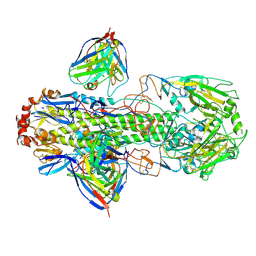
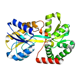
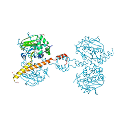
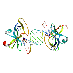
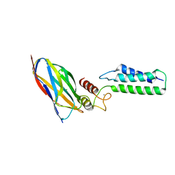 | | The Cohesin-Dockerin Complex of NagJ and NagH from Clostridium perfringens | | 分子名称: | CALCIUM ION, CHLORIDE ION, Hyalurononglucosaminidase, ... | | 著者 | Adams, J.J, Boraston, A, Smith, S.P. | | 登録日 | 2007-02-26 | | 公開日 | 2008-05-06 | | 最終更新日 | 2024-02-21 | | 実験手法 | X-RAY DIFFRACTION (1.6 Å) | | 主引用文献 | Structural basis of Clostridium perfringens toxin complex formation.
Proc.Natl.Acad.Sci.Usa, 105, 2008
|
|
2P55
 | |
2P54
 | | a crystal structure of PPAR alpha bound with SRC1 peptide and GW735 | | 分子名称: | 2-METHYL-2-(4-{[({4-METHYL-2-[4-(TRIFLUOROMETHYL)PHENYL]-1,3-THIAZOL-5-YL}CARBONYL)AMINO]METHYL}PHENOXY)PROPANOIC ACID, Nuclear receptor coactivator 1, Peroxisome proliferator-activated receptor alpha | | 著者 | Xu, R.X, Xu, H.E, Sierra, M.L, Montana, V.G, Lambert, M.H, Pianetti, P.M. | | 登録日 | 2007-03-14 | | 公開日 | 2007-04-24 | | 最終更新日 | 2024-04-03 | | 実験手法 | X-RAY DIFFRACTION (1.79 Å) | | 主引用文献 | Substituted 2-[(4-Aminomethyl)phenoxy]-2-methylpropionic Acid PPAR Agonists. 1.Discovery of a Novel Series of Potent HDLc Raising Agents.
J.Med.Chem., 50, 2007
|
|
2PG2
 | |
2O68
 | |
2PJY
 | |
2Q11
 | | Structure of BACE complexed to compound 1 | | 分子名称: | 3-(2-AMINO-6-BENZOYLQUINAZOLIN-3(4H)-YL)-N-CYCLOHEXYL-N-METHYLPROPANAMIDE, Beta-secretase 1 | | 著者 | Sharff, A.J. | | 登録日 | 2007-05-23 | | 公開日 | 2007-08-14 | | 最終更新日 | 2021-10-20 | | 実験手法 | X-RAY DIFFRACTION (2.4 Å) | | 主引用文献 | 2-Amino-3,4-dihydroquinazolines as inhibitors of BACE-1 (beta-Site APP cleaving enzyme): Use of structure based design to convert a micromolar hit into a nanomolar lead.
J.Med.Chem., 50, 2007
|
|
2Q15
 | | Structure of BACE complexed to compound 3a | | 分子名称: | (4S)-4-(2-AMINO-6-PHENOXYQUINAZOLIN-3(4H)-YL)-N,4-DICYCLOHEXYL-N-METHYLBUTANAMIDE, Beta-secretase 1 | | 著者 | Sharff, A.J. | | 登録日 | 2007-05-23 | | 公開日 | 2007-08-14 | | 最終更新日 | 2021-10-20 | | 実験手法 | X-RAY DIFFRACTION (2.4 Å) | | 主引用文献 | 2-Amino-3,4-dihydroquinazolines as inhibitors of BACE-1 (beta-Site APP cleaving enzyme): Use of structure based design to convert a micromolar hit into a nanomolar lead.
J.Med.Chem., 50, 2007
|
|
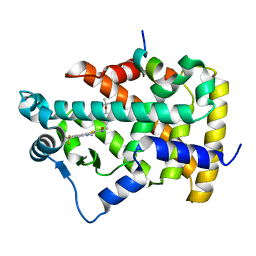
3IHR
 | | Crystal Structure of Uch37 | | 分子名称: | FORMIC ACID, SODIUM ION, Ubiquitin carboxyl-terminal hydrolase isozyme L5 | | 著者 | Burgie, E.S, Bingman, C.A, Phillips Jr, G.N, Center for Eukaryotic Structural Genomics (CESG) | | 登録日 | 2009-07-30 | | 公開日 | 2009-08-11 | | 最終更新日 | 2023-07-26 | | 実験手法 | X-RAY DIFFRACTION (2.95 Å) | | 主引用文献 | Structural characterization of human Uch37.
Proteins, 80, 2012
|
|
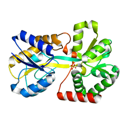
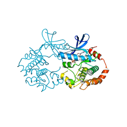
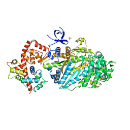
3KOO
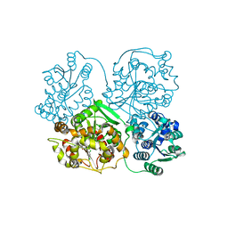 | | Crystal Structure of soluble epoxide Hydrolase | | 分子名称: | Epoxide hydrolase 2, N-(2,4-dichlorobenzyl)-4-(pyrimidin-2-yloxy)piperidine-1-carboxamide | | 著者 | Farrow, N.A. | | 登録日 | 2009-11-13 | | 公開日 | 2010-04-28 | | 最終更新日 | 2024-02-21 | | 実験手法 | X-RAY DIFFRACTION (2.791 Å) | | 主引用文献 | Optimization of piperidyl-ureas as inhibitors of soluble epoxide hydrolase.
Bioorg.Med.Chem.Lett., 20, 2010
|
|
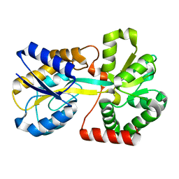
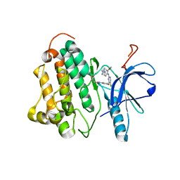
3I1Y
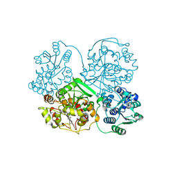 | | Crystal Structure of soluble epoxide Hydrolase | | 分子名称: | Epoxide hydrolase 2, N-(3,3-diphenylpropyl)pyridine-3-carboxamide | | 著者 | Farrow, N.A. | | 登録日 | 2009-06-28 | | 公開日 | 2009-10-13 | | 最終更新日 | 2024-02-21 | | 実験手法 | X-RAY DIFFRACTION (2.469 Å) | | 主引用文献 | Structure-based optimization of arylamides as inhibitors of soluble epoxide hydrolase.
J.Med.Chem., 52, 2009
|
|
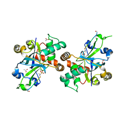
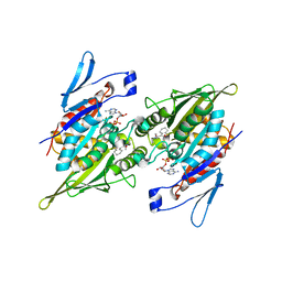
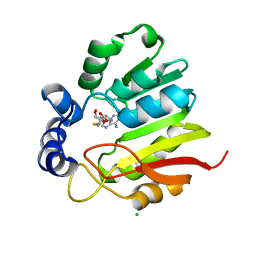
3I28
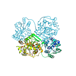 | | Crystal Structure of soluble epoxide Hydrolase | | 分子名称: | 4-cyano-N-{(3S)-3-(4-fluorophenyl)-3-[4-(methylsulfonyl)phenyl]propyl}benzamide, Epoxide hydrolase 2 | | 著者 | Farrow, N.A. | | 登録日 | 2009-06-29 | | 公開日 | 2009-10-13 | | 最終更新日 | 2024-02-21 | | 実験手法 | X-RAY DIFFRACTION (1.95 Å) | | 主引用文献 | Structure-based optimization of arylamides as inhibitors of soluble epoxide hydrolase.
J.Med.Chem., 52, 2009
|
|
3LRR
 | |
3L9I
 | | Myosin VI nucleotide-free (mdinsert2) L310G mutant crystal structure | | 分子名称: | ACETATE ION, CALCIUM ION, Calmodulin, ... | | 著者 | Pylypenko, O, Song, L, Sweeney, L.H, Houdusse, A. | | 登録日 | 2010-01-05 | | 公開日 | 2010-12-08 | | 最終更新日 | 2023-11-01 | | 実験手法 | X-RAY DIFFRACTION (2.2 Å) | | 主引用文献 | Role of insert i of myosin VI in modulating nucleotide affinity
To be Published
|
|
3IKA
 | |
3LCC
 | |
1T14
 | |
8GGJ
 | | Locally refined cryoEM structure of receptor from beta-2-adrenergic receptor in complex with GTP-bound Gs heterotrimer (transition intermediate #2 of 20) | | 分子名称: | (5R,6R)-6-(methylamino)-5,6,7,8-tetrahydronaphthalene-1,2,5-triol, Beta-2 adrenergic receptor | | 著者 | Papasergi-Scott, M.M, Skiniotis, G. | | 登録日 | 2023-03-08 | | 公開日 | 2024-03-06 | | 最終更新日 | 2024-06-05 | | 実験手法 | ELECTRON MICROSCOPY (3.5 Å) | | 主引用文献 | Time-resolved cryo-EM of G-protein activation by a GPCR.
Nature, 629, 2024
|
|
8GGW
 | | Locally refined cryoEM structure of receptor from beta-2-adrenergic receptor in complex with GTP-bound Gs heterotrimer (transition intermediate #15 of 20) | | 分子名称: | (5R,6R)-6-(methylamino)-5,6,7,8-tetrahydronaphthalene-1,2,5-triol, Beta-2 adrenergic receptor | | 著者 | Papasergi-Scott, M.M, Skiniotis, G. | | 登録日 | 2023-03-08 | | 公開日 | 2024-03-06 | | 最終更新日 | 2024-06-05 | | 実験手法 | ELECTRON MICROSCOPY (3.6 Å) | | 主引用文献 | Time-resolved cryo-EM of G-protein activation by a GPCR.
Nature, 629, 2024
|
|
8GDZ
 | | CryoEM structure of beta-2-adrenergic receptor in complex with nucleotide-free Gs heterotrimer (#1 of 20) | | 分子名称: | (5R,6R)-6-(methylamino)-5,6,7,8-tetrahydronaphthalene-1,2,5-triol, Beta-2 adrenergic receptor, Guanine nucleotide-binding protein G(I)/G(S)/G(O) subunit gamma-2, ... | | 著者 | Papasergi-Scott, M.M, Skiniotis, G. | | 登録日 | 2023-03-06 | | 公開日 | 2024-03-06 | | 最終更新日 | 2024-06-12 | | 実験手法 | ELECTRON MICROSCOPY (3.5 Å) | | 主引用文献 | Time-resolved cryo-EM of G-protein activation by a GPCR.
Nature, 629, 2024
|
|
8GE1
 | | CryoEM structure of beta-2-adrenergic receptor in complex with nucleotide-free Gs heterotrimer (#2 of 20) | | 分子名称: | (5R,6R)-6-(methylamino)-5,6,7,8-tetrahydronaphthalene-1,2,5-triol, Beta-2 adrenergic receptor, Guanine nucleotide-binding protein G(I)/G(S)/G(O) subunit gamma-2, ... | | 著者 | Papasergi-Scott, M.M, Skiniotis, G. | | 登録日 | 2023-03-06 | | 公開日 | 2024-03-06 | | 最終更新日 | 2024-06-12 | | 実験手法 | ELECTRON MICROSCOPY (3.4 Å) | | 主引用文献 | Time-resolved cryo-EM of G-protein activation by a GPCR.
Nature, 629, 2024
|
|


