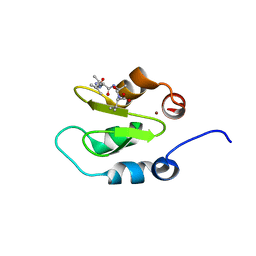
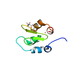
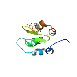
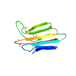
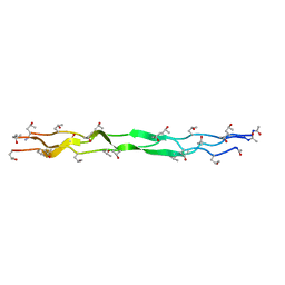
2LZJ
 
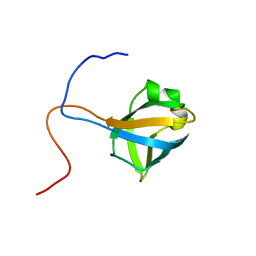 | | Refined solution structure and dynamics of First Catalytic Cysteine Half-domain from mouse E1 enzyme | | 分子名称: | Ubiquitin-like modifier-activating enzyme 1 | | 著者 | Jaremko, M, Jaremko, L, Nowakowski, M, Szczepanowski, R.H, Filipek, R, Wojciechowski, M, Bochtler, M, Ejchart, A. | | 登録日 | 2012-10-03 | | 公開日 | 2013-09-18 | | 最終更新日 | 2024-05-15 | | 実験手法 | SOLUTION NMR | | 主引用文献 | NMR structural studies of the first catalytic half-domain of ubiquitin activating enzyme.
J.Struct.Biol., 185, 2014
|
|
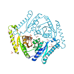
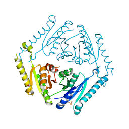
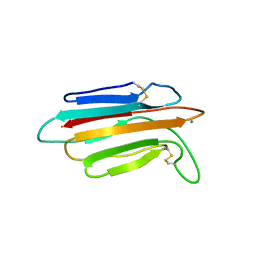
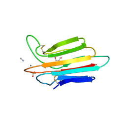
4J48
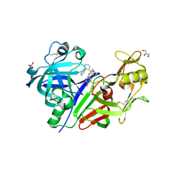
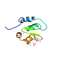 | | Crystal structure of XIAP-BIR2 domain with AMRV bound | | 分子名称: | E3 ubiquitin-protein ligase XIAP, GLYCEROL, PEPTIDE (ALA-MET-ARG-VAL), ... | | 著者 | Gosu, R. | | 登録日 | 2013-02-06 | | 公開日 | 2013-09-25 | | 最終更新日 | 2023-09-20 | | 実験手法 | X-RAY DIFFRACTION (2.1 Å) | | 主引用文献 | The structure of XIAP BIR2: understanding the selectivity of the BIR domains.
Acta Crystallogr.,Sect.D, 69, 2013
|
|
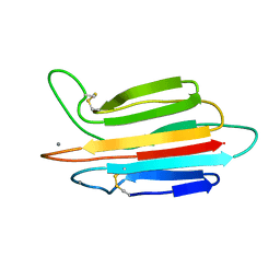
4J47
 | |
4J3Y
 | |
4J44
 | |
4J45
 | |
4J46
 | |
4R2K
 | |
4R2J
 | |
4R2L
 | |
4R2M
 | |
6OVG
 | |
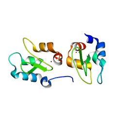
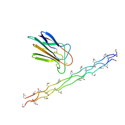
4LMF
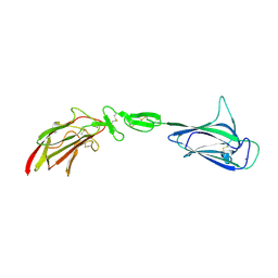 | | C1s CUB1-EGF-CUB2 | | 分子名称: | CALCIUM ION, Complement C1s subcomponent heavy chain, SODIUM ION | | 著者 | Wallis, R, Venkatraman Girija, U, Moody, P.C.E, Marshall, J.E. | | 登録日 | 2013-07-10 | | 公開日 | 2013-08-07 | | 最終更新日 | 2018-01-24 | | 実験手法 | X-RAY DIFFRACTION (2.921 Å) | | 主引用文献 | Structural basis of the C1q/C1s interaction and its central role in assembly of the C1 complex of complement activation.
Proc.Natl.Acad.Sci.USA, 110, 2013
|
|
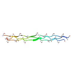
4LOS
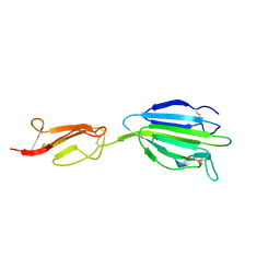 | | C1s CUB2-CCP1 | | 分子名称: | CALCIUM ION, Complement C1s subcomponent heavy chain | | 著者 | Wallis, R, Venkatraman Girija, U, Moody, P.C.E, Marshall, J.E, Gingras, A.R. | | 登録日 | 2013-07-13 | | 公開日 | 2013-08-07 | | 最終更新日 | 2013-09-04 | | 実験手法 | X-RAY DIFFRACTION (1.996 Å) | | 主引用文献 | Structural basis of the C1q/C1s interaction and its central role in assembly of the C1 complex of complement activation.
Proc.Natl.Acad.Sci.USA, 110, 2013
|
|
4LOT
 | |
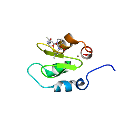
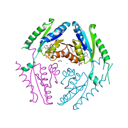
3POJ
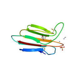 | | Crystal structure of MASP-1 CUB2 domain bound to Ethylamine | | 分子名称: | 2-AMINO-2-HYDROXYMETHYL-PROPANE-1,3-DIOL, CALCIUM ION, ETHANAMINE, ... | | 著者 | Gingras, A.R, Moody, P.C.E, Wallis, R. | | 登録日 | 2010-11-22 | | 公開日 | 2011-08-24 | | 最終更新日 | 2023-09-06 | | 実験手法 | X-RAY DIFFRACTION (1.451 Å) | | 主引用文献 | Structural Basis of Mannan-Binding Lectin Recognition by Its Associated Serine Protease MASP-1: Implications for Complement Activation.
Structure, 19, 2011
|
|
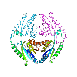
3POF
 | | Crystal structure of MASP-1 CUB2 domain bound to Ca2+ | | 分子名称: | 2-AMINO-2-HYDROXYMETHYL-PROPANE-1,3-DIOL, CALCIUM ION, Mannan-binding lectin serine protease 1, ... | | 著者 | Gingras, A.R, Moody, P.C.E, Wallis, R. | | 登録日 | 2010-11-22 | | 公開日 | 2011-08-24 | | 最終更新日 | 2023-09-06 | | 実験手法 | X-RAY DIFFRACTION (1.501 Å) | | 主引用文献 | Structural Basis of Mannan-Binding Lectin Recognition by Its Associated Serine Protease MASP-1: Implications for Complement Activation.
Structure, 19, 2011
|
|
3POB
 | |
3POG
 | |
3PON
 | |
3POD
 | |
3POI
 | | Crystal structure of MASP-1 CUB2 domain bound to Methylamine | | 分子名称: | 2-AMINO-2-HYDROXYMETHYL-PROPANE-1,3-DIOL, CALCIUM ION, METHYLAMINE, ... | | 著者 | Gingras, A.R, Moody, P.C.E, Wallis, R. | | 登録日 | 2010-11-22 | | 公開日 | 2011-08-24 | | 最終更新日 | 2011-11-30 | | 実験手法 | X-RAY DIFFRACTION (1.701 Å) | | 主引用文献 | Structural Basis of Mannan-Binding Lectin Recognition by Its Associated Serine Protease MASP-1: Implications for Complement Activation.
Structure, 19, 2011
|
|
3POE
 | |
5VHE
 | | DHX36 in complex with the c-Myc G-quadruplex | | 分子名称: | DEAH (Asp-Glu-Ala-His) box polypeptide 36, DNA (5'-D(*AP*GP*GP*GP*TP*GP*GP*GP*TP*AP*GP*GP*GP*TP*GP*GP*GP*TP*TP*TP*TP*TP*TP*T)-3'), POTASSIUM ION | | 著者 | Chen, M, Ferre-D'Amare, A. | | 登録日 | 2017-04-13 | | 公開日 | 2018-06-13 | | 最終更新日 | 2023-10-04 | | 実験手法 | X-RAY DIFFRACTION (3.793 Å) | | 主引用文献 | Structural basis of G-quadruplex unfolding by the DEAH/RHA helicase DHX36.
Nature, 558, 2018
|
|
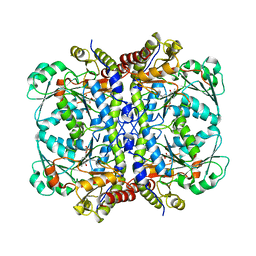
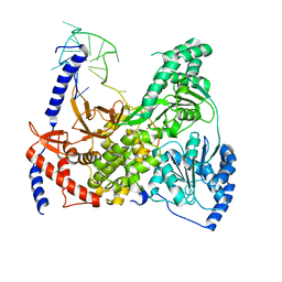
3Q3T
 | | Alkyl Amine Renin Inhibitors: Filling S1 from S3 | | 分子名称: | 2-acetamido-2-deoxy-beta-D-glucopyranose, 2-acetamido-2-deoxy-beta-D-glucopyranose-(1-4)-2-acetamido-2-deoxy-beta-D-glucopyranose, GLYCEROL, ... | | 著者 | Wu, Z, McKeever, B. | | 登録日 | 2010-12-22 | | 公開日 | 2011-08-03 | | 最終更新日 | 2023-09-13 | | 実験手法 | X-RAY DIFFRACTION (2.6 Å) | | 主引用文献 | Biphenyl/diphenyl ether renin inhibitors: Filling the S1 pocket of renin via the S3 pocket.
Bioorg.Med.Chem.Lett., 21, 2011
|
|


