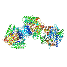
3E9N
 
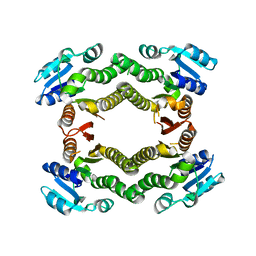 | | Crystal structure of a putative short-chain dehydrogenase/reductase from Corynebacterium glutamicum | | 分子名称: | PUTATIVE SHORT-CHAIN DEHYDROGENASE/REDUCTASE | | 著者 | Bonanno, J.B, Gilmore, M, Bain, K.T, Hu, S, Romero, R, Smith, D, Wasserman, S, Sauder, J.M, Burley, S.K, Almo, S.C, New York SGX Research Center for Structural Genomics (NYSGXRC) | | 登録日 | 2008-08-22 | | 公開日 | 2008-09-02 | | 最終更新日 | 2024-02-21 | | 実験手法 | X-RAY DIFFRACTION (2.4 Å) | | 主引用文献 | Crystal structure of a putative short-chain dehydrogenase/reductase from Corynebacterium glutamicum
To be Published
|
|
1ELA
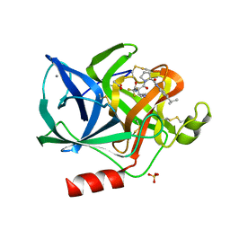 | | Analogous inhibitors of elastase do not always bind analogously | | 分子名称: | 6-ammonio-N-(trifluoroacetyl)-L-norleucyl-N-[4-(1-methylethyl)phenyl]-L-prolinamide, ACETIC ACID, CALCIUM ION, ... | | 著者 | Mattos, C, Rasmussen, B, Ding, X, Petsko, G.A, Ringe, D. | | 登録日 | 1993-12-07 | | 公開日 | 1994-04-30 | | 最終更新日 | 2024-06-05 | | 実験手法 | X-RAY DIFFRACTION (2 Å) | | 主引用文献 | Analogous inhibitors of elastase do not always bind analogously.
Nat.Struct.Biol., 1, 1994
|
|
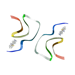
2LVR
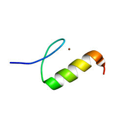 | | Solution structure of Miz-1 zinc finger 8 | | 分子名称: | ZINC ION, Zinc finger and BTB domain-containing protein 17 | | 著者 | Bedard, M, Maltais, L, Beaulieu, M, Bernard, D, Lavigne, P. | | 登録日 | 2012-07-10 | | 公開日 | 2012-07-25 | | 最終更新日 | 2024-05-15 | | 実験手法 | SOLUTION NMR | | 主引用文献 | NMR structure note: solution structure of human Miz-1 zinc fingers 8 to 10.
J.Biomol.Nmr, 54, 2012
|
|
7YNQ
 | |
2LVB
 | | Solution NMR Structure DE NOVO DESIGNED PFK fold PROTEIN, Northeast Structural Genomics Consortium (NESG) Target OR250 | | 分子名称: | DE NOVO DESIGNED PFK fold PROTEIN | | 著者 | Liu, G, Koga, N, Koga, R, Xiao, R, Hamilton, K, Kohan, E, Acton, T.B, Kornhaber, G, Everett, J.K, Baker, D, Montelione, G.T, Northeast Structural Genomics Consortium (NESG) | | 登録日 | 2012-06-30 | | 公開日 | 2012-08-08 | | 最終更新日 | 2024-05-15 | | 実験手法 | SOLUTION NMR | | 主引用文献 | Principles for designing ideal protein structures.
Nature, 491, 2012
|
|
3H74
 | | Crystal structure of pyridoxal kinase from Lactobacillus plantarum | | 分子名称: | GLYCEROL, Pyridoxal kinase, SULFATE ION | | 著者 | Bagaria, A, Kumaran, D, Burley, S.K, Swaminathan, S, New York SGX Research Center for Structural Genomics (NYSGXRC) | | 登録日 | 2009-04-24 | | 公開日 | 2009-05-26 | | 最終更新日 | 2021-02-10 | | 実験手法 | X-RAY DIFFRACTION (1.3 Å) | | 主引用文献 | Crystal structure of pyridoxal kinase from Lactobacillus plantarum
To be Published
|
|
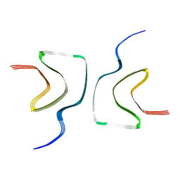
2LVT
 | | Solution structure of Miz-1 zinc finger 9 | | 分子名称: | ZINC ION, Zinc finger and BTB domain-containing protein 17 | | 著者 | Bedard, M, Maltais, L, Beaulieu, M, Bernard, D, Lavigne, P. | | 登録日 | 2012-07-11 | | 公開日 | 2012-07-25 | | 最終更新日 | 2024-05-15 | | 実験手法 | SOLUTION NMR | | 主引用文献 | NMR structure note: solution structure of human Miz-1 zinc fingers 8 to 10.
J.Biomol.Nmr, 54, 2012
|
|
7YNT
 | |
6RK7
 | | Inter-dimeric interface controls function and stability of S-methionine adenosyltransferase from U. urealiticum | | 分子名称: | CHLORIDE ION, Methionine adenosyltransferase, S-ADENOSYLMETHIONINE | | 著者 | Shahar, A, Zarivach, R, Bershtein, S, Kleiner, D, Shmulevich, F. | | 登録日 | 2019-04-30 | | 公開日 | 2019-09-25 | | 最終更新日 | 2024-01-24 | | 実験手法 | X-RAY DIFFRACTION (1.8 Å) | | 主引用文献 | The interdimeric interface controls function and stability of Ureaplasma urealiticum methionine S-adenosyltransferase.
J.Mol.Biol., 431, 2019
|
|
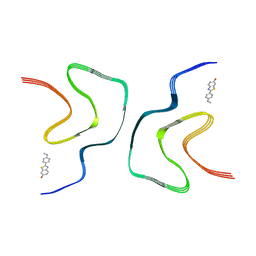
7YNM
 | | ThT-bound alpha-synuclein fibrils conformation 1 | | 分子名称: | 2-[4-(dimethylamino)phenyl]-3,6-dimethyl-1,3-benzothiazol-3-ium, Alpha-synuclein | | 著者 | Tao, Y.Q, Zhao, Q.Y, Liu, C, Li, D. | | 登録日 | 2022-07-31 | | 公開日 | 2023-08-02 | | 最終更新日 | 2024-07-03 | | 実験手法 | ELECTRON MICROSCOPY (2.9 Å) | | 主引用文献 | ThT-bound alpha-synuclein fibrils conformation 1
To Be Published
|
|
7YNR
 | |
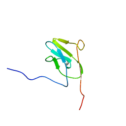
1E8B
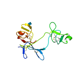 | | Solution structure of 6F11F22F2, a compact three-module fragment of the gelatin-binding domain of human fibronectin | | 分子名称: | 2-acetamido-2-deoxy-beta-D-glucopyranose, FIBRONECTIN | | 著者 | Pickford, A.R, Smith, S.P, Staunton, D, Boyd, J, Campbell, I.D. | | 登録日 | 2000-09-18 | | 公開日 | 2000-10-15 | | 最終更新日 | 2020-07-29 | | 実験手法 | SOLUTION NMR | | 主引用文献 | The Hairpin Structure of the (6)F1(1)F2(2)F2 Fragment from Human Fibronectin Enhances Gelatin Binding
Embo J., 20, 2001
|
|
1ELU
 | | COMPLEX BETWEEN THE CYSTINE C-S LYASE C-DES AND ITS REACTION PRODUCT CYSTEINE PERSULFIDE. | | 分子名称: | 2-[(3-HYDROXY-2-METHYL-5-PHOSPHONOOXYMETHYL-PYRIDIN-4-YLMETHYL)-AMINO]-PROPIONIC ACID, L-CYSTEINE/L-CYSTINE C-S LYASE, POTASSIUM ION, ... | | 著者 | Clausen, T, Kaiser, J.T, Steegborn, C, Huber, R, Kessler, D. | | 登録日 | 2000-03-14 | | 公開日 | 2000-04-19 | | 最終更新日 | 2024-02-07 | | 実験手法 | X-RAY DIFFRACTION (1.55 Å) | | 主引用文献 | Crystal structure of the cystine C-S lyase from Synechocystis: stabilization of cysteine persulfide for FeS cluster biosynthesis.
Proc.Natl.Acad.Sci.USA, 97, 2000
|
|
7YNL
 | | EB-bound alpha-synuclein fibrils | | 分子名称: | 4-azanyl-6-[[4-[4-[(~{E})-(8-azanyl-1-oxidanyl-5,7-disulfo-naphthalen-2-yl)diazenyl]-3-methyl-phenyl]-2-methyl-phenyl]diazenyl]-5-oxidanyl-naphthalene-1,3-disulfonic acid, Alpha-synuclein | | 著者 | Tao, Y.Q, Zhao, Q.Y, Liu, C, Li, D. | | 登録日 | 2022-07-31 | | 公開日 | 2023-08-02 | | 最終更新日 | 2024-07-03 | | 実験手法 | ELECTRON MICROSCOPY (2.6 Å) | | 主引用文献 | EB-bound alpha-synuclein fibrils
To Be Published
|
|
6RO9
 | | Human spectrin SH3 domain D48G, E7V, K60V | | 分子名称: | Spectrin alpha, non-erythrocytic 1 | | 著者 | Reverter, D, Navarro, S, Ventura, S. | | 登録日 | 2019-05-10 | | 公開日 | 2020-06-03 | | 最終更新日 | 2024-01-24 | | 実験手法 | X-RAY DIFFRACTION (1.814 Å) | | 主引用文献 | Human spectrin SH3 domain D48G, E7V, K60V
To Be Published
|
|
2M57
 | |
6ROA
 | | Crystal structure of V57G mutant of human cystatin C | | 分子名称: | Cystatin-C | | 著者 | Orlikowska, M, Behrendt, I, Borek, D, Otwinowski, Z, Skowron, P, Szymanska, A. | | 登録日 | 2019-05-10 | | 公開日 | 2019-08-07 | | 最終更新日 | 2024-01-24 | | 実験手法 | X-RAY DIFFRACTION (2.65 Å) | | 主引用文献 | NMR and crystallographic structural studies of the extremely stable monomeric variant of human cystatin C with single amino acid substitution.
Febs J., 287, 2020
|
|
7YNP
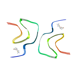 | | BF227-bound alpha-synuclein fibrils | | 分子名称: | 5-[(~{E})-2-(6-methoxy-1,3-benzoxazol-2-yl)ethenyl]-~{N},~{N}-dimethyl-1,3-thiazol-2-amine, Alpha-synuclein | | 著者 | Tao, Y.Q, Zhao, Q.Y, Liu, C, Li, D. | | 登録日 | 2022-07-31 | | 公開日 | 2023-08-02 | | 最終更新日 | 2024-07-03 | | 実験手法 | ELECTRON MICROSCOPY (2.8 Å) | | 主引用文献 | BF227-bound alpha-synuclein fibrils
To Be Published
|
|
6ROI
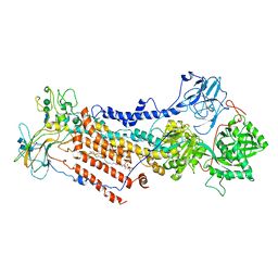 | | Cryo-EM structure of the partially activated Drs2p-Cdc50p | | 分子名称: | (2R)-1-{[(R)-hydroxy{[(1R,2R,3R,4R,5S,6R)-2,3,5,6-tetrahydroxy-4-(phosphonooxy)cyclohexyl]oxy}phosphoryl]oxy}-3-(octadecanoyloxy)propan-2-yl (5Z,8Z,11Z,14Z)-icosa-5,8,11,14-tetraenoate, 2-acetamido-2-deoxy-beta-D-glucopyranose, 2-acetamido-2-deoxy-beta-D-glucopyranose-(1-4)-2-acetamido-2-deoxy-beta-D-glucopyranose, ... | | 著者 | Timcenko, M, Lyons, J.A, Januliene, D, Ulstrup, J.J, Dieudonne, T, Montigny, C, Ash, M.R, Karlsen, J.L, Boesen, T, Kuhlbrandt, W, Lenoir, G, Moeller, A, Nissen, P. | | 登録日 | 2019-05-13 | | 公開日 | 2019-07-03 | | 最終更新日 | 2020-07-29 | | 実験手法 | ELECTRON MICROSCOPY (3.7 Å) | | 主引用文献 | Structure and autoregulation of a P4-ATPase lipid flippase.
Nature, 571, 2019
|
|
3H6I
 | |
2LU3
 | |
3HAP
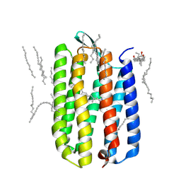 | | Crystal structure of bacteriorhodopsin mutant L111A crystallized from bicelles | | 分子名称: | 3-[(3-CHOLAMIDOPROPYL)DIMETHYLAMMONIO]-1-PROPANESULFONATE, Bacteriorhodopsin, DECANE, ... | | 著者 | Joh, N.H, Yang, D, Bowie, J.U. | | 登録日 | 2009-05-02 | | 公開日 | 2009-09-22 | | 最終更新日 | 2021-10-13 | | 実験手法 | X-RAY DIFFRACTION (1.6 Å) | | 主引用文献 | Similar energetic contributions of packing in the core of membrane and water-soluble proteins.
J.Am.Chem.Soc., 131, 2009
|
|
3HBB
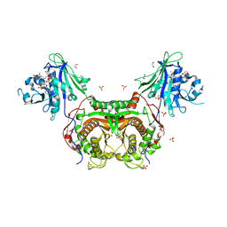 | | Structures of dihydrofolate reductase-thymidylate synthase of Trypanosoma cruzi in the folate-free state and in complex with two antifolate drugs, trimetrexate and methotrexate | | 分子名称: | 1,2-ETHANEDIOL, Dihydrofolate reductase-thymidylate synthase, NADP NICOTINAMIDE-ADENINE-DINUCLEOTIDE PHOSPHATE, ... | | 著者 | Schormann, N, Senkovich, O, Chattopadhyay, D. | | 登録日 | 2009-05-04 | | 公開日 | 2009-05-19 | | 最終更新日 | 2023-09-06 | | 実験手法 | X-RAY DIFFRACTION (3 Å) | | 主引用文献 | Structures of dihydrofolate reductase-thymidylate synthase of Trypanosoma cruzi in the folate-free state and in complex with two antifolate drugs, trimetrexate and methotrexate.
Acta Crystallogr.,Sect.D, 65, 2009
|
|
3VWC
 | |
3EDZ
 | | Crystal structure of catalytic domain of TACE with hydroxamate inhibitor | | 分子名称: | ADAM 17, CITRIC ACID, N-{(2R)-2-[2-(hydroxyamino)-2-oxoethyl]-4-methylpentanoyl}-3-methyl-L-valyl-N-(2-aminoethyl)-L-alaninamide, ... | | 著者 | Mazzola, R.D, Zhu, Z, Sinning, L, McKittrick, B, Lavey, B, Spitler, J, Kozlowski, J, Neng-Yang, S, Zhou, G, Guo, Z, Orth, P, Madison, V, Sun, J, Lundell, D, Niu, X. | | 登録日 | 2008-09-03 | | 公開日 | 2008-09-23 | | 最終更新日 | 2021-10-20 | | 実験手法 | X-RAY DIFFRACTION (1.9 Å) | | 主引用文献 | Discovery of novel hydroxamates as highly potent tumor necrosis factor-alpha converting enzyme inhibitors. Part II: optimization of the S3' pocket.
Bioorg.Med.Chem.Lett., 18, 2008
|
|


