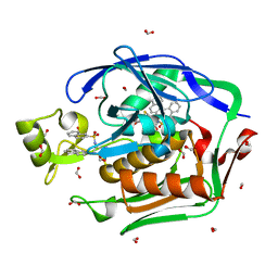
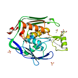
5DRO
 
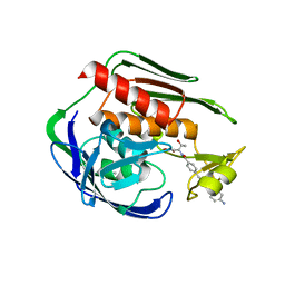 | | Structure of the Aquifex aeolicus LpxC/LPC-011 Complex | | 分子名称: | 4-[4-(4-aminophenyl)buta-1,3-diyn-1-yl]-N-[(2S,3R)-3-hydroxy-1-(hydroxyamino)-1-oxobutan-2-yl]benzamide, ACETATE ION, DIMETHYL SULFOXIDE, ... | | 著者 | Lee, C.-J, Najeeb, J, Zhou, P. | | 登録日 | 2015-09-16 | | 公開日 | 2016-03-09 | | 最終更新日 | 2023-09-27 | | 実験手法 | X-RAY DIFFRACTION (2.01 Å) | | 主引用文献 | Drug design from the cryptic inhibitor envelope.
Nat Commun, 7, 2016
|
|
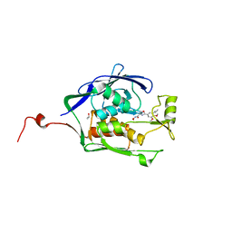
5DRP
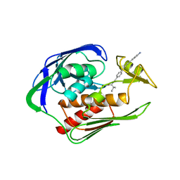 | | Structure of the AaLpxC/LPC-023 Complex | | 分子名称: | CHLORIDE ION, DIMETHYL SULFOXIDE, N~2~-{4-[4-(4-aminophenyl)buta-1,3-diyn-1-yl]benzoyl}-N-hydroxy-L-isoleucinamide, ... | | 著者 | Najeeb, J, Lee, C.-J, Zhou, P. | | 登録日 | 2015-09-16 | | 公開日 | 2016-03-09 | | 最終更新日 | 2023-09-27 | | 実験手法 | X-RAY DIFFRACTION (1.889 Å) | | 主引用文献 | Drug design from the cryptic inhibitor envelope.
Nat Commun, 7, 2016
|
|
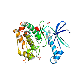
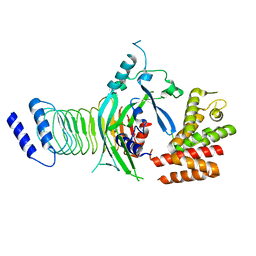
5DRR
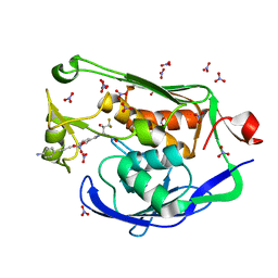 | | Crystal structure of the Pseudomonas aeruginosa LpxC/LPC-058 complex | | 分子名称: | 4-[4-(4-aminophenyl)buta-1,3-diyn-1-yl]-N-[(2S,3S)-4,4-difluoro-3-hydroxy-1-(hydroxyamino)-3-methyl-1-oxobutan-2-yl]benzamide, NITRATE ION, UDP-3-O-[3-hydroxymyristoyl] N-acetylglucosamine deacetylase, ... | | 著者 | Lee, C.-J, Najeeb, J, Zhou, P. | | 登録日 | 2015-09-16 | | 公開日 | 2016-03-09 | | 最終更新日 | 2023-09-27 | | 実験手法 | X-RAY DIFFRACTION (1.59 Å) | | 主引用文献 | Drug design from the cryptic inhibitor envelope.
Nat Commun, 7, 2016
|
|
4S1G
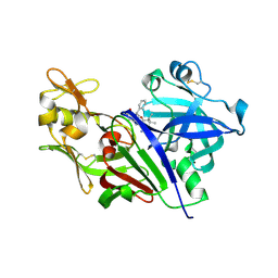 | |
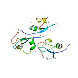
4ISA
 | | Crystal Structure of the Escherichia coli LpxC/BB-78485 complex | | 分子名称: | (2R)-N-hydroxy-3-naphthalen-2-yl-2-[(naphthalen-2-ylsulfonyl)amino]propanamide, (4S,5S)-1,2-DITHIANE-4,5-DIOL, FORMIC ACID, ... | | 著者 | Lee, C.-J, Zhou, P. | | 登録日 | 2013-01-16 | | 公開日 | 2013-10-30 | | 最終更新日 | 2024-02-28 | | 実験手法 | X-RAY DIFFRACTION (1.8 Å) | | 主引用文献 | Structural Basis of the Promiscuous Inhibitor Susceptibility of Escherichia coli LpxC.
Acs Chem.Biol., 9, 2014
|
|
4IS9
 | | Crystal Structure of the Escherichia coli LpxC/L-161,240 complex | | 分子名称: | (4R)-2-(3,4-dimethoxy-5-propylphenyl)-N-hydroxy-4,5-dihydro-1,3-oxazole-4-carboxamide, ISOPROPYL ALCOHOL, SODIUM ION, ... | | 著者 | Lee, C.-J, Zhou, P. | | 登録日 | 2013-01-16 | | 公開日 | 2013-10-30 | | 最終更新日 | 2024-02-28 | | 実験手法 | X-RAY DIFFRACTION (2.13 Å) | | 主引用文献 | Structural Basis of the Promiscuous Inhibitor Susceptibility of Escherichia coli LpxC.
Acs Chem.Biol., 9, 2014
|
|
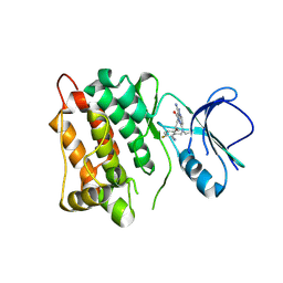
6CMJ
 | | Human CAMKK2 with GSK650393 | | 分子名称: | 1,2-ETHANEDIOL, 2-(2-methylpropyl)-4-(5-phenyl-1H-pyrrolo[2,3-b]pyridin-3-yl)benzoic acid, Calcium/calmodulin-dependent protein kinase kinase 2, ... | | 著者 | Williams, S.P, Reid, R.A, Price, D.J, Drewry, D.H. | | 登録日 | 2018-03-05 | | 公開日 | 2018-04-04 | | 最終更新日 | 2018-05-16 | | 実験手法 | X-RAY DIFFRACTION (2.4 Å) | | 主引用文献 | An orally available, brain-penetrant CAMKK2 inhibitor reduces food intake in rodent model.
Bioorg. Med. Chem. Lett., 28, 2018
|
|
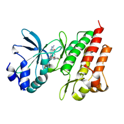
4LG8
 | | Crystal structure of PRPF19 WD40 repeats | | 分子名称: | Pre-mRNA-processing factor 19, SODIUM ION, UNKNOWN ATOM OR ION | | 著者 | Xu, C, Tempel, W, He, H, Dobrovetsky, E, Seitova, A, Bountra, C, Arrowsmith, C.H, Edwards, A.M, Min, J, Structural Genomics Consortium (SGC) | | 登録日 | 2013-06-27 | | 公開日 | 2013-08-07 | | 最終更新日 | 2023-09-20 | | 実験手法 | X-RAY DIFFRACTION (1.89 Å) | | 主引用文献 | Crystal structure of the WD40 domain of human PRPF19.
Biochem. Biophys. Res. Commun., 493, 2017
|
|
4O62
 | | CW-type zinc finger of ZCWPW2 in complex with the amino terminus of histone H3 | | 分子名称: | Histone H3.3, UNKNOWN ATOM OR ION, ZINC ION, ... | | 著者 | Liu, Y, Tempel, W, Dong, A, Loppnau, P, Bountra, C, Weigelt, J, Arrowsmith, C.H, Edwards, A.M, Min, J, Structural Genomics Consortium (SGC) | | 登録日 | 2013-12-20 | | 公開日 | 2014-03-26 | | 最終更新日 | 2016-06-08 | | 実験手法 | X-RAY DIFFRACTION (1.78 Å) | | 主引用文献 | Family-wide Characterization of Histone Binding Abilities of Human CW Domain-containing Proteins.
J.Biol.Chem., 291, 2016
|
|
4MQY
 | | Crystal Structure of the Escherichia coli LpxC/LPC-138 complex | | 分子名称: | 4-[4-(4-aminophenyl)buta-1,3-diyn-1-yl]-N-[(2S,3R)-3-hydroxy-2-methyl-1-nitroso-1-oxobutan-2-yl]benzamide, 4-ethynyl-N-[(1S,2R)-2-hydroxy-1-(oxocarbamoyl)propyl]benzamide, DIMETHYL SULFOXIDE, ... | | 著者 | Lee, C.-J, Najeeb, J, Zhou, P. | | 登録日 | 2013-09-17 | | 公開日 | 2013-10-23 | | 最終更新日 | 2024-02-28 | | 実験手法 | X-RAY DIFFRACTION (2.005 Å) | | 主引用文献 | Structural Basis of the Promiscuous Inhibitor Susceptibility of Escherichia coli LpxC.
Acs Chem.Biol., 9, 2014
|
|
7FJ2
 | |
6D7J
 | |
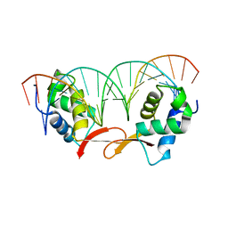
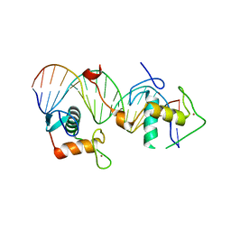
8J54
 | | Crystal structure of RXR/DR2 complex | | 分子名称: | DNA (5'-D(*CP*AP*TP*GP*AP*CP*CP*TP*AP*CP*TP*GP*AP*CP*CP*TP*AP*G)-3'), DNA (5'-D(*CP*TP*AP*GP*GP*TP*CP*AP*GP*TP*AP*GP*GP*TP*CP*AP*TP*G)-3'), Retinoic acid receptor RXR, ... | | 著者 | Chen, Y, Jiang, L. | | 登録日 | 2023-04-21 | | 公開日 | 2024-01-17 | | 最終更新日 | 2024-04-17 | | 実験手法 | X-RAY DIFFRACTION (2.72 Å) | | 主引用文献 | Structural characterization of the DNA binding mechanism of retinoic acid-related orphan receptor gamma.
Structure, 32, 2024
|
|
7WJQ
 | |
8K79
 | |
8K78
 | |
7XVN
 | |
8INK
 | |
8IE3
 | |
8IDT
 | |
8INF
 | |
8INE
 | |
8IDY
 | |
8IR3
 | |
8IPY
 | |


