7RB7
 
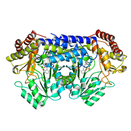
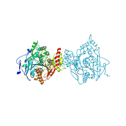 | | Room temperature structure of hAChE in complex with substrate analog 4K-TMA and MMB4 oxime | | 分子名称: | 1,1'-methylenebis{4-[(E)-(hydroxyimino)methyl]pyridin-1-ium}, 4,4-DIHYDROXY-N,N,N-TRIMETHYLPENTAN-1-AMINIUM, Acetylcholinesterase | | 著者 | Kovalevsky, A, Gerlits, O, Radic, Z. | | 登録日 | 2021-07-05 | | 公開日 | 2021-09-22 | | 最終更新日 | 2023-10-18 | | 実験手法 | X-RAY DIFFRACTION (2.6 Å) | | 主引用文献 | Room temperature crystallography of human acetylcholinesterase bound to a substrate analogue 4K-TMA: Towards a neutron structure
Curr Res Struct Biol, 3, 2021
|
|
8SSJ
 | |
8SUI
 | |
8SSY
 | |
8SUJ
 | |
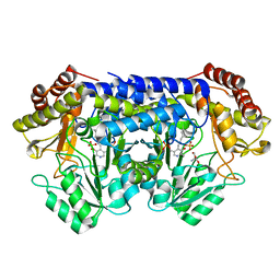
6BBS
 | | Joint X-ray/neutron structure of human carbonic anhydrase II in complex with brinzolamide | | 分子名称: | (+)-4-ETHYLAMINO-3,4-DIHYDRO-2-(METHOXY)PROPYL-2H-THIENO[3,2-E]-1,2-THIAZINE-6-SULFONAMIDE-1,1-DIOXIDE, Carbonic anhydrase 2, ZINC ION | | 著者 | Kovalevsky, A, Aggarwal, M, McKenna, R. | | 登録日 | 2017-10-19 | | 公開日 | 2018-02-28 | | 最終更新日 | 2024-03-13 | | 実験手法 | NEUTRON DIFFRACTION (2 Å), X-RAY DIFFRACTION | | 主引用文献 | "To Be or Not to Be" Protonated: Atomic Details of Human Carbonic Anhydrase-Clinical Drug Complexes by Neutron Crystallography and Simulation.
Structure, 26, 2018
|
|
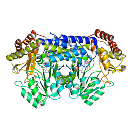
6BCC
 | | Joint X-ray/neutron structure of human carbonic anhydrase II in complex with ethoxzolamide | | 分子名称: | 6-ethoxy-1,3-benzothiazole-2-sulfonamide, Carbonic anhydrase 2, ZINC ION | | 著者 | Kovalevsky, A, McKenna, R, Aggarwal, M. | | 登録日 | 2017-10-20 | | 公開日 | 2018-02-28 | | 最終更新日 | 2023-10-04 | | 実験手法 | NEUTRON DIFFRACTION (1.8 Å), X-RAY DIFFRACTION | | 主引用文献 | "To Be or Not to Be" Protonated: Atomic Details of Human Carbonic Anhydrase-Clinical Drug Complexes by Neutron Crystallography and Simulation.
Structure, 26, 2018
|
|
6BC9
 | | Joint X-ray/neutron structure of human carbonic anhydrase II in complex with dorzolamide | | 分子名称: | (4S-TRANS)-4-(ETHYLAMINO)-5,6-DIHYDRO-6-METHYL-4H-THIENO(2,3-B)THIOPYRAN-2-SULFONAMIDE-7,7-DIOXIDE, Carbonic anhydrase 2, ZINC ION | | 著者 | Kovalevsky, A, McKenna, R, Aggarwal, M. | | 登録日 | 2017-10-20 | | 公開日 | 2018-02-28 | | 最終更新日 | 2023-10-04 | | 実験手法 | NEUTRON DIFFRACTION (1.8 Å), X-RAY DIFFRACTION | | 主引用文献 | "To Be or Not to Be" Protonated: Atomic Details of Human Carbonic Anhydrase-Clinical Drug Complexes by Neutron Crystallography and Simulation.
Structure, 26, 2018
|
|
6BQ8
 | | Joint X-ray/neutron structure of PKG II CNB-B domain in complex with 8-pCPT-cGMP | | 分子名称: | (4S)-2-METHYL-2,4-PENTANEDIOL, 2-(~2~H_2_)amino-8-[(4-chlorophenyl)sulfanyl]-9-[(2S,4aR,6R,7R,7aS)-2-hydroxy-7-(~2~H)hydroxy-2-oxotetrahydro-2H,4H-2lambda~5~-furo[3,2-d][1,3,2]dioxaphosphinin-6-yl](~2~H)-1,9-dihydro-6H-purin-6-one, STRONTIUM ION, ... | | 著者 | Kim, C, Kovalevsky, A, Gerlits, O. | | 登録日 | 2017-11-27 | | 公開日 | 2018-03-21 | | 最終更新日 | 2024-04-03 | | 実験手法 | NEUTRON DIFFRACTION (2 Å), X-RAY DIFFRACTION | | 主引用文献 | Neutron Crystallography Detects Differences in Protein Dynamics: Structure of the PKG II Cyclic Nucleotide Binding Domain in Complex with an Activator.
Biochemistry, 57, 2018
|
|
6D4L
 | |
5T8H
 | |
6EXY
 | | Neutron crystal structure of perdeuterated galectin-3C in complex with glycerol | | 分子名称: | GLYCEROL, Galectin-3 | | 著者 | Manzoni, F, Schrader, T.E, Ostermann, A, Oksanen, E, Logan, D.T. | | 登録日 | 2017-11-10 | | 公開日 | 2018-09-12 | | 最終更新日 | 2024-05-01 | | 実験手法 | NEUTRON DIFFRACTION (1.1 Å), X-RAY DIFFRACTION | | 主引用文献 | Elucidation of Hydrogen Bonding Patterns in Ligand-Free, Lactose- and Glycerol-Bound Galectin-3C by Neutron Crystallography to Guide Drug Design.
J. Med. Chem., 61, 2018
|
|
5VJZ
 | | Joint X-ray/neutron structure of aspartate aminotransferase with alpha-methyl-aspartate at pH 7.5 | | 分子名称: | 2-[(3-HYDROXY-2-METHYL-5-PHOSPHONOOXYMETHYL-PYRIDIN-4-YLMETHYL)-AMINO]-2-METHYL-SUCCINIC ACID, Aspartate aminotransferase, cytoplasmic | | 著者 | Dajnowicz, S, Kovalevsky, A.Y, Mueser, T.C. | | 登録日 | 2017-04-20 | | 公開日 | 2017-11-01 | | 最終更新日 | 2022-03-16 | | 実験手法 | NEUTRON DIFFRACTION (2 Å), X-RAY DIFFRACTION | | 主引用文献 | Direct visualization of critical hydrogen atoms in a pyridoxal 5'-phosphate enzyme.
Nat Commun, 8, 2017
|
|
5VK7
 | |
4DUO
 | | Room-temperature X-ray structure of D-Xylose Isomerase in complex with 2Mg2+ ions and xylitol at pH 7.7 | | 分子名称: | MAGNESIUM ION, Xylitol, Xylose isomerase | | 著者 | Kovalevsky, A.Y, Hanson, L, Langan, P. | | 登録日 | 2012-02-22 | | 公開日 | 2012-08-29 | | 最終更新日 | 2024-02-28 | | 実験手法 | X-RAY DIFFRACTION (2 Å) | | 主引用文献 | Inhibition of D-xylose isomerase by polyols: atomic details by joint X-ray/neutron crystallography.
Acta Crystallogr.,Sect.D, 68, 2012
|
|
4DVO
 | | Room-temperature joint X-ray/neutron structure of D-xylose isomerase in complex with 2Ni2+ and per-deuterated D-sorbitol at pH 5.9 | | 分子名称: | NICKEL (II) ION, Xylose isomerase, sorbitol | | 著者 | Kovalevsky, A.Y, Hanson, L, Langan, P. | | 登録日 | 2012-02-23 | | 公開日 | 2012-08-29 | | 最終更新日 | 2024-02-28 | | 実験手法 | NEUTRON DIFFRACTION (2 Å), X-RAY DIFFRACTION | | 主引用文献 | Inhibition of D-xylose isomerase by polyols: atomic details by joint X-ray/neutron crystallography.
Acta Crystallogr.,Sect.D, 68, 2012
|
|
3KBS
 | |
3KBV
 | |
3KBM
 | |
3KCJ
 | |
3KBW
 | |
3KCL
 | |
3KBJ
 | |
3KCO
 | |
3KBN
 | |


