1ONA
 
 | | CO-CRYSTALS OF CONCANAVALIN A WITH METHYL-3,6-DI-O-(ALPHA-D-MANNOPYRANOSYL)-ALPHA-D-MANNOPYRANOSIDE | | 分子名称: | CALCIUM ION, CONCANAVALIN A, MANGANESE (II) ION, ... | | 著者 | Bouckaert, J, Maes, D, Poortmans, F, Wyns, L, Loris, R. | | 登録日 | 1996-07-07 | | 公開日 | 1997-09-17 | | 最終更新日 | 2024-05-22 | | 実験手法 | X-RAY DIFFRACTION (2.35 Å) | | 主引用文献 | A structure of the complex between concanavalin A and methyl-3,6-di-O-(alpha-D-mannopyranosyl)-alpha-D-mannopyranoside reveals two binding modes.
J.Biol.Chem., 271, 1996
|
|
5KLI
 | | Rhodobacter sphaeroides bc1 with stigmatellin and antimycin | | 分子名称: | (1R)-2-{[(R)-(2-AMINOETHOXY)(HYDROXY)PHOSPHORYL]OXY}-1-[(DODECANOYLOXY)METHYL]ETHYL (9Z)-OCTADEC-9-ENOATE, (2R,3S,6S,7R,8R)-3-{[3-(FORMYLAMINO)-2-HYDROXYBENZOYL]AMINO}-8-HEXYL-2,6-DIMETHYL-4,9-DIOXO-1,5-DIOXONAN-7-YL (2S)-2-METHYLBUTANOATE, Cytochrome b, ... | | 著者 | Xia, D, Esser, L, Zhou, F, Tang, W.K, Yu, C.A. | | 登録日 | 2016-06-24 | | 公開日 | 2016-10-12 | | 最終更新日 | 2023-09-27 | | 実験手法 | X-RAY DIFFRACTION (2.996 Å) | | 主引用文献 | Hydrogen Bonding to the Substrate Is Not Required for Rieske Iron-Sulfur Protein Docking to the Quinol Oxidation Site of Complex III.
J.Biol.Chem., 291, 2016
|
|
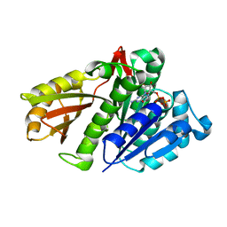
2RJG
 | | Crystal structure of biosynthetic alaine racemase from Escherichia coli | | 分子名称: | Alanine racemase, PYRIDOXAL-5'-PHOSPHATE, SULFATE ION | | 著者 | Wu, D, Hu, T, Zhang, L, Jiang, H, Shen, X. | | 登録日 | 2007-10-15 | | 公開日 | 2008-07-08 | | 最終更新日 | 2023-11-15 | | 実験手法 | X-RAY DIFFRACTION (2.4 Å) | | 主引用文献 | Residues Asp164 and Glu165 at the substrate entryway function potently in substrate orientation of alanine racemase from E. coli: Enzymatic characterization with crystal structure analysis
Protein Sci., 17, 2008
|
|
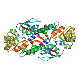
5KNX
 | | Crystal structure of E. coli hypoxanthine phosphoribosyltransferase in complexed with {[(2-[(Hypoxanthin-9H-yl)methyl]propane-1,3-diyl)bis(oxy)]bis- (methylene)}diphosphonic Acid | | 分子名称: | Hypoxanthine-guanine phosphoribosyltransferase, MAGNESIUM ION, [2-[(6-oxidanylidene-1~{H}-purin-9-yl)methyl]-3-(phosphonomethoxy)propoxy]methylphosphonic acid | | 著者 | Eng, W.S, Keough, D.T, Hockova, D, Janeba, Z, Guddat, L.W. | | 登録日 | 2016-06-28 | | 公開日 | 2017-07-19 | | 最終更新日 | 2024-03-06 | | 実験手法 | X-RAY DIFFRACTION (2.4 Å) | | 主引用文献 | Crystal Structures of Acyclic Nucleoside Phosphonates in Complex with Escherichia coli Hypoxanthine Phosphoribosyltransferase
Chemistryselect, 1, 2016
|
|
1P6Y
 | | T4 LYSOZYME CORE REPACKING MUTANT M120Y/TA | | 分子名称: | 2-HYDROXYETHYL DISULFIDE, CHLORIDE ION, LYSOZYME, ... | | 著者 | Mooers, B.H, Datta, D, Baase, W.A, Zollars, E.S, Mayo, S.L, Matthews, B.W. | | 登録日 | 2003-04-30 | | 公開日 | 2003-10-07 | | 最終更新日 | 2023-08-16 | | 実験手法 | X-RAY DIFFRACTION (1.54 Å) | | 主引用文献 | Repacking the Core of T4 Lysozyme by Automated Design
J.Mol.Biol., 332, 2003
|
|
2RGX
 | | Crystal Structure of Adenylate Kinase from Aquifex Aeolicus in complex with Ap5A | | 分子名称: | Adenylate kinase, BIS(ADENOSINE)-5'-PENTAPHOSPHATE, ZINC ION | | 著者 | Thai, V, Wolf-Watz, M, Fenn, T, Pozharski, E, Wilson, M.A, Petsko, G.A, Kern, D. | | 登録日 | 2007-10-05 | | 公開日 | 2007-12-18 | | 最終更新日 | 2023-08-30 | | 実験手法 | X-RAY DIFFRACTION (1.9 Å) | | 主引用文献 | Intrinsic motions along an enzymatic reaction trajectory.
Nature, 450, 2007
|
|
1OOE
 | | Structural Genomics of Caenorhabditis elegans : Dihydropteridine reductase | | 分子名称: | 2-(N-MORPHOLINO)-ETHANESULFONIC ACID, Dihydropteridine reductase | | 著者 | Symersky, J, Li, S, Nagy, L, Qiu, S, Lin, G, Tsao, J, Luo, D, Carson, M, DeLucas, L, Luo, M, Southeast Collaboratory for Structural Genomics (SECSG) | | 登録日 | 2003-03-03 | | 公開日 | 2003-03-18 | | 最終更新日 | 2023-08-16 | | 実験手法 | X-RAY DIFFRACTION (1.65 Å) | | 主引用文献 | Structural genomics of Caenorhabditis elegans: structure of dihydropteridine reductase.
Proteins, 53, 2003
|
|
2RH6
 | |
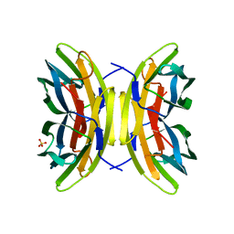
1QJZ
 | | Three Dimensional Structure of Physalis Mottle Virus : Implications for the Viral Assembly | | 分子名称: | COAT PROTEIN | | 著者 | Krishna, S.S, Hiremath, C.N, Munshi, S.K, Prahadeeswaran, D, Sastri, M, Savithri, H.S, Murthy, M.R.N. | | 登録日 | 1999-07-07 | | 公開日 | 1999-07-08 | | 最終更新日 | 2023-12-13 | | 実験手法 | X-RAY DIFFRACTION (3.8 Å) | | 主引用文献 | Three Dimensional Structure of Physalis Movirus: Implications for the Viral Assembly
J.Mol.Biol., 289, 1999
|
|
1OP8
 | | Crystal Structure of Human Granzyme A | | 分子名称: | Granzyme A, SULFATE ION | | 著者 | Hink-Schauer, C, Estebanez-Perpina, E, Bode, W, Jenne, D. | | 登録日 | 2003-03-05 | | 公開日 | 2003-07-01 | | 最終更新日 | 2011-07-13 | | 実験手法 | X-RAY DIFFRACTION (2.5 Å) | | 主引用文献 | Crystal structure of the apoptosis-inducing human granzyme A dimer
NAT.STRUCT.BIOL., 10, 2003
|
|
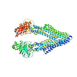
5KOY
 | | Mouse pgp 34 linker deleted bound with ATP | | 分子名称: | ADENOSINE-5'-TRIPHOSPHATE, Multidrug resistance protein 1A | | 著者 | Xia, D, Esser, L, Zhou, F. | | 登録日 | 2016-07-01 | | 公開日 | 2016-12-14 | | 最終更新日 | 2023-10-04 | | 実験手法 | X-RAY DIFFRACTION (3.85 Å) | | 主引用文献 | Structures of the Multidrug Transporter P-glycoprotein Reveal Asymmetric ATP Binding and the Mechanism of Polyspecificity.
J. Biol. Chem., 292, 2017
|
|
5KKQ
 | | Homo sapiens CCCTC-binding factor (CTCF) ZnF3-7 and DNA complex structure | | 分子名称: | DNA (5'-D(*GP*CP*CP*AP*GP*CP*AP*GP*GP*GP*GP*GP*CP*GP*CP*TP*A)-3'), DNA (5'-D(*TP*AP*GP*CP*GP*CP*CP*CP*CP*CP*TP*GP*CP*TP*GP*GP*C)-3'), Transcriptional repressor CTCF, ... | | 著者 | Hashimoto, H, Wang, D, Cheng, X. | | 登録日 | 2016-06-22 | | 公開日 | 2017-05-24 | | 最終更新日 | 2024-03-06 | | 実験手法 | X-RAY DIFFRACTION (1.744 Å) | | 主引用文献 | Structural Basis for the Versatile and Methylation-Dependent Binding of CTCF to DNA.
Mol. Cell, 66, 2017
|
|
5KLV
 | | Structure of bos taurus cytochrome bc1 with fenamidone inhibited | | 分子名称: | (2R)-3-{[(S)-(2-aminoethoxy)(hydroxy)phosphoryl]oxy}-2-(tetradecanoyloxy)propyl octadecanoate, (5S)-5-methyl-2-(methylsulfanyl)-5-phenyl-3-(phenylamino)-3,5-dihydro-4H-imidazol-4-one, 1,2-DIHEXANOYL-SN-GLYCERO-3-PHOSPHOETHANOLAMINE, ... | | 著者 | Xia, D, Esser, L, Zhou, F, Zhou, Y, Xiao, Y, Tang, W.K, Yu, C.A, Qin, Z. | | 登録日 | 2016-06-25 | | 公開日 | 2016-10-12 | | 最終更新日 | 2023-09-27 | | 実験手法 | X-RAY DIFFRACTION (2.652 Å) | | 主引用文献 | Hydrogen Bonding to the Substrate Is Not Required for Rieske Iron-Sulfur Protein Docking to the Quinol Oxidation Site of Complex III.
J.Biol.Chem., 291, 2016
|
|
2ROG
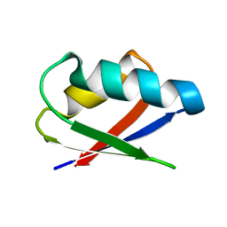 | | Solution structure of Thermus thermophilus HB8 TTHA1718 protein in living E. coli cells | | 分子名称: | Heavy metal binding protein | | 著者 | Sakakibara, D, Sasaki, A, Ikeya, T, Hamatsu, J, Koyama, H, Mishima, M, Mikawa, T, Waelchli, M, Smith, B.O, Shirakawa, M, Guentert, P, Ito, Y. | | 登録日 | 2008-03-21 | | 公開日 | 2009-03-03 | | 最終更新日 | 2024-05-29 | | 実験手法 | SOLUTION NMR | | 主引用文献 | Protein structure determination in living cells by in-cell NMR spectroscopy
Nature, 458, 2009
|
|
1OS1
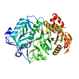 | | Structure of Phosphoenolpyruvate Carboxykinase complexed with ATP,Mg, Ca and pyruvate. | | 分子名称: | ADENOSINE-5'-TRIPHOSPHATE, CALCIUM ION, MAGNESIUM ION, ... | | 著者 | Sudom, A, Walters, R, Pastushok, L, Goldie, D, Prasad, L, Delbaere, L.T, Goldie, H. | | 登録日 | 2003-03-18 | | 公開日 | 2003-09-23 | | 最終更新日 | 2023-11-15 | | 実験手法 | X-RAY DIFFRACTION (1.8 Å) | | 主引用文献 | Mechanisms of activation of phosphoenolpyruvate carboxykinase from Escherichia coli by Ca2+ and of desensitization by trypsin.
J.BACTERIOL., 185, 2003
|
|
2R6R
 | | Aquifex aeolicus FtsZ | | 分子名称: | Cell division protein ftsZ, GUANOSINE-5'-DIPHOSPHATE | | 著者 | Trambaiolo, D, Oliva, M.A, Lowe, J. | | 登録日 | 2007-09-06 | | 公開日 | 2007-10-16 | | 最終更新日 | 2023-08-30 | | 実験手法 | X-RAY DIFFRACTION (1.7 Å) | | 主引用文献 | Structural insights into the conformational variability of FtsZ.
J.Mol.Biol., 373, 2007
|
|
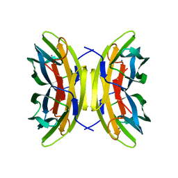
1QAP
 | | QUINOLINIC ACID PHOSPHORIBOSYLTRANSFERASE WITH BOUND QUINOLINIC ACID | | 分子名称: | QUINOLINIC ACID, QUINOLINIC ACID PHOSPHORIBOSYLTRANSFERASE | | 著者 | Eads, J.C, Ozturk, D, Wexler, T.B, Grubmeyer, C, Sacchettini, J.C. | | 登録日 | 1996-09-20 | | 公開日 | 1997-03-12 | | 最終更新日 | 2024-02-14 | | 実験手法 | X-RAY DIFFRACTION (2.8 Å) | | 主引用文献 | A new function for a common fold: the crystal structure of quinolinic acid phosphoribosyltransferase.
Structure, 5, 1997
|
|
1OUS
 | | Lecb (PA-LII) calcium-free | | 分子名称: | SULFATE ION, hypothetical protein LecB | | 著者 | Loris, R, Tielker, D, Jaeger, K.-E, Wyns, L. | | 登録日 | 2003-03-25 | | 公開日 | 2003-09-09 | | 最終更新日 | 2024-03-13 | | 実験手法 | X-RAY DIFFRACTION (1.2 Å) | | 主引用文献 | Structural Basis of Carbohydrate Recognition by the Lectin LecB from Pseudomonas aeruginosa
J.MOL.BIOL., 331, 2003
|
|
5KPD
 | | Mouse pgp 34 linker deleted double EQ mutant | | 分子名称: | Multidrug resistance protein 1A | | 著者 | Xia, D, Esser, L, Zhou, F. | | 登録日 | 2016-07-03 | | 公開日 | 2016-11-30 | | 最終更新日 | 2023-10-04 | | 実験手法 | X-RAY DIFFRACTION (3.35 Å) | | 主引用文献 | Structures of the Multidrug Transporter P-glycoprotein Reveal Asymmetric ATP Binding and the Mechanism of Polyspecificity.
J. Biol. Chem., 292, 2017
|
|
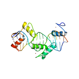
1OOA
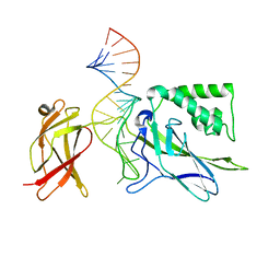 | | CRYSTAL STRUCTURE OF NF-kB(p50)2 COMPLEXED TO A HIGH-AFFINITY RNA APTAMER | | 分子名称: | Nuclear factor NF-kappa-B p105 subunit, RNA aptamer | | 著者 | Huang, D.B, Vu, D, Cassiday, L.A, Zimmerman, J.M, Maher III, L.J, Ghosh, G. | | 登録日 | 2003-03-03 | | 公開日 | 2003-07-22 | | 最終更新日 | 2023-08-16 | | 実験手法 | X-RAY DIFFRACTION (2.45 Å) | | 主引用文献 | Crystal structure of NF-kappaB (p50)2 complexed to a high-affinity RNA aptamer.
Proc.Natl.Acad.Sci.USA, 100, 2003
|
|
1QKU
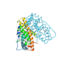 | | WILD TYPE ESTROGEN NUCLEAR RECEPTOR LIGAND BINDING DOMAIN COMPLEXED WITH ESTRADIOL | | 分子名称: | ESTRADIOL, ESTRADIOL RECEPTOR | | 著者 | Ruff, M, Gangloff, M, Eiler, S, Duclaud, S, Wurtz, J.M, Moras, D. | | 登録日 | 1999-08-05 | | 公開日 | 2000-08-18 | | 最終更新日 | 2024-05-08 | | 実験手法 | X-RAY DIFFRACTION (3.2 Å) | | 主引用文献 | Crystal structure of a mutant hERalpha ligand-binding domain reveals key structural features for the mechanism of partial agonism.
J. Biol. Chem., 276, 2001
|
|
1OVS
 | | LecB (PA-LII) in complex with core trimannoside | | 分子名称: | CALCIUM ION, alpha-D-mannopyranose, alpha-D-mannopyranose-(1-3)-alpha-D-mannopyranose, ... | | 著者 | Loris, R, Tielker, D, Jaeger, K.-E, Wyns, L. | | 登録日 | 2003-03-27 | | 公開日 | 2003-09-09 | | 最終更新日 | 2024-03-13 | | 実験手法 | X-RAY DIFFRACTION (1.75 Å) | | 主引用文献 | Structural Basis of Carbohydrate Recognition by the Lectin LecB from Pseudomonas aeruginosa
J.MOL.BIOL., 331, 2003
|
|
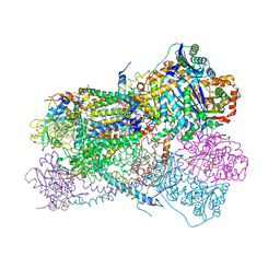
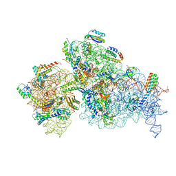
7NAU
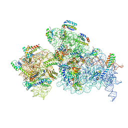 | | Bacterial 30S ribosomal subunit assembly complex state C (Consensus Refinement) | | 分子名称: | 16S rRNA, 30S ribosomal protein S10, 30S ribosomal protein S11, ... | | 著者 | Schedlbauer, A, Iturrioz, I, Ochoa-Lizarralde, B, Diercks, T, Kaminishi, T, Capuni, R, Astigarraga, E, Gil-Carton, D, Fucini, P, Connell, S. | | 登録日 | 2021-01-25 | | 公開日 | 2021-12-08 | | 最終更新日 | 2024-04-24 | | 実験手法 | ELECTRON MICROSCOPY (3.78 Å) | | 主引用文献 | A conserved rRNA switch is central to decoding site maturation on the small ribosomal subunit.
Sci Adv, 7, 2021
|
|
7NAV
 | | Bacterial 30S ribosomal subunit assembly complex state D (Consensus refinement) | | 分子名称: | 16S rRNA, 30S ribosomal protein S10, 30S ribosomal protein S11, ... | | 著者 | Schedlbauer, A, Iturrioz, I, Ochoa-Lizarralde, B, Diercks, T, Kaminishi, T, Capuni, R, Astigarraga, E, Gil-Carton, D, Fucini, P, Connell, S. | | 登録日 | 2021-01-25 | | 公開日 | 2021-12-08 | | 最終更新日 | 2024-04-24 | | 実験手法 | ELECTRON MICROSCOPY (4.8 Å) | | 主引用文献 | A conserved rRNA switch is central to decoding site maturation on the small ribosomal subunit.
Sci Adv, 7, 2021
|
|
5KRS
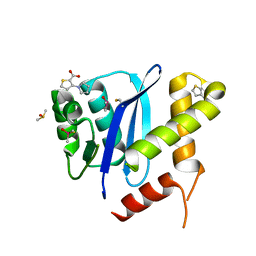 | | HIV-1 Integrase Catalytic Core Domain in Complex with an Allosteric Inhibitor, 3-(1H-pyrrol-1-yl)-2-thiophenecarboxylic acid | | 分子名称: | 3-pyrrol-1-ylthiophene-2-carboxylic acid, DIMETHYL SULFOXIDE, Integrase | | 著者 | Patel, D, Bauman, J.D, Arnold, E. | | 登録日 | 2016-07-07 | | 公開日 | 2016-09-28 | | 最終更新日 | 2022-03-23 | | 実験手法 | X-RAY DIFFRACTION (1.7 Å) | | 主引用文献 | A New Class of Allosteric HIV-1 Integrase Inhibitors Identified by Crystallographic Fragment Screening of the Catalytic Core Domain.
J.Biol.Chem., 291, 2016
|
|


