3HS3
 
 | |
3BWI
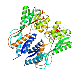
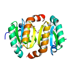
 | | Crystal structure of the catalytic domain of botulinum neurotoxin serotype A with an acetate ion bound at the active site | | 分子名称: | ACETATE ION, Botulinum neurotoxin A light chain, SULFATE ION, ... | | 著者 | Kumaran, D, Rawat, R, Swaminathan, S. | | 登録日 | 2008-01-09 | | 公開日 | 2008-04-22 | | 最終更新日 | 2023-08-30 | | 実験手法 | X-RAY DIFFRACTION (1.7 Å) | | 主引用文献 | Structure- and Substrate-based Inhibitor Design for Clostridium botulinum Neurotoxin Serotype A
J.Biol.Chem., 283, 2008
|
|
3IQ0
 | |
3C88
 | |
3BT3
 | |
6K1T
 | |
1RVH
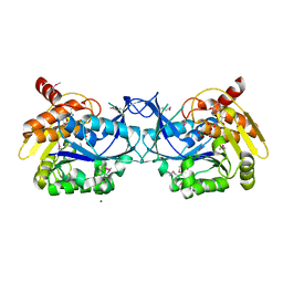
 | | SOLUTION STRUCTURE OF THE DNA DODECAMER GCAAAATTTTGC | | 分子名称: | 5'-D(*GP*CP*AP*AP*AP*AP*TP*TP*TP*TP*GP*C)-3' | | 著者 | Stefl, R, Wu, H, Ravindranathan, S, Sklenar, V, Feigon, J. | | 登録日 | 2003-12-13 | | 公開日 | 2004-02-10 | | 最終更新日 | 2024-05-22 | | 実験手法 | SOLUTION NMR | | 主引用文献 | DNA A-tract bending in three dimensions: Solving the dA4T4 vs. dT4A4 conundrum.
Proc.Natl.Acad.Sci.USA, 101, 2004
|
|
4HGV
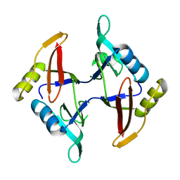
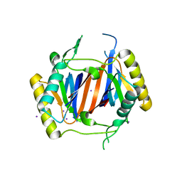
 | | Crystal structure of a fumarate hydratase | | 分子名称: | Fumarate hydratase class II, SULFATE ION | | 著者 | Eswaramoorthy, S, Evans, B, Foti, R, Gizzi, A, Hillerich, B, Kar, A, Lafleur, J, Seidel, R, Villigas, G, Zencheck, W, Almo, S.C, Swaminathan, S, New York Structural Genomics Research Consortium (NYSGRC) | | 登録日 | 2012-10-08 | | 公開日 | 2012-10-31 | | 実験手法 | X-RAY DIFFRACTION (2.09 Å) | | 主引用文献 | Crystal structure of a fumarate hydratase
To be Published
|
|
1RRM
 | |
3LJS
 | |
3LM4
 | | Crystal Structure of 2,3-Dihydroxy Biphenyl dioxygenase from Rhodococcus sp. (strain RHA1) | | 分子名称: | (2Z,4E)-2-HYDROXY-6-OXO-6-PHENYLHEXA-2,4-DIENOIC ACID, Catechol 2,3-dioxygenase, FE (III) ION, ... | | 著者 | Syed Ibrahim, B, Kumaran, D, Burley, S.K, Swaminathan, S, New York SGX Research Center for Structural Genomics (NYSGXRC) | | 登録日 | 2010-01-29 | | 公開日 | 2010-02-23 | | 最終更新日 | 2021-02-10 | | 実験手法 | X-RAY DIFFRACTION (1.8 Å) | | 主引用文献 | Crystal Structure of 2,3-Dihydroxy Biphenyl dioxygenase from Rhodococcus sp. (strain RHA1)
To be Published
|
|
7LUG
 | | Crystal structure of the pnRFP B30Y mutant | | 分子名称: | PHOSPHATE ION, Red Fluorescent pnRFP B30Y mutant | | 著者 | Huang, M, Ng, H.L, Pang, Y, Zhang, S, Fan, Y, Yeh, H, Xiong, Y, Li, X, Ai, H. | | 登録日 | 2021-02-22 | | 公開日 | 2022-03-16 | | 最終更新日 | 2023-11-15 | | 実験手法 | X-RAY DIFFRACTION (1.95 Å) | | 主引用文献 | Development, Characterization, and Structural Analysis of a Genetically Encoded Red Fluorescent Peroxynitrite Biosensor
To Be Published
|
|
7LQO
 | | Crystal structure of a genetically encoded red fluorescent peroxynitrite biosensor, pnRFP | | 分子名称: | PHOSPHATE ION, red fluorescent peroxynitrite biosensor pnRFP | | 著者 | Huang, M, Ng, H.L, Pang, Y, Zhang, S, Fan, Y, Yeh, H, Xiong, Y, Li, X, Ai, H. | | 登録日 | 2021-02-14 | | 公開日 | 2022-03-16 | | 最終更新日 | 2023-11-15 | | 実験手法 | X-RAY DIFFRACTION (2.1 Å) | | 主引用文献 | Development, Characterization, and Structural Analysis of a Genetically Encoded Red Fluorescent Peroxynitrite Biosensor
To Be Published
|
|
3C8B
 | |
1NJR
 | | Crystal structure of yeast ymx7, an ADP-ribose-1''-monophosphatase | | 分子名称: | 32.1 kDa protein in ADH3-RCA1 intergenic region, Xylitol | | 著者 | Kumaran, D, Eswaramoorthy, S, Studier, F.W, Swaminathan, S, Burley, S.K, New York SGX Research Center for Structural Genomics (NYSGXRC) | | 登録日 | 2003-01-02 | | 公開日 | 2004-08-17 | | 最終更新日 | 2021-02-03 | | 実験手法 | X-RAY DIFFRACTION (1.9 Å) | | 主引用文献 | Structure and mechanism of ADP-ribose-1''-monophosphatase (Appr-1''-pase), a ubiquitous cellular processing enzyme
Protein Sci., 14, 2005
|
|
1NKQ
 | | Crystal structure of yeast ynq8, a fumarylacetoacetate hydrolase family protein | | 分子名称: | ACETIC ACID, CALCIUM ION, Hypothetical 28.8 kDa protein in PSD1-SKO1 intergenic region, ... | | 著者 | Eswaramoorthy, S, Kumaran, D, Daniels, B, Studier, F.W, Swaminathan, S, Burley, S.K, New York SGX Research Center for Structural Genomics (NYSGXRC) | | 登録日 | 2003-01-03 | | 公開日 | 2004-06-15 | | 最終更新日 | 2021-02-03 | | 実験手法 | X-RAY DIFFRACTION (2.2 Å) | | 主引用文献 | Crtystal Structure of Yeast Hypothetical Protein YNQ8_YEAST
To be Published
|
|
3HZ4
 | |
3C89
 | |
3LMU
 | | Crystal structure of DTD from Plasmodium falciparum | | 分子名称: | D-tyrosyl-tRNA(Tyr) deacylase, IODIDE ION | | 著者 | Manickam, Y, Bhatt, T.K, Khan, S, Sharma, A. | | 登録日 | 2010-02-01 | | 公開日 | 2010-03-02 | | 最終更新日 | 2024-03-20 | | 実験手法 | X-RAY DIFFRACTION (3.3 Å) | | 主引用文献 | Structure of D-tyrosyl-tRNATyr deacylase using home-source Cu Kalpha and moderate-quality iodide-SAD data: structural polymorphism and HEPES-bound enzyme states
Acta Crystallogr.,Sect.D, 66, 2010
|
|
1XJI
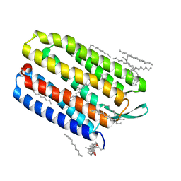 | | Bacteriorhodopsin crystallized in bicelles at room temperature | | 分子名称: | 3-[(3-CHOLAMIDOPROPYL)DIMETHYLAMMONIO]-1-PROPANESULFONATE, Bacteriorhodopsin, DECANE, ... | | 著者 | Faham, S, Boulting, G.L, Massey, E.A, Yohannan, S, Yang, D, Bowie, J.U. | | 登録日 | 2004-09-23 | | 公開日 | 2005-04-19 | | 最終更新日 | 2023-08-23 | | 実験手法 | X-RAY DIFFRACTION (2.2 Å) | | 主引用文献 | Crystallization of bacteriorhodopsin from bicelle formulations at room temperature
Protein Sci., 14, 2005
|
|
3LDT
 | |
3LHL
 | | Crystal structure of a putative agmatinase from Clostridium difficile | | 分子名称: | (4S)-2-METHYL-2,4-PENTANEDIOL, MANGANESE (II) ION, PHOSPHATE ION, ... | | 著者 | Palani, K, Burley, S.K, Swaminathan, S, New York SGX Research Center for Structural Genomics (NYSGXRC) | | 登録日 | 2010-01-22 | | 公開日 | 2010-02-23 | | 最終更新日 | 2021-02-10 | | 実験手法 | X-RAY DIFFRACTION (2.3 Å) | | 主引用文献 | Crystal structure of a putative agmatinase from Clostridium difficile
To be Published
|
|
3LKI
 | |
1TH3
 | | Crystal structure of NADPH depleted bovine live catalase complexed with cyanide | | 分子名称: | CYANIDE ION, Catalase, PROTOPORPHYRIN IX CONTAINING FE | | 著者 | Sugadev, R, Balasundaresan, D, Ponnuswamy, M.N, Kumaran, D, Swaminathan, S, Sekar, K. | | 登録日 | 2004-06-01 | | 公開日 | 2005-07-05 | | 最終更新日 | 2023-08-23 | | 実験手法 | X-RAY DIFFRACTION (2.8 Å) | | 主引用文献 | The crystal structure of bovine liver catalase
TO BE PUBLISHED
|
|
1SRU
 | | Crystal structure of full length E. coli SSB protein | | 分子名称: | Single-strand binding protein | | 著者 | Savvides, S.N, Raghunathan, S, Fuetterer, K, Kozlov, A.G, Lohman, T.M, Waksman, G. | | 登録日 | 2004-03-23 | | 公開日 | 2004-08-03 | | 最終更新日 | 2024-02-14 | | 実験手法 | X-RAY DIFFRACTION (3.3 Å) | | 主引用文献 | The C-terminal domain of full-length E. coli SSB is disordered even when bound to DNA.
Protein Sci., 13, 2004
|
|


