Loading
PDBj
メニューPDBj@FacebookPDBj@TwitterPDBj@YouTubewwPDB FoundationwwPDB
RCSB PDBPDBeBMRBAdv. SearchSearch help
Search by PDB author
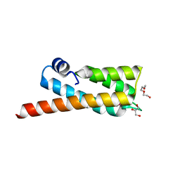
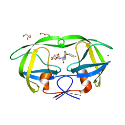
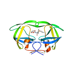
2F80
DownloadVisualize
BU of 2f80 by Molmil
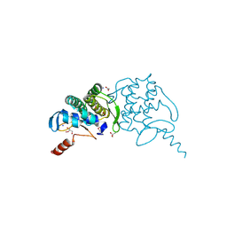
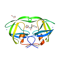
HIV-1 Protease mutant D30N complexed with inhibitor TMC114
分子名称: (3R,3AS,6AR)-HEXAHYDROFURO[2,3-B]FURAN-3-YL(1S,2R)-3-[[(4-AMINOPHENYL)SULFONYL](ISOBUTYL)AMINO]-1-BENZYL-2-HYDROXYPROPYLCARBAMATE, CHLORIDE ION, POL POLYPROTEIN, ...
著者Kovalevsky, A.Y, Weber, I.T.
登録日2005-12-01
公開日2006-03-07
最終更新日2024-02-14
実験手法X-RAY DIFFRACTION (1.45 Å)
主引用文献Effectiveness of Nonpeptide Clinical Inhibitor TMC-114 on HIV-1 Protease with Highly Drug Resistant Mutations D30N, I50V, and L90M.
J.Med.Chem., 49, 2006
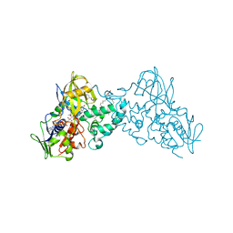
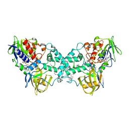
7DRT
DownloadVisualize
BU of 7drt by Molmil
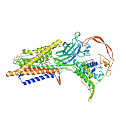
Human Wntless in complex with Wnt3a
分子名称: 1,2-DIOLEOYL-SN-GLYCERO-3-PHOSPHOCHOLINE, 1-O-OCTADECYL-SN-GLYCERO-3-PHOSPHOCHOLINE, 2-acetamido-2-deoxy-beta-D-glucopyranose-(1-4)-2-acetamido-2-deoxy-beta-D-glucopyranose, ...
著者Zhong, Q, Zhao, Y, Ye, F, Xiao, Z, Huang, G, Zhang, Y, Lu, P, Xu, W, Zhou, Q, Ma, D.
登録日2020-12-29
公開日2021-07-14
最終更新日2024-10-30
実験手法ELECTRON MICROSCOPY (2.2 Å)
主引用文献Cryo-EM structure of human Wntless in complex with Wnt3a.
Nat Commun, 12, 2021
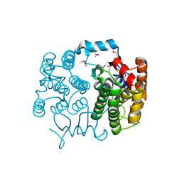
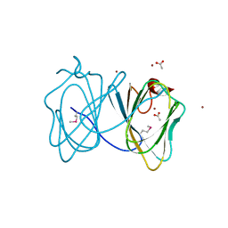
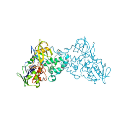
1JV4
DownloadVisualize
BU of 1jv4 by Molmil
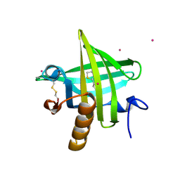
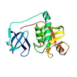
Crystal structure of recombinant major mouse urinary protein (rmup) at 1.75 A resolution
分子名称: 2-(SEC-BUTYL)THIAZOLE, CADMIUM ION, Major urinary protein 2
著者Kuser, P.R, Franzoni, L, Ferrari, E, Spisni, A, Polikarpov, I.
登録日2001-08-28
公開日2001-12-05
最終更新日2024-10-30
実験手法X-RAY DIFFRACTION (1.75 Å)
主引用文献The X-ray structure of a recombinant major urinary protein at 1.75 A resolution. A comparative study of X-ray and NMR-derived structures.
Acta Crystallogr.,Sect.D, 57, 2001
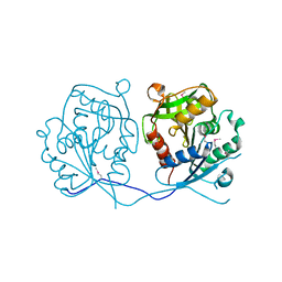
1CRZ
DownloadVisualize
BU of 1crz by Molmil
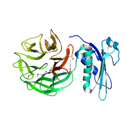
CRYSTAL STRUCTURE OF THE E. COLI TOLB PROTEIN
分子名称: TOLB PROTEIN
著者Abergel, C, Bouveret, E, Claverie, J.-M, Brown, K, Rigal, A, Lazdunski, C, Benedetti, H.
登録日1999-08-16
公開日2000-08-16
最終更新日2024-10-09
実験手法X-RAY DIFFRACTION (1.95 Å)
主引用文献Structure of the Escherichia coli TolB protein determined by MAD methods at 1.95 A resolution.
Structure Fold.Des., 7, 1999
3RG6
DownloadVisualize
BU of 3rg6 by Molmil
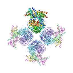
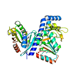
Crystal structure of a chaperone-bound assembly intermediate of form I Rubisco
分子名称: RbcX protein, Ribulose bisphosphate carboxylase large chain
著者Bracher, A, Starling-Windhof, A, Hartl, F.U, Hayer-Hartl, M.
登録日2011-04-07
公開日2011-07-20
最終更新日2023-09-13
実験手法X-RAY DIFFRACTION (3.2 Å)
主引用文献Crystal structure of a chaperone-bound assembly intermediate of form I Rubisco.
Nat.Struct.Mol.Biol., 18, 2011
2OOC
DownloadVisualize
BU of 2ooc by Molmil
Crystal structure of Histidine Phosphotransferase ShpA (NP_419930.1) from Caulobacter crescentus at 1.52 A resolution
分子名称: GLYCEROL, Histidine phosphotransferase, TETRAETHYLENE GLYCOL
著者Joint Center for Structural Genomics (JCSG)
登録日2007-01-25
公開日2007-02-06
最終更新日2024-10-30
実験手法X-RAY DIFFRACTION (1.52 Å)
主引用文献Crystal structure of histidine phosphotransfer protein ShpA, an essential regulator of stalk biogenesis in Caulobacter crescentus.
J.Mol.Biol., 390, 2009
3NG7
DownloadVisualize
BU of 3ng7 by Molmil
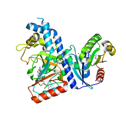
Complex of dithionite-reduced 6-hydroxy-L-nicotine oxidase with substrate bound at active site and inhibitor at exit cavity
分子名称: (1R)-2-{[(S)-(2-aminoethoxy)(hydroxy)phosphoryl]oxy}-1-[(pentadecanoyloxy)methyl]ethyl (12E)-hexadeca-9,12-dienoate, 5-[(2R)-1-methylpyrrolidin-2-yl]pyridin-2-ol, 5-[(2S)-1-methylpyrrolidin-2-yl]pyridin-2-ol, ...
著者Kachalova, G.S, Bartunik, H.D.
登録日2010-06-11
公開日2011-03-16
最終更新日2023-11-01
実験手法X-RAY DIFFRACTION (1.95 Å)
主引用文献Crystallographic snapshots of the complete reaction cycle of nicotine degradation by an amine oxidase of the monoamine oxidase (MAO) family
Proc.Natl.Acad.Sci.USA, 108, 2011
2FNO
DownloadVisualize
BU of 2fno by Molmil
Crystal structure of a glutathione s-transferase (atu5508) from agrobacterium tumefaciens str. c58 at 2.00 A resolution
分子名称: AGR_pAT_752p, THIOCYANATE ION
著者Joint Center for Structural Genomics (JCSG)
登録日2006-01-11
公開日2006-02-14
最終更新日2023-01-25
実験手法X-RAY DIFFRACTION (2 Å)
主引用文献Comparative structural analysis of a novel glutathioneS-transferase (ATU5508) from Agrobacterium tumefaciens at 2.0 A resolution.
Proteins, 65, 2006
2GHR
DownloadVisualize
BU of 2ghr by Molmil
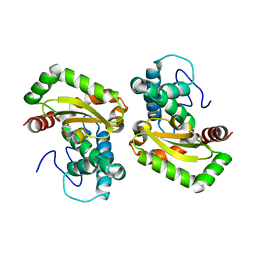
Crystal structure of homoserine o-succinyltransferase (NP_981826.1) from Bacillus cereus ATCC 10987 at 2.40 A resolution
分子名称: Homoserine O-succinyltransferase, SULFATE ION
著者Joint Center for Structural Genomics (JCSG)
登録日2006-03-27
公開日2006-04-11
最終更新日2023-01-25
実験手法X-RAY DIFFRACTION (2.4 Å)
主引用文献Crystal structure of homoserine O-succinyltransferase from Bacillus cereus at 2.4 A resolution
Proteins, 68, 2007
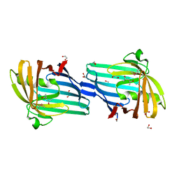
3H0N
DownloadVisualize
BU of 3h0n by Molmil
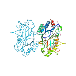
Crystal structure of a duf1470 family protein (jann_2411) from jannaschia sp. ccs1 at 1.45 A resolution
分子名称: ACETATE ION, GLYCEROL, NICKEL (II) ION, ...
著者Joint Center for Structural Genomics (JCSG)
登録日2009-04-09
公開日2009-04-21
最終更新日2023-02-01
実験手法X-RAY DIFFRACTION (1.45 Å)
主引用文献The structure of Jann_2411 (DUF1470) from Jannaschia sp. at 1.45  resolution reveals a new fold (the ABATE domain) and suggests its possible role as a transcription regulator.
Acta Crystallogr.,Sect.F, 66, 2010
2HBW
DownloadVisualize
BU of 2hbw by Molmil
Crystal structure of a putative endopeptidase (ava_3396) from anabaena variabilis atcc 29413 at 1.05 A resolution
分子名称: ACETATE ION, NLP/P60 protein, UNKNOWN LIGAND
著者Joint Center for Structural Genomics (JCSG)
登録日2006-06-14
公開日2006-08-08
最終更新日2023-01-25
実験手法X-RAY DIFFRACTION (1.05 Å)
主引用文献Structural Basis of Murein Peptide Specificity of a gamma-D-Glutamyl-L-Diamino Acid Endopeptidase.
Structure, 17, 2009
7DN8
DownloadVisualize
BU of 7dn8 by Molmil
Crystal structure of Salmonella effector SopF in complex with ARF1
分子名称: ADP-ribosylation factor 1, GUANOSINE-5'-DIPHOSPHATE, MAGNESIUM ION, ...
著者Ding, J, Shao, F.
登録日2020-12-09
公開日2021-12-15
最終更新日2024-05-29
実験手法X-RAY DIFFRACTION (2.6084 Å)
主引用文献ARF GTPases activate Salmonella effector SopF to ADP-ribosylate host V-ATPase and inhibit endomembrane damage-induced autophagy.
Nat.Struct.Mol.Biol., 29, 2022
7DN9
DownloadVisualize
BU of 7dn9 by Molmil
Crystal structure of Salmonella effector in complex with NAD and host co-factor ARF1
分子名称: ADP-ribosylation factor 1, GUANOSINE-5'-DIPHOSPHATE, MAGNESIUM ION, ...
著者Ding, J, Shao, F.
登録日2020-12-09
公開日2021-12-15
最終更新日2024-05-29
実験手法X-RAY DIFFRACTION (3.29 Å)
主引用文献ARF GTPases activate Salmonella effector SopF to ADP-ribosylate host V-ATPase and inhibit endomembrane damage-induced autophagy.
Nat.Struct.Mol.Biol., 29, 2022
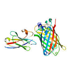
7E53
DownloadVisualize
BU of 7e53 by Molmil
Crystal structure of sfGFP complexed with the nanobody nb2 at 2.2 Angstron resolution
分子名称: Green fluorescent protein, Green fluorescent protein's nanobody nb2
著者Ding, Y, Zhong, P.Y.
登録日2021-02-16
公開日2021-10-13
最終更新日2023-11-29
実験手法X-RAY DIFFRACTION (2.21 Å)
主引用文献Structural insights into two distinct nanobodies recognizing the same epitope of green fluorescent protein.
Biochem.Biophys.Res.Commun., 565, 2021
3H50
DownloadVisualize
BU of 3h50 by Molmil
CRYSTAL STRUCTURE OF A TETRACENOMYCIN POLYKETIDE SYNTHESIS PROTEIN (TCMJ) FROM XANTHOMONAS CAMPESTRIS PV. CAMPESTRIS AT 1.60 A RESOLUTION
分子名称: ACETATE ION, Tetracenomycin polyketide synthesis protein, ZINC ION
著者Joint Center for Structural Genomics (JCSG)
登録日2009-04-21
公開日2009-05-05
最終更新日2024-10-30
実験手法X-RAY DIFFRACTION (1.6 Å)
主引用文献Conformational changes associated with the binding of zinc acetate at the putative active site of XcTcmJ, a cupin from Xanthomonas campestris pv. campestris.
Acta Crystallogr.,Sect.F, 66, 2010
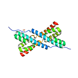
3HYB
DownloadVisualize
BU of 3hyb by Molmil
Crystal structure of RbcX from Anabaena, crystal form II
分子名称: RbcX protein, SULFATE ION
著者Bracher, A, Liu, C.
登録日2009-06-22
公開日2010-01-19
最終更新日2023-09-06
実験手法X-RAY DIFFRACTION (2.3 Å)
主引用文献Coupled chaperone action in folding and assembly of hexadecameric Rubisco.
Nature, 463, 2010
3EQX
DownloadVisualize
BU of 3eqx by Molmil
CRYSTAL STRUCTURE OF A FIC FAMILY PROTEIN (SO_4266) FROM SHEWANELLA ONEIDENSIS AT 1.6 A RESOLUTION
分子名称: FIC DOMAIN CONTAINING TRANSCRIPTIONAL REGULATOR, TRIETHYLENE GLYCOL
著者Joint Center for Structural Genomics (JCSG)
登録日2008-10-01
公開日2008-10-14
最終更新日2023-02-01
実験手法X-RAY DIFFRACTION (1.6 Å)
主引用文献Crystal structure of the Fic (Filamentation induced by cAMP) family protein SO4266 (gi|24375750) from Shewanella oneidensis MR-1 at 1.6 A resolution.
Proteins, 75, 2009
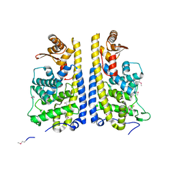
3K7M
DownloadVisualize
BU of 3k7m by Molmil
Crystal structure of 6-hydroxy-L-nicotine oxidase from Arthrobacter nicotinovorans
分子名称: (1R)-2-{[(S)-(2-aminoethoxy)(hydroxy)phosphoryl]oxy}-1-[(pentadecanoyloxy)methyl]ethyl (12E)-hexadeca-9,12-dienoate, 6-hydroxy-L-nicotine oxidase, FLAVIN-ADENINE DINUCLEOTIDE
著者Bourenkov, G.P, Kachalova, G.S, Bartunik, H.D.
登録日2009-10-13
公開日2010-01-19
最終更新日2024-02-21
実験手法X-RAY DIFFRACTION (1.95 Å)
主引用文献Crystal Structure Analysis of Free and Substrate-Bound 6-Hydroxy-l-Nicotine Oxidase from Arthrobacter nicotinovorans.
J.Mol.Biol., 396, 2010
3K7T
DownloadVisualize
BU of 3k7t by Molmil
Crystal structure of apo-form 6-hydroxy-L-nicotine oxidase, crystal form P3121
分子名称: (1R)-2-{[(S)-(2-aminoethoxy)(hydroxy)phosphoryl]oxy}-1-[(pentadecanoyloxy)methyl]ethyl (12E)-hexadeca-9,12-dienoate, 6-hydroxy-L-nicotine oxidase, FLAVIN-ADENINE DINUCLEOTIDE
著者Bourenkov, G.P, Kachalova, G.S, Bartunik, H.D.
登録日2009-10-13
公開日2010-01-19
最終更新日2023-09-06
実験手法X-RAY DIFFRACTION (2.85 Å)
主引用文献Crystal Structure Analysis of Free and Substrate-Bound 6-Hydroxy-l-Nicotine Oxidase from Arthrobacter nicotinovorans.
J.Mol.Biol., 396, 2010
2F81
DownloadVisualize
BU of 2f81 by Molmil
HIV-1 Protease mutant L90M complexed with inhibitor TMC114
分子名称: (3R,3AS,6AR)-HEXAHYDROFURO[2,3-B]FURAN-3-YL(1S,2R)-3-[[(4-AMINOPHENYL)SULFONYL](ISOBUTYL)AMINO]-1-BENZYL-2-HYDROXYPROPYLCARBAMATE, CHLORIDE ION, GLYCEROL, ...
著者Kovalevsky, A.Y, Weber, I.T.
登録日2005-12-01
公開日2006-03-07
最終更新日2024-02-14
実験手法X-RAY DIFFRACTION (1.25 Å)
主引用文献Effectiveness of Nonpeptide Clinical Inhibitor TMC-114 on HIV-1 Protease with Highly Drug Resistant Mutations D30N, I50V, and L90M.
J.Med.Chem., 49, 2006
2H1T
DownloadVisualize
BU of 2h1t by Molmil
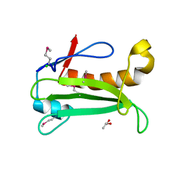
Crystal structure of a duf1089 family protein (pa1994) from pseudomonas aeruginosa at 1.80 A resolution
分子名称: (4S)-2-METHYL-2,4-PENTANEDIOL, 1,2-ETHANEDIOL, Hypothetical protein
著者Joint Center for Structural Genomics (JCSG)
登録日2006-05-16
公開日2006-05-30
最終更新日2023-01-25
実験手法X-RAY DIFFRACTION (1.8 Å)
主引用文献The structure of the first representative of Pfam family PF06475 reveals a new fold with possible involvement in glycolipid metabolism.
Acta Crystallogr.,Sect.F, 66, 2010
4F2N
DownloadVisualize
BU of 4f2n by Molmil
Crystal structure of iron superoxide dismutase from Leishmania major
分子名称: FE (II) ION, Superoxide dismutase
著者Seattle Structural Genomics Center for Infectious Disease (SSGCID)
登録日2012-05-08
公開日2012-05-16
最終更新日2023-09-13
実験手法X-RAY DIFFRACTION (1.85 Å)
主引用文献Iron superoxide dismutases in eukaryotic pathogens: new insights from Apicomplexa and Trypanosoma structures.
Acta Crystallogr F Struct Biol Commun, 71, 2015
2IAY
DownloadVisualize
BU of 2iay by Molmil
Crystal structure of a duf1831 family protein (lp2179) from lactobacillus plantarum at 1.20 A resolution
分子名称: CHLORIDE ION, GLYCEROL, Hypothetical protein
著者Joint Center for Structural Genomics (JCSG)
登録日2006-09-08
公開日2006-10-10
最終更新日2023-01-25
実験手法X-RAY DIFFRACTION (1.2 Å)
主引用文献Structure of LP2179, the first representative of Pfam family PF08866, suggests a new fold with a role in amino-acid metabolism.
Acta Crystallogr.,Sect.F, 66, 2010
2HAG
DownloadVisualize
BU of 2hag by Molmil
Crystal structure of a putative dyp-type peroxidase protein (so_0740) from shewanella oneidensis at 2.75 A resolution
分子名称: Melanin biosynthesis protein TyrA, putative
著者Joint Center for Structural Genomics (JCSG)
登録日2006-06-12
公開日2006-08-08
最終更新日2023-01-25
実験手法X-RAY DIFFRACTION (2.75 Å)
主引用文献Crystal structures of two novel dye-decolorizing peroxidases reveal a beta-barrel fold with a conserved heme-binding motif.
Proteins, 69, 2007
2HB3
DownloadVisualize
BU of 2hb3 by Molmil
Wild-type HIV-1 Protease in complex with potent inhibitor GRL06579
分子名称: (3AS,5R,6AR)-HEXAHYDRO-2H-CYCLOPENTA[B]FURAN-5-YL (2S,3S)-3-HYDROXY-4-(4-(HYDROXYMETHYL)-N-ISOBUTYLPHENYLSULFONAMIDO)-1-PHENYLBUTAN-2-YLCARBAMATE, CHLORIDE ION, GLYCEROL, ...
著者Kovalevsky, A.Y, Weber, I.T.
登録日2006-06-13
公開日2006-08-29
最終更新日2024-02-14
実験手法X-RAY DIFFRACTION (1.35 Å)
主引用文献Structure-Based Design of Novel HIV-1 Protease Inhibitors To Combat Drug Resistance.
J.Med.Chem., 49, 2006

226707

件を2024-10-30に公開中

PDB statisticsPDBj update infoContact PDBjnumon