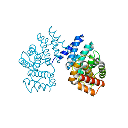
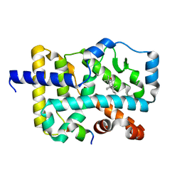
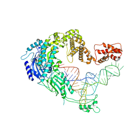
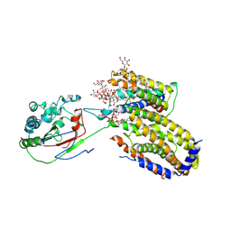
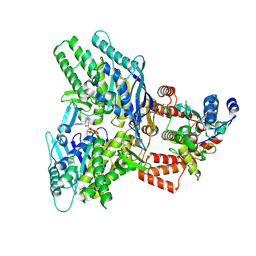
7D8C
 
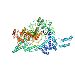 | | Crystal structure of the Cas12i1-crRNA binary complex | | 分子名称: | 12i1, CITRIC ACID, RNA (3-MER), ... | | 著者 | Zhang, B, Luo, D.Y, Li, Y, OuYang, S.Y. | | 登録日 | 2020-10-07 | | 公開日 | 2021-05-19 | | 最終更新日 | 2023-11-29 | | 実験手法 | X-RAY DIFFRACTION (3.6 Å) | | 主引用文献 | Mechanistic insights into the R-loop formation and cleavage in CRISPR-Cas12i1.
Nat Commun, 12, 2021
|
|
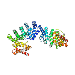
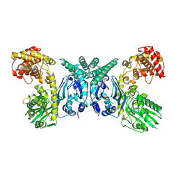
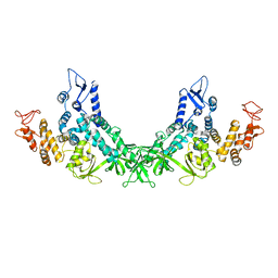
7D3J
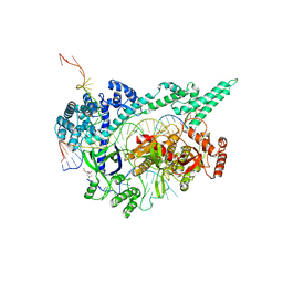 | | Crystal structure of the Cas12i1 R-loop complex after target DNA cleavage | | 分子名称: | 12i1-WT, CITRIC ACID, DNA (23-MER), ... | | 著者 | Zhang, B, Luo, D.Y, Li, Y, OuYang, S.Y. | | 登録日 | 2020-09-19 | | 公開日 | 2021-05-19 | | 最終更新日 | 2023-11-29 | | 実験手法 | X-RAY DIFFRACTION (2.45 Å) | | 主引用文献 | Mechanistic insights into the R-loop formation and cleavage in CRISPR-Cas12i1.
Nat Commun, 12, 2021
|
|
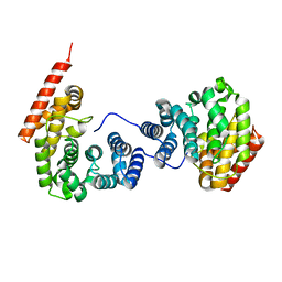
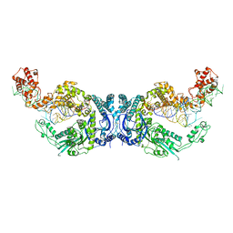
7EU9
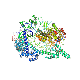 | | Crystal structure of the selenomethionine(SeMet)-derived Cas12i1 R-loop complex before target DNA cleavage | | 分子名称: | CITRIC ACID, Cas12i1 D647A mutant, DNA (24-MER), ... | | 著者 | Zhang, B, Luo, D.Y, Li, Y, OuYang, S.Y. | | 登録日 | 2021-05-16 | | 公開日 | 2021-05-26 | | 最終更新日 | 2021-06-23 | | 実験手法 | X-RAY DIFFRACTION (2.35 Å) | | 主引用文献 | Mechanistic insights into the R-loop formation and cleavage in CRISPR-Cas12i1.
Nat Commun, 12, 2021
|
|
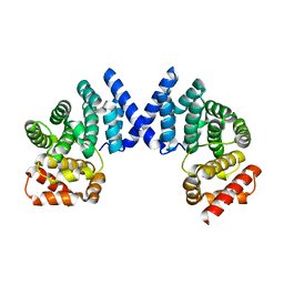
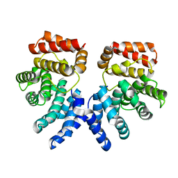
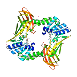
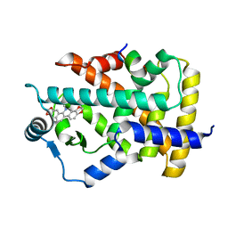
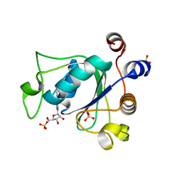
7EP0
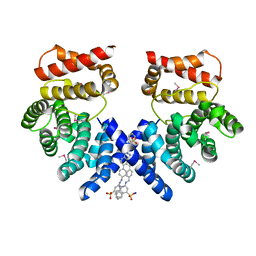 | | Crystal structure of ZYG11B bound to GSTE degron | | 分子名称: | Protein zyg-11 homolog B, sodium 3,3'-(1E,1'E)-biphenyl-4,4'-diylbis(diazene-2,1-diyl)bis(4-aminonaphthalene-1-sulfonate) | | 著者 | Yan, X, Li, Y. | | 登録日 | 2021-04-26 | | 公開日 | 2021-07-14 | | 最終更新日 | 2021-09-01 | | 実験手法 | X-RAY DIFFRACTION (2.16 Å) | | 主引用文献 | Molecular basis for recognition of Gly/N-degrons by CRL2 ZYG11B and CRL2 ZER1 .
Mol.Cell, 81, 2021
|
|
7EP3
 | |
7EP2
 | |
7EP5
 | |
7EP1
 | |
7EP4
 | |
8I2N
 | |
7DNO
 | | Characterization of Peptide Ligands Against WDR5 Isolated Using Phage Display Technique | | 分子名称: | CYS-ARG-THR-LEU-PRO-PHE, WD repeat-containing protein 5 | | 著者 | Cao, J, Cao, D, Xiong, B, Li, Y, Fan, T. | | 登録日 | 2020-12-10 | | 公開日 | 2021-02-10 | | 最終更新日 | 2023-11-29 | | 実験手法 | X-RAY DIFFRACTION (2.03 Å) | | 主引用文献 | Phage-Display Based Discovery and Characterization of Peptide Ligands against WDR5.
Molecules, 26, 2021
|
|
7W3Q
 | | Crystal structure of RORgamma in complex with natural inverse agonist | | 分子名称: | (3S,5R,6S,8R,9R,10R,12R,13R,14R,17S)-4,4,8,10,14-pentamethyl-17-[(2R)-6-methyl-2-oxidanyl-hept-5-en-2-yl]-2,3,5,6,7,9,11,12,13,15,16,17-dodecahydro-1H-cyclopenta[a]phenanthrene-3,6,12-triol, Nuclear receptor ROR-gamma, Peptide from Nuclear receptor coactivator 2 | | 著者 | Tian, S.Y, Li, Y. | | 登録日 | 2021-11-25 | | 公開日 | 2022-12-07 | | 最終更新日 | 2023-11-29 | | 実験手法 | X-RAY DIFFRACTION (2 Å) | | 主引用文献 | Crystal structure of RORgamma in complex with natural inverse agonist
To Be Published
|
|
7W3P
 | | Crystal structure of RORgamma in complex with natural inverse agonist | | 分子名称: | (3S,5R,8R,9R,10R,12R,13R,14R,17S)-4,4,8,10,14-pentamethyl-17-[(2R)-2,6,6-trimethyloxan-2-yl]-2,3,5,6,7,9,11,12,13,15,16,17-dodecahydro-1H-cyclopenta[a]phenanthrene-3,12-diol, Nuclear receptor ROR-gamma, Peptide from Nuclear receptor coactivator 2 | | 著者 | Tian, S.Y, Li, Y. | | 登録日 | 2021-11-25 | | 公開日 | 2022-12-07 | | 最終更新日 | 2023-11-29 | | 実験手法 | X-RAY DIFFRACTION (1.77 Å) | | 主引用文献 | Crystal structure of RORgamma in complex with natural inverse agonist
To Be Published
|
|
7U9I
 | | Co-crystal structure of human CARM1 in complex with MT556 inhibitor | | 分子名称: | 7-[5-S-(4-{[(4-ethylpyridin-3-yl)methyl]amino}butyl)-5-thio-beta-D-ribofuranosyl]-7H-pyrrolo[2,3-d]pyrimidin-4-amine, Histone-arginine methyltransferase CARM1, UNKNOWN ATOM OR ION | | 著者 | Zeng, H, Perveen, S, Dong, A, Hutchinson, A, Seitova, A, Gibson, E, Hajian, T, Li, Y, Gao, Y.D, Schneider, S, Siliphaivanh, P, Sloman, D, Nicholson, B, Fischer, C, Hicks, J, Vedadi, M, Brown, P.J, Arrowsmith, C.H, Edwards, A.M, Halabelian, L, Structural Genomics Consortium (SGC) | | 登録日 | 2022-03-10 | | 公開日 | 2023-01-18 | | 最終更新日 | 2023-10-25 | | 実験手法 | X-RAY DIFFRACTION (2 Å) | | 主引用文献 | Co-crystal structure of human CARM1 in complex with MT556 inhibitor
To Be Published
|
|
7UW7
 | | Crystal structure of the Human TRIP12 WWE domain (isoform 2) in complex with ADP | | 分子名称: | ADENOSINE-5'-DIPHOSPHATE, Isoform 2 of E3 ubiquitin-protein ligase TRIP12, UNKNOWN ATOM OR ION | | 著者 | Kimani, S, Dong, A, Li, Y, Arrowsmith, C.H, Edwards, A.M, Halabelian, L, Structural Genomics Consortium (SGC) | | 登録日 | 2022-05-02 | | 公開日 | 2022-06-08 | | 最終更新日 | 2023-10-18 | | 実験手法 | X-RAY DIFFRACTION (1.17 Å) | | 主引用文献 | Crystal structure of the Human TRIP12 WWE domain (isoform 2) in complex with ADP
To Be Published
|
|
7UNK
 | | Structure of Importin-4 bound to the H3-H4-ASF1 histone-histone chaperone complex | | 分子名称: | Histone H3, Histone H4, Histone chaperone, ... | | 著者 | Bernardes, N.E, Chook, Y.M, Fung, H.Y.J, Chen, Z, Li, Y. | | 登録日 | 2022-04-11 | | 公開日 | 2022-09-21 | | 最終更新日 | 2024-06-12 | | 実験手法 | ELECTRON MICROSCOPY (3.45 Å) | | 主引用文献 | Structure of IMPORTIN-4 bound to the H3-H4-ASF1 histone-histone chaperone complex.
Proc.Natl.Acad.Sci.USA, 119, 2022
|
|
7XJZ
 | | Cryo-EM strucrture of Oryza sativa plastid glycyl-tRNA synthetase in complex with tRNA (tRNA binding state) | | 分子名称: | Glycine--tRNA ligase, tRNA(gly) | | 著者 | Yu, Z, Wu, Z, Li, Y, Lu, G, Lin, J. | | 登録日 | 2022-04-19 | | 公開日 | 2023-05-03 | | 最終更新日 | 2024-07-03 | | 実験手法 | ELECTRON MICROSCOPY (3.9 Å) | | 主引用文献 | Structural basis of a two-step tRNA recognition mechanism for plastid glycyl-tRNA synthetase.
Nucleic Acids Res., 51, 2023
|
|
7XJY
 | | Cryo-EM structure of Oryza sativa plastid glycyl-tRNA synthetase (apo form) | | 分子名称: | Glycine--tRNA ligase | | 著者 | Yu, Z, Wu, Z, Li, Y, Lu, G, Lin, J. | | 登録日 | 2022-04-19 | | 公開日 | 2023-05-03 | | 最終更新日 | 2024-07-03 | | 実験手法 | ELECTRON MICROSCOPY (3.6 Å) | | 主引用文献 | Structural basis of a two-step tRNA recognition mechanism for plastid glycyl-tRNA synthetase.
Nucleic Acids Res., 51, 2023
|
|
7XK1
 | | Cryo-EM structure of Oryza sativa plastid glycyl-tRNA synthetase in complex with two tRNAs (both in tRNA binding states) | | 分子名称: | Glycine--tRNA ligase, tRNA(gly) | | 著者 | Yu, Z, Wu, Z, Li, Y, Lu, G, Lin, J. | | 登録日 | 2022-04-19 | | 公開日 | 2023-05-03 | | 最終更新日 | 2023-05-24 | | 実験手法 | ELECTRON MICROSCOPY (4.3 Å) | | 主引用文献 | Structural basis of a two-step tRNA recognition mechanism for plastid glycyl-tRNA synthetase.
Nucleic Acids Res., 51, 2023
|
|
7C6Q
 | | Novel natural PPARalpha agonist with a unique binding mode | | 分子名称: | 13-methyl[1,3]benzodioxolo[5,6-c][1,3]dioxolo[4,5-i]phenanthridin-13-ium, LYS-ILE-LEU-HIS-ARG-LEU-LEU-GLN, Peroxisome proliferator-activated receptor alpha | | 著者 | Tian, S.Y, Wang, R, Zheng, W.L, Li, Y. | | 登録日 | 2020-05-22 | | 公開日 | 2021-05-26 | | 最終更新日 | 2023-11-29 | | 実験手法 | X-RAY DIFFRACTION (2.76 Å) | | 主引用文献 | Structural Basis for PPARs Activation by The Dual PPAR alpha / gamma Agonist Sanguinarine: A Unique Mode of Ligand Recognition.
Molecules, 26, 2021
|
|
7ETW
 | | Cryo-EM structure of Scap/Insig complex in the present of digitonin. | | 分子名称: | 2-acetamido-2-deoxy-beta-D-glucopyranose-(1-4)-2-acetamido-2-deoxy-beta-D-glucopyranose, Digitonin, Insulin-induced gene 2 protein, ... | | 著者 | Yan, R, Cao, P, Song, W, Li, Y, Wang, T, Qian, H, Yan, C, Yan, N. | | 登録日 | 2021-05-14 | | 公開日 | 2021-06-23 | | 最終更新日 | 2022-03-02 | | 実験手法 | ELECTRON MICROSCOPY (4.1 Å) | | 主引用文献 | Structural basis for sterol sensing by Scap and Insig
Cell Rep, 35, 2021
|
|
7F52
 | |
6ZCN
 | | Crystal structure of YTHDC1 with m6A | | 分子名称: | N6-METHYLADENOSINE-5'-MONOPHOSPHATE, SULFATE ION, YTHDC1 | | 著者 | Bedi, R.K, Huang, D, Wiedmer, L, Caflisch, A. | | 登録日 | 2020-06-11 | | 公開日 | 2020-07-29 | | 最終更新日 | 2024-01-24 | | 実験手法 | X-RAY DIFFRACTION (1.6 Å) | | 主引用文献 | Atomistic and Thermodynamic Analysis of N6-Methyladenosine (m 6 A) Recognition by the Reader Domain of YTHDC1.
J Chem Theory Comput, 17, 2021
|
|
6ZCM
 | | Crystal structure of YTHDC1 with compound DHU_DC1_180 | | 分子名称: | 6-[[cyclopropyl-[(7-methoxy-1,3-benzodioxol-5-yl)methyl]amino]methyl]-1~{H}-pyrimidine-2,4-dione, SULFATE ION, YTHDC1 | | 著者 | Bedi, R.K, Huang, D, Wiedmer, L, Caflisch, A. | | 登録日 | 2020-06-11 | | 公開日 | 2020-07-29 | | 最終更新日 | 2024-01-24 | | 実験手法 | X-RAY DIFFRACTION (1.24 Å) | | 主引用文献 | Atomistic and Thermodynamic Analysis of N6-Methyladenosine (m 6 A) Recognition by the Reader Domain of YTHDC1.
J Chem Theory Comput, 17, 2021
|
|
8EE7
 | |


