2BE1
 
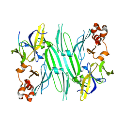 | | Structure of the compact lumenal domain of yeast Ire1 | | 分子名称: | Serine/threonine-protein kinase/endoribonuclease IRE1, peptide | | 著者 | Credle, J.J, Finer-Moore, J.S, Papa, F.R, Stroud, R.M, Walter, P. | | 登録日 | 2005-10-21 | | 公開日 | 2005-12-13 | | 最終更新日 | 2024-04-03 | | 実験手法 | X-RAY DIFFRACTION (2.983 Å) | | 主引用文献 | Inaugural Article: On the mechanism of sensing unfolded protein in the endoplasmic reticulum
Proc.Natl.Acad.Sci.Usa, 102, 2005
|
|
1FFH
 
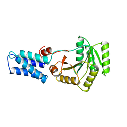 | | N AND GTPASE DOMAINS OF THE SIGNAL SEQUENCE RECOGNITION PROTEIN FFH FROM THERMUS AQUATICUS | | 分子名称: | FFH, MAGNESIUM ION | | 著者 | Freymann, D.M, Keenan, R.J, Stroud, R.M, Walter, P. | | 登録日 | 1996-12-30 | | 公開日 | 1997-12-31 | | 最終更新日 | 2024-02-07 | | 実験手法 | X-RAY DIFFRACTION (2.05 Å) | | 主引用文献 | Structure of the conserved GTPase domain of the signal recognition particle.
Nature, 385, 1997
|
|
1Z9O
 
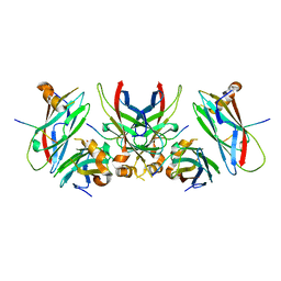 | | 1.9 Angstrom Crystal Structure of the Rat VAP-A MSP Homology Domain in Complex with the Rat ORP1 FFAT Motif | | 分子名称: | Oxysterol binding protein, Vesicle-associated membrane protein-associated protein A | | 著者 | Kaiser, S.E, Brickner, J.H, Reilein, A.R, Fenn, T.D, Walter, P, Brunger, A.T. | | 登録日 | 2005-04-03 | | 公開日 | 2005-07-19 | | 最終更新日 | 2023-08-23 | | 実験手法 | X-RAY DIFFRACTION (1.9 Å) | | 主引用文献 | Structural basis of FFAT motif-mediated ER targeting
Structure, 13, 2005
|
|
1RJ9
 
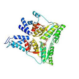 | | Structure of the heterodimer of the conserved GTPase domains of the Signal Recognition Particle (Ffh) and Its Receptor (FtsY) | | 分子名称: | MAGNESIUM ION, PHOSPHOMETHYLPHOSPHONIC ACID GUANYLATE ESTER, Signal Recognition Protein, ... | | 著者 | Egea, P.F, Shan, S.O, Napetschnig, J, Savage, D.F, Walter, P, Stroud, R.M. | | 登録日 | 2003-11-18 | | 公開日 | 2004-01-27 | | 最終更新日 | 2024-04-03 | | 実験手法 | X-RAY DIFFRACTION (1.9 Å) | | 主引用文献 | Substrate twinning activates the signal recognition particle and its receptor
Nature, 427, 2004
|
|
1Z9L
 
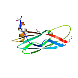 | | 1.7 Angstrom Crystal Structure of the Rat VAP-A MSP Homology Domain | | 分子名称: | Vesicle-associated membrane protein-associated protein A | | 著者 | Kaiser, S.E, Brickner, J.H, Reilein, A.R, Fenn, T.D, Walter, P, Brunger, A.T. | | 登録日 | 2005-04-03 | | 公開日 | 2005-07-19 | | 最終更新日 | 2011-07-13 | | 実験手法 | X-RAY DIFFRACTION (1.7 Å) | | 主引用文献 | Structural basis of FFAT motif-mediated ER targeting
Structure, 13, 2005
|
|
6URC
 
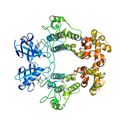 | | Crystal structure of IRE1a in complex with compound 18 | | 分子名称: | 2-chloro-N-(6-methyl-5-{[3-(2-{[(3S)-piperidin-3-yl]amino}pyrimidin-4-yl)pyridin-2-yl]oxy}naphthalen-1-yl)benzene-1-sulfonamide, GLYCEROL, Serine/threonine-protein kinase/endoribonuclease IRE1 | | 著者 | Wallweber, H.H, Wang, W. | | 登録日 | 2019-10-23 | | 公開日 | 2019-11-06 | | 最終更新日 | 2023-10-11 | | 実験手法 | X-RAY DIFFRACTION (2.2 Å) | | 主引用文献 | Disruption of IRE1 alpha through its kinase domain attenuates multiple myeloma.
Proc. Natl. Acad. Sci. U.S.A., 116, 2019
|
|
3KL4
 
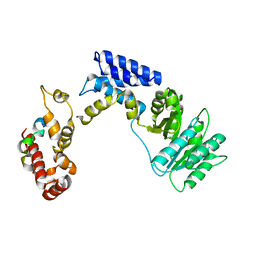 | | Recognition of a signal peptide by the signal recognition particle | | 分子名称: | Signal peptide of yeast dipeptidyl aminopeptidase B, Signal recognition 54 kDa protein | | 著者 | Janda, C.Y, Nagai, K, Li, J, Oubridge, C. | | 登録日 | 2009-11-06 | | 公開日 | 2010-03-31 | | 最終更新日 | 2024-02-21 | | 実験手法 | X-RAY DIFFRACTION (3.5 Å) | | 主引用文献 | Recognition of a signal peptide by the signal recognition particle.
Nature, 465, 2010
|
|
2Q9C
 
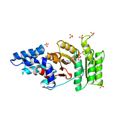 | | Structure of FTSY:GMPPNP with MGCL Complex | | 分子名称: | Cell division protein ftsY, PHOSPHOAMINOPHOSPHONIC ACID-GUANYLATE ESTER, SULFATE ION | | 著者 | Reyes, C.L, Stroud, R.M. | | 登録日 | 2007-06-12 | | 公開日 | 2007-07-03 | | 最終更新日 | 2024-02-21 | | 実験手法 | X-RAY DIFFRACTION (2.2 Å) | | 主引用文献 | X-ray Structures of the Signal Recognition Particle Receptor Reveal Targeting Cycle Intermediates.
Plos One, 2, 2007
|
|
2Q9B
 
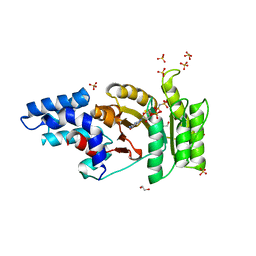 | | Structure of FTSY:GMPPNP Complex | | 分子名称: | 1,2-ETHANEDIOL, Cell division protein ftsY, PHOSPHOAMINOPHOSPHONIC ACID-GUANYLATE ESTER, ... | | 著者 | Reyes, C.L, Stroud, R.M. | | 登録日 | 2007-06-12 | | 公開日 | 2007-07-03 | | 最終更新日 | 2024-02-21 | | 実験手法 | X-RAY DIFFRACTION (2.3 Å) | | 主引用文献 | X-ray Structures of the Signal Recognition Particle Receptor Reveal Targeting Cycle Intermediates.
Plos One, 2, 2007
|
|
2Q9A
 
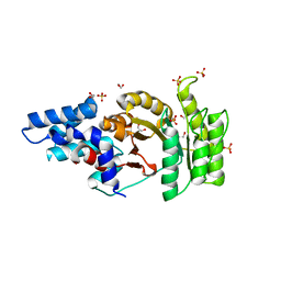 | | Structure of Apo FTSY | | 分子名称: | 1,2-ETHANEDIOL, Cell division protein ftsY, SULFATE ION | | 著者 | Reyes, C.L, Stroud, R.M. | | 登録日 | 2007-06-12 | | 公開日 | 2007-07-03 | | 最終更新日 | 2024-02-21 | | 実験手法 | X-RAY DIFFRACTION (2.24 Å) | | 主引用文献 | X-ray Structures of the Signal Recognition Particle Receptor Reveal Targeting Cycle Intermediates.
Plos One, 2, 2007
|
|
8TQO
 
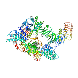 | | Eukaryotic translation initiation factor 2B tetramer | | 分子名称: | Translation initiation factor eIF-2B subunit beta, Translation initiation factor eIF-2B subunit delta, Translation initiation factor eIF-2B subunit epsilon, ... | | 著者 | Wang, L, Lawrence, R, Sangwan, S, Anand, A, Shoemaker, S, Deal, A, Marqusee, S, Watler, P. | | 登録日 | 2023-08-08 | | 公開日 | 2023-12-06 | | 最終更新日 | 2024-04-10 | | 実験手法 | ELECTRON MICROSCOPY (3.1 Å) | | 主引用文献 | A helical fulcrum in eIF2B coordinates allosteric regulation of stress signaling.
Nat.Chem.Biol., 20, 2024
|
|
8TQZ
 
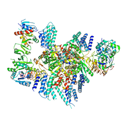 | | Eukaryotic translation initiation factor 2B with a mutation (L516A) in the delta subunit | | 分子名称: | Translation initiation factor eIF-2B subunit alpha, Translation initiation factor eIF-2B subunit beta, Translation initiation factor eIF-2B subunit delta, ... | | 著者 | Wang, L, Lawrence, R, Sangwan, S, Anand, A, Shoemaker, S, Deal, A, Marqusee, S, Watler, P. | | 登録日 | 2023-08-08 | | 公開日 | 2023-12-06 | | 最終更新日 | 2024-04-10 | | 実験手法 | ELECTRON MICROSCOPY (2.9 Å) | | 主引用文献 | A helical fulcrum in eIF2B coordinates allosteric regulation of stress signaling.
Nat.Chem.Biol., 20, 2024
|
|
1LS1
 
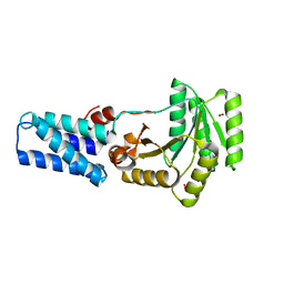 | |
6O85
 
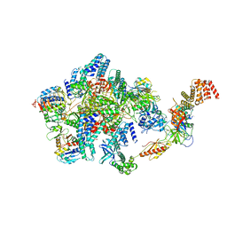 | | Electron cryo-microscopy of the eukaryotic translation initiation factor 2B bound to eukaryotic translation initiation factor 2 from Homo sapiens | | 分子名称: | 2-(4-chloranylphenoxy)-~{N}-[4-[2-(4-chloranylphenoxy)ethanoylamino]cyclohexyl]ethanamide, Eukaryotic translation initiation factor 2 subunit 1, Eukaryotic translation initiation factor 2 subunit 3, ... | | 著者 | Nguyen, H.C, Kenner, L.R, Frost, A.S. | | 登録日 | 2019-03-08 | | 公開日 | 2019-05-15 | | 最終更新日 | 2024-03-13 | | 実験手法 | ELECTRON MICROSCOPY (3.03 Å) | | 主引用文献 | eIF2B-catalyzed nucleotide exchange and phosphoregulation by the integrated stress response.
Science, 364, 2019
|
|
6O81
 
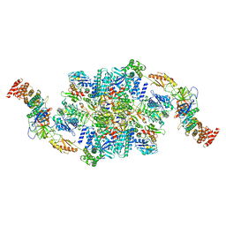 | | Electron cryo-microscopy of the eukaryotic translation initiation factor 2B bound to translation initiation factor 2 from Homo sapiens | | 分子名称: | 2-(4-chloranylphenoxy)-~{N}-[4-[2-(4-chloranylphenoxy)ethanoylamino]cyclohexyl]ethanamide, Eukaryotic translation initiation factor 2 subunit 1, Eukaryotic translation initiation factor 2 subunit 3, ... | | 著者 | Nguyen, H, Kenner, L, Frost, A. | | 登録日 | 2019-03-08 | | 公開日 | 2019-05-15 | | 最終更新日 | 2024-03-20 | | 実験手法 | ELECTRON MICROSCOPY (3.21 Å) | | 主引用文献 | eIF2B-catalyzed nucleotide exchange and phosphoregulation by the integrated stress response.
Science, 364, 2019
|
|
6O9Z
 
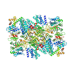 | | Electron cryo-microscopy of the eukaryotic translation initiation factor 2B bound to eukaryotic translation initiation factor 2 from Homo sapiens | | 分子名称: | Eukaryotic translation initiation factor 2 subunit 1, Translation initiation factor eIF-2B subunit alpha, Translation initiation factor eIF-2B subunit beta, ... | | 著者 | Nguyen, H.C, Kenner, L.R, Frost, A.S. | | 登録日 | 2019-03-15 | | 公開日 | 2019-05-15 | | 最終更新日 | 2024-10-30 | | 実験手法 | ELECTRON MICROSCOPY (3.03 Å) | | 主引用文献 | eIF2B-catalyzed nucleotide exchange and phosphoregulation by the integrated stress response.
Science, 364, 2019
|
|
3FAR
 
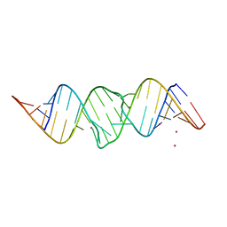 | |
2FCZ
 
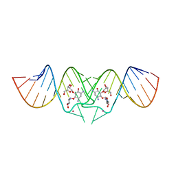 | | HIV-1 DIS kissing-loop in complex with ribostamycin | | 分子名称: | HIV-1 DIS RNA, POTASSIUM ION, RIBOSTAMYCIN | | 著者 | Ennifar, E, Paillart, J.C, Marquet, R, Dumas, P. | | 登録日 | 2005-12-13 | | 公開日 | 2006-05-16 | | 最終更新日 | 2023-08-30 | | 実験手法 | X-RAY DIFFRACTION (2.01 Å) | | 主引用文献 | Targeting the dimerization initiation site of HIV-1 RNA with aminoglycosides: from crystal to cell.
Nucleic Acids Res., 34, 2006
|
|
2FCY
 
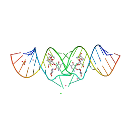 | | HIV-1 DIS kissing-loop in complex with Neomycin | | 分子名称: | CHLORIDE ION, HIV-1 DIS RNA, NEOMYCIN, ... | | 著者 | Ennifar, E, Paillart, J.C, Marquet, R, Dumas, P. | | 登録日 | 2005-12-13 | | 公開日 | 2006-05-16 | | 最終更新日 | 2023-08-30 | | 実験手法 | X-RAY DIFFRACTION (2.2 Å) | | 主引用文献 | Targeting the dimerization initiation site of HIV-1 RNA with aminoglycosides: from crystal to cell.
Nucleic Acids Res., 34, 2006
|
|
1CQL
 
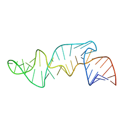 | |
2FD0
 
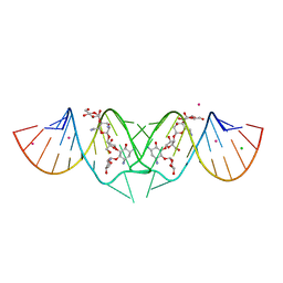 | | HIV-1 DIS kissing-loop in complex with lividomycin | | 分子名称: | (2R,3S,4S,5S,6R)-2-((2S,3S,4R,5R,6R)-5-AMINO-2-(AMINOMETHYL)-6-((2R,3S,4R,5S)-5-((1R,2R,3S,5R,6S)-3,5-DIAMINO-2-((2S,3R ,5S,6R)-3-AMINO-5-HYDROXY-6-(HYDROXYMETHYL)-TETRAHYDRO-2H-PYRAN-2-YLOXY)-6-HYDROXYCYCLOHEXYLOXY)-4-HYDROXY-2-(HYDROXYMET HYL)-TETRAHYDROFURAN-3-YLOXY)-4-HYDROXY-TETRAHYDRO-2H-PYRAN-3-YLOXY)-6-(HYDROXYMETHYL)-TETRAHYDRO-2H-PYRAN-3,4,5-TRIOL, CHLORIDE ION, HIV-1 DIS RNA, ... | | 著者 | Ennifar, E, Paillart, J.C, Marquet, R, Dumas, P. | | 登録日 | 2005-12-13 | | 公開日 | 2006-05-16 | | 最終更新日 | 2023-08-30 | | 実験手法 | X-RAY DIFFRACTION (1.8 Å) | | 主引用文献 | Targeting the dimerization initiation site of HIV-1 RNA with aminoglycosides: from crystal to cell.
Nucleic Acids Res., 34, 2006
|
|
2FCX
 
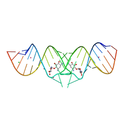 | | HIV-1 DIS kissing-loop in complex with neamine | | 分子名称: | (1R,2R,3S,4R,6S)-4,6-diamino-2,3-dihydroxycyclohexyl 2,6-diamino-2,6-dideoxy-alpha-D-glucopyranoside, CHLORIDE ION, HIV-1 DIS RNA, ... | | 著者 | Ennifar, E, Paillart, J.C, Marquet, R, Dumas, P. | | 登録日 | 2005-12-13 | | 公開日 | 2006-05-16 | | 最終更新日 | 2024-02-14 | | 実験手法 | X-RAY DIFFRACTION (2 Å) | | 主引用文献 | Targeting the dimerization initiation site of HIV-1 RNA with aminoglycosides: from crystal to cell.
Nucleic Acids Res., 34, 2006
|
|
2BR2
 
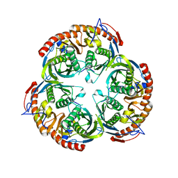 | | RNase PH core of the archaeal exosome | | 分子名称: | CHLORIDE ION, EXOSOME COMPLEX EXONUCLEASE 1, EXOSOME COMPLEX EXONUCLEASE 2 | | 著者 | lorentzen, E, Fribourg, S, Conti, E. | | 登録日 | 2005-04-30 | | 公開日 | 2005-06-06 | | 最終更新日 | 2023-12-13 | | 実験手法 | X-RAY DIFFRACTION (2.8 Å) | | 主引用文献 | The Archaeal Exosome Core is a Hexameric Ring Structure with Three Catalytic Subunits.
Nat.Struct.Mol.Biol., 12, 2005
|
|
5W0T
 
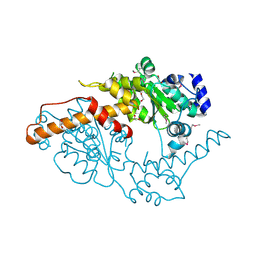 | |
2J45
 
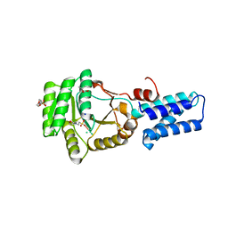 | | Water structure of T. Aquaticus Ffh NG Domain At 1.1A Resolution | | 分子名称: | (4R)-2-METHYLPENTANE-2,4-DIOL, 1,2-ETHANEDIOL, 2-(N-MORPHOLINO)-ETHANESULFONIC ACID, ... | | 著者 | Freymann, D.M, Ramirez, U.D. | | 登録日 | 2006-08-24 | | 公開日 | 2006-11-30 | | 最終更新日 | 2023-12-13 | | 実験手法 | X-RAY DIFFRACTION (1.14 Å) | | 主引用文献 | Analysis of Protein Hydration in Ultra-High Resolution Structures of the Srp Gtpase Ffh
Acta Crystallogr.,Sect.D, 62, 2006
|
|
