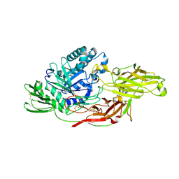
3VUD
 
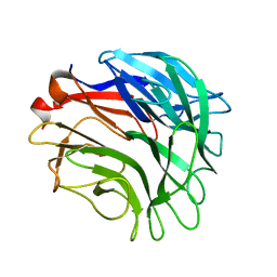
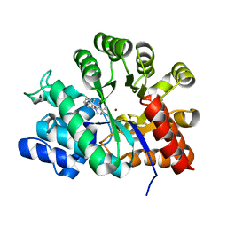
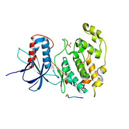 | | Crystal structure of a cysteine-deficient mutant M1 in MAP kinase JNK1 | | 分子名称: | Mitogen-activated protein kinase 8, Peptide from C-Jun-amino-terminal kinase-interacting protein 1, SULFATE ION | | 著者 | Nakaniwa, T, Kinoshita, T, Inoue, T. | | 登録日 | 2012-06-28 | | 公開日 | 2013-02-13 | | 最終更新日 | 2024-03-20 | | 実験手法 | X-RAY DIFFRACTION (3.5 Å) | | 主引用文献 | Seven cysteine-deficient mutants depict the interplay between thermal and chemical stabilities of individual cysteine residues in mitogen-activated protein kinase c-Jun N-terminal kinase 1
Biochemistry, 51, 2012
|
|
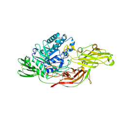
3VUM
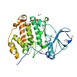
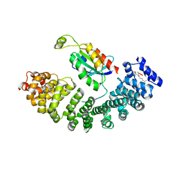
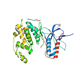 | | Crystal structure of a cysteine-deficient mutant M7 in MAP kinase JNK1 | | 分子名称: | 2-AMINO-2-HYDROXYMETHYL-PROPANE-1,3-DIOL, Mitogen-activated protein kinase 8, Peptide from C-Jun-amino-terminal kinase-interacting protein 1, ... | | 著者 | Nakaniwa, T, Kinoshita, T, Inoue, T. | | 登録日 | 2012-07-02 | | 公開日 | 2013-02-13 | | 最終更新日 | 2024-03-20 | | 実験手法 | X-RAY DIFFRACTION (2.69 Å) | | 主引用文献 | Seven cysteine-deficient mutants depict the interplay between thermal and chemical stabilities of individual cysteine residues in mitogen-activated protein kinase c-Jun N-terminal kinase 1
Biochemistry, 51, 2012
|
|
3VUL
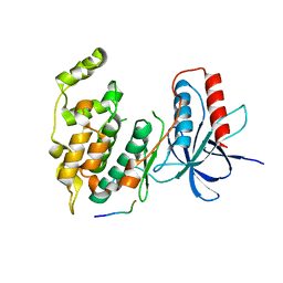 | |
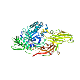
3VUK
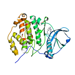
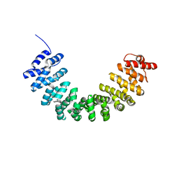
 | | Crystal structure of a cysteine-deficient mutant M5 in MAP kinase JNK1 | | 分子名称: | Mitogen-activated protein kinase 8, Peptide from C-Jun-amino-terminal kinase-interacting protein 1, SULFATE ION | | 著者 | Nakaniwa, T, Kinoshita, T, Inoue, T. | | 登録日 | 2012-07-02 | | 公開日 | 2013-02-13 | | 最終更新日 | 2024-03-20 | | 実験手法 | X-RAY DIFFRACTION (2.95 Å) | | 主引用文献 | Seven cysteine-deficient mutants depict the interplay between thermal and chemical stabilities of individual cysteine residues in mitogen-activated protein kinase c-Jun N-terminal kinase 1
Biochemistry, 51, 2012
|
|
7M6F
 | |
7M6G
 | |
7M6I
 | |
7M6E
 | |
7M6H
 | |
7M6D
 | |
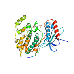
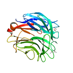
3VUI
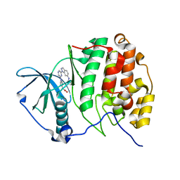
 | | Crystal structure of a cysteine-deficient mutant M2 in MAP kinase JNK1 | | 分子名称: | Mitogen-activated protein kinase 8, Peptide from C-Jun-amino-terminal kinase-interacting protein 1, SULFATE ION | | 著者 | Nakaniwa, T, Kinoshita, T, Inoue, T. | | 登録日 | 2012-06-28 | | 公開日 | 2013-02-13 | | 最終更新日 | 2024-03-20 | | 実験手法 | X-RAY DIFFRACTION (2.8 Å) | | 主引用文献 | Seven cysteine-deficient mutants depict the interplay between thermal and chemical stabilities of individual cysteine residues in mitogen-activated protein kinase c-Jun N-terminal kinase 1
Biochemistry, 51, 2012
|
|
1MMJ
 | | Porcine pancreatic elastase complexed with a potent peptidyl inhibitor, FR136706 | | 分子名称: | 2-[4-[[(S)-1-[[(S)-2-[[(RS)-3,3,3-TRIFLUORO-1-ISOPROPYL-2-OXOPROPYL]AMINOCARBONYL]PYRROLIDIN-1-YL-]CARBONYL]-2-METHYLPROPYL]AMINOCARBONYL]BENZOYLAMINO]ACETIC ACID, CALCIUM ION, SULFATE ION, ... | | 著者 | Kinoshita, T. | | 登録日 | 2002-09-04 | | 公開日 | 2002-12-23 | | 最終更新日 | 2017-10-11 | | 実験手法 | X-RAY DIFFRACTION (2.2 Å) | | 主引用文献 | True interaction mode of porcine pancreatic elastase with FR136706, a
potent peptidyl inhibitor
Bioorg.Med.Chem.Lett., 13, 2003
|
|
6IK5
 | | Crystal structure of tomato beta-galactosidase (TBG) 4 in complex with galactose | | 分子名称: | 2-acetamido-2-deoxy-beta-D-glucopyranose, 2-acetamido-2-deoxy-beta-D-glucopyranose-(1-4)-2-acetamido-2-deoxy-beta-D-glucopyranose, Beta-galactosidase, ... | | 著者 | Matsuyama, K, Nakae, S, Igarashi, K, Tada, T, Ishimaru, M. | | 登録日 | 2018-10-15 | | 公開日 | 2018-11-28 | | 最終更新日 | 2023-11-22 | | 実験手法 | X-RAY DIFFRACTION (1.82 Å) | | 主引用文献 | Substrate-recognition mechanism of tomato beta-galactosidase 4 using X-ray crystallography and docking simulation.
Planta, 252, 2020
|
|
6IK7
 | | Crystal structure of tomato beta-galactosidase (TBG) 4 in complex with beta-1,3-galactobiose | | 分子名称: | 2-acetamido-2-deoxy-beta-D-glucopyranose, 2-acetamido-2-deoxy-beta-D-glucopyranose-(1-4)-2-acetamido-2-deoxy-beta-D-glucopyranose, Beta-galactosidase, ... | | 著者 | Matsuyama, K, Nakae, S, Igarashi, K, Tada, T, Ishimaru, M. | | 登録日 | 2018-10-15 | | 公開日 | 2018-11-28 | | 最終更新日 | 2023-11-22 | | 実験手法 | X-RAY DIFFRACTION (3.1 Å) | | 主引用文献 | Substrate-recognition mechanism of tomato beta-galactosidase 4 using X-ray crystallography and docking simulation.
Planta, 252, 2020
|
|
6IK8
 | | Crystal structure of tomato beta-galactosidase (TBG) 4 in complex with beta-1,6-galactobiose | | 分子名称: | 2-acetamido-2-deoxy-beta-D-glucopyranose, 2-acetamido-2-deoxy-beta-D-glucopyranose-(1-4)-2-acetamido-2-deoxy-beta-D-glucopyranose, Beta-galactosidase, ... | | 著者 | Matsuyama, K, Nakae, S, Igarashi, K, Tada, T, Ishimaru, M. | | 登録日 | 2018-10-15 | | 公開日 | 2018-11-28 | | 最終更新日 | 2023-11-22 | | 実験手法 | X-RAY DIFFRACTION (2.8 Å) | | 主引用文献 | Substrate-recognition mechanism of tomato beta-galactosidase 4 using X-ray crystallography and docking simulation.
Planta, 252, 2020
|
|
6IK6
 | | Crystal structure of Tomato beta-galactosidase (TBG) 4 with beta-1,4-galactobiose | | 分子名称: | 2-acetamido-2-deoxy-beta-D-glucopyranose, 2-acetamido-2-deoxy-beta-D-glucopyranose-(1-4)-2-acetamido-2-deoxy-beta-D-glucopyranose, Beta-galactosidase, ... | | 著者 | Matsuyama, K, Nakae, S, Igarashi, K, Tada, T, Ishimaru, M. | | 登録日 | 2018-10-15 | | 公開日 | 2018-11-28 | | 最終更新日 | 2023-11-22 | | 実験手法 | X-RAY DIFFRACTION (2.791 Å) | | 主引用文献 | Substrate-recognition mechanism of tomato beta-galactosidase 4 using X-ray crystallography and docking simulation.
Planta, 252, 2020
|
|
3A71
 | |
3A72
 | |
3AT4
 | | Crystal structure of CK2alpha with pyradine derivertive | | 分子名称: | Casein kinase II subunit alpha, [1-(6-{6-[(1-methylethyl)amino]-1H-indazol-1-yl}pyrazin-2-yl)-1H-pyrrol-3-yl]acetic acid | | 著者 | Kinoshita, T. | | 登録日 | 2010-12-23 | | 公開日 | 2011-11-09 | | 最終更新日 | 2023-11-01 | | 実験手法 | X-RAY DIFFRACTION (2.2 Å) | | 主引用文献 | A detailed thermodynamic profile of cyclopentyl and isopropyl derivatives binding to CK2 kinase
Mol.Cell.Biochem., 356, 2011
|
|
3AT3
 | | Crystal structure of CK2alpha with pyradine derivative | | 分子名称: | (1-{6-[6-(cyclopentylamino)-1H-indazol-1-yl]pyrazin-2-yl}-1H-pyrrol-3-yl)acetic acid, Casein kinase II subunit alpha | | 著者 | Kinoshita, T. | | 登録日 | 2010-12-23 | | 公開日 | 2011-11-09 | | 最終更新日 | 2023-11-01 | | 実験手法 | X-RAY DIFFRACTION (2.6 Å) | | 主引用文献 | A detailed thermodynamic profile of cyclopentyl and isopropyl derivatives binding to CK2 kinase
Mol.Cell.Biochem., 356, 2011
|
|
3AT2
 | | Crystal structure of CK2alpha | | 分子名称: | 1,2-ETHANEDIOL, Casein kinase II subunit alpha | | 著者 | Kinoshita, T. | | 登録日 | 2010-12-23 | | 公開日 | 2011-11-09 | | 最終更新日 | 2023-11-01 | | 実験手法 | X-RAY DIFFRACTION (1.6 Å) | | 主引用文献 | A detailed thermodynamic profile of cyclopentyl and isopropyl derivatives binding to CK2 kinase
Mol.Cell.Biochem., 356, 2011
|
|
2Z7G
 | |
5ZHX
 | |
5XGC
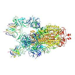
 | | Crystal structure of SmgGDS-558 | | 分子名称: | Rap1 GTPase-GDP dissociation stimulator 1 | | 著者 | Shimizu, H, Toma-Fukai, S, Shimizu, T. | | 登録日 | 2017-04-13 | | 公開日 | 2017-06-28 | | 最終更新日 | 2024-03-27 | | 実験手法 | X-RAY DIFFRACTION (2.1 Å) | | 主引用文献 | Structure-based analysis of the guanine nucleotide exchange factor SmgGDS reveals armadillo-repeat motifs and key regions for activity and GTPase binding
J. Biol. Chem., 292, 2017
|
|
1MG8
 | | NMR structure of ubiquitin-like domain in murine Parkin | | 分子名称: | Parkin | | 著者 | Tashiro, M, Okubo, S, Shimotakahara, S, Hatanaka, H, Yasuda, H, Kainosho, M, Yokoyama, S, Shindo, H, RIKEN Structural Genomics/Proteomics Initiative (RSGI) | | 登録日 | 2002-08-15 | | 公開日 | 2003-04-08 | | 最終更新日 | 2024-05-29 | | 実験手法 | SOLUTION NMR | | 主引用文献 | NMR structure of ubiquitin-like domain in PARKIN: Gene product of familial Parkinson's disease.
J.Biomol.NMR, 25, 2003
|
|


