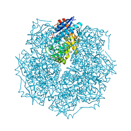
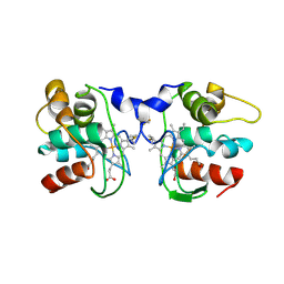
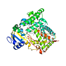
2P1J
 
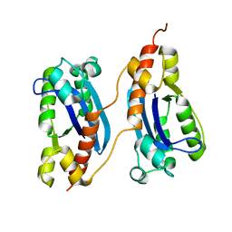 | | Crystal structure of a polC-type DNA polymerase III exonuclease domain from Thermotoga maritima | | 分子名称: | DNA polymerase III polC-type | | 著者 | Bonanno, J.B, Rutter, M, Bain, K.T, Izuka, M, Sridhar, V, Smith, D, Wasserman, S, Sauder, J.M, Burley, S.K, Almo, S.C, New York SGX Research Center for Structural Genomics (NYSGXRC) | | 登録日 | 2007-03-05 | | 公開日 | 2007-03-20 | | 最終更新日 | 2024-02-21 | | 実験手法 | X-RAY DIFFRACTION (2.5 Å) | | 主引用文献 | Crystal structure of a polC-type DNA polymerase III exonuclease domain from Thermotoga maritima
To be Published
|
|
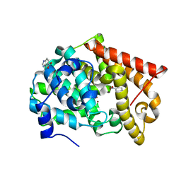
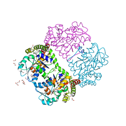
2Q01
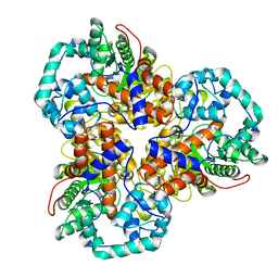 | | Crystal structure of glucuronate isomerase from Caulobacter crescentus | | 分子名称: | POTASSIUM ION, Uronate isomerase | | 著者 | Patskovsky, Y, Bonanno, J, Sridhar, V, Sauder, J.M, Freeman, J, Powell, A, Koss, J, Groshong, C, Gheyi, T, Wasserman, S.R, Raushel, F, Burley, S.K, Almo, S.C, New York SGX Research Center for Structural Genomics (NYSGXRC) | | 登録日 | 2007-05-18 | | 公開日 | 2007-05-29 | | 最終更新日 | 2023-08-30 | | 実験手法 | X-RAY DIFFRACTION (2.34 Å) | | 主引用文献 | Crystal Structure of Glucuronate Isomerase from Caulobacter crescentus.
To be Published
|
|
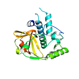
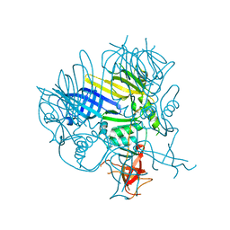
2NWI
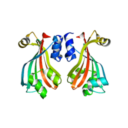 | | Crystal structure of protein AF1396 from Archaeoglobus fulgidus, Pfam DUF98 | | 分子名称: | Hypothetical protein | | 著者 | Bonanno, J.B, Dickey, M, Bain, K.T, Adams, J, Sridhar, V, Wasserman, S, Smith, D, Sauder, J.M, Burley, S.K, Almo, S.C, New York SGX Research Center for Structural Genomics (NYSGXRC) | | 登録日 | 2006-11-14 | | 公開日 | 2006-11-21 | | 最終更新日 | 2023-12-27 | | 実験手法 | X-RAY DIFFRACTION (2.2 Å) | | 主引用文献 | Crystal structure of hypothetical protein O28875 from Archaeoglobus fulgidus
To be Published
|
|
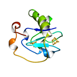
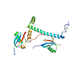
2O3A
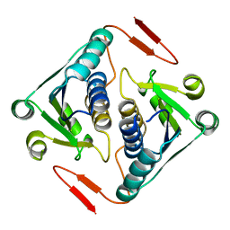 | | Crystal structure of a protein AF_0751 from Archaeoglobus fulgidus | | 分子名称: | UPF0106 protein AF_0751 | | 著者 | Bonanno, J.B, Dickey, M, Bain, K.T, Wu, B, Slocombe, A, Sridhar, V, Sauder, J.M, Burley, S.K, Almo, S.C, New York SGX Research Center for Structural Genomics (NYSGXRC) | | 登録日 | 2006-12-01 | | 公開日 | 2006-12-26 | | 最終更新日 | 2023-12-27 | | 実験手法 | X-RAY DIFFRACTION (2.2 Å) | | 主引用文献 | Crystal structure of a protein AF_0751 from Archaeoglobus fulgidus
To be Published
|
|
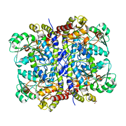
2OO6
 | | Crystal structure of putative L-alanine-DL-glutamate epimerase from Burkholderia xenovorans strain LB400 | | 分子名称: | Putative L-alanine-DL-glutamate epimerase, SULFATE ION | | 著者 | Bonanno, J.B, Dickey, M, Bain, K.T, Wu, B, Sridhar, V, Freeman, J, Smyth, L, Atwell, S, Sauder, J.M, Burley, S.K, Almo, S.C, New York SGX Research Center for Structural Genomics (NYSGXRC) | | 登録日 | 2007-01-25 | | 公開日 | 2007-02-06 | | 最終更新日 | 2023-12-27 | | 実験手法 | X-RAY DIFFRACTION (1.8 Å) | | 主引用文献 | Crystal structure of putative L-alanine-DL-glutamate epimerase from Burkholderia xenovorans strain LB400
To be Published
|
|
2P1G
 | | Crystal structure of a putative xylanase from Bacteroides fragilis | | 分子名称: | Putative xylanase | | 著者 | Bonanno, J.B, Freeman, J, Bain, K.T, Zhang, F, Sridhar, V, Smith, D, Wasserman, S, Sauder, J.M, Burley, S.K, Almo, S.C, New York SGX Research Center for Structural Genomics (NYSGXRC) | | 登録日 | 2007-03-05 | | 公開日 | 2007-03-20 | | 最終更新日 | 2024-02-21 | | 実験手法 | X-RAY DIFFRACTION (1.8 Å) | | 主引用文献 | Crystal structure of a putative xylanase from Bacteroides fragilis
To be Published
|
|
1AXQ
 | | FERRICYANIDE OXIDIZED FDI | | 分子名称: | FE3-S4 CLUSTER, FERREDOXIN, IRON/SULFUR CLUSTER | | 著者 | Stout, C.D. | | 登録日 | 1997-10-17 | | 公開日 | 1998-01-28 | | 最終更新日 | 2024-05-22 | | 実験手法 | X-RAY DIFFRACTION (2.1 Å) | | 主引用文献 | Crystal structures of ferricyanide-oxidized [Fe-S] clusters in Azotobacter vinelandii ferredoxin I.
J.Biol.Inorg.Chem., 3, 1998
|
|
8UUI
 | | X-ray structure of Interleukin-23 in complex with peptide 23-446 | | 分子名称: | Interleukin-12 subunit beta, Interleukin-23 subunit alpha, alpha-D-mannopyranose-(1-3)-beta-D-mannopyranose-(1-4)-2-acetamido-2-deoxy-beta-D-glucopyranose-(1-4)-2-acetamido-2-deoxy-beta-D-glucopyranose, ... | | 著者 | Joseph, J.S, Sridhar, V.S, Liu, M, Hu, Y, Gatchalian, J. | | 登録日 | 2023-11-01 | | 公開日 | 2025-07-09 | | 実験手法 | X-RAY DIFFRACTION (2.43 Å) | | 主引用文献 | Identification of an Induced Orthosteric Pocket in IL-23: A New Avenue for Non-biological Therapeutic Targeting.
Acs Chem.Biol., 2025
|
|
1PG8
 | | Crystal Structure of L-methionine alpha-, gamma-lyase | | 分子名称: | DI(HYDROXYETHYL)ETHER, Methionine gamma-lyase, PYRIDOXAL-5'-PHOSPHATE, ... | | 著者 | Allen, T.W, Sridhar, V, Prasad, G.S, Han, Q, Xu, M, Tan, Y, Hoffman, R.M, Ramaswamy, S. | | 登録日 | 2003-05-28 | | 公開日 | 2004-06-08 | | 最終更新日 | 2023-08-16 | | 実験手法 | X-RAY DIFFRACTION (2.68 Å) | | 主引用文献 |
To Be Published
|
|
1FOC
 | | Cytochrome C557: improperly folded thermus thermophilus C552 | | 分子名称: | CYTOCHROME RC557, HEME C | | 著者 | McRee, D.E, Williams, P.A, Fee, J.A, Bren, K.L. | | 登録日 | 2000-08-27 | | 公開日 | 2000-11-08 | | 最終更新日 | 2024-12-25 | | 実験手法 | X-RAY DIFFRACTION (3 Å) | | 主引用文献 | Recombinant cytochrome rC557 obtained from Escherichia coli cells expressing a truncated Thermus thermophilus cycA gene. Heme inversion in an improperly matured protein
J.Biol.Chem., 276, 2001
|
|
4MSA
 | | Crystal structure of PDE10A2 with fragment ZT0449 (5-nitro-1H-benzimidazole) | | 分子名称: | 5-nitro-1H-benzimidazole, NICKEL (II) ION, cAMP and cAMP-inhibited cGMP 3',5'-cyclic phosphodiesterase 10A | | 著者 | Sridar, V, Badger, J, Logan, C, Chie-Leon, B, Nienaber, V. | | 登録日 | 2013-09-18 | | 公開日 | 2014-05-14 | | 最終更新日 | 2023-09-20 | | 実験手法 | X-RAY DIFFRACTION (1.62 Å) | | 主引用文献 | Identification and optimization of PDE10A inhibitors using fragment-based screening by nanocalorimetry and X-ray crystallography.
J Biomol Screen, 19, 2014
|
|
2FNO
 | |
1G3O
 | | CRYSTAL STRUCTURE OF V19E MUTANT OF FERREDOXIN I | | 分子名称: | 7FE FERREDOXIN I, FE3-S4 CLUSTER, IRON/SULFUR CLUSTER | | 著者 | Stout, C.D, Burgess, B.K, Bonagura, C.A, Jung, Y.S. | | 登録日 | 2000-10-24 | | 公開日 | 2000-11-06 | | 最終更新日 | 2023-08-09 | | 実験手法 | X-RAY DIFFRACTION (1.65 Å) | | 主引用文献 | Azotobacter vinelandii ferredoxin I: a sequence and structure comparison approach to alteration of [4Fe-4S]2+/+ reduction potential
J.Biol.Chem., 277, 2002
|
|
1G6B
 | | CRYSTAL STRUCTURE OF P47S MUTANT OF FERREDOXIN I | | 分子名称: | 7FE FERREDOXIN I, FE3-S4 CLUSTER, IRON/SULFUR CLUSTER | | 著者 | Stout, C.D, Burgess, B.K, Bonagura, C.A, Jung, Y.S. | | 登録日 | 2000-11-03 | | 公開日 | 2000-11-22 | | 最終更新日 | 2023-08-09 | | 実験手法 | X-RAY DIFFRACTION (1.9 Å) | | 主引用文献 | Azotobacter vinelandii ferredoxin I: a sequence and structure comparison approach to alteration of [4Fe-4S]2+/+ reduction potential.
J.Biol.Chem., 277, 2002
|
|
2OG9
 | |
1N6B
 | | Microsomal Cytochrome P450 2C5/3LVdH Complex with a dimethyl derivative of sulfaphenazole | | 分子名称: | 4-METHYL-N-METHYL-N-(2-PHENYL-2H-PYRAZOL-3-YL)BENZENESULFONAMIDE, Cytochrome P450 2C5, PROTOPORPHYRIN IX CONTAINING FE, ... | | 著者 | Wester, M.R, Johnson, E.F, Marques-Soares, C, Dansette, P.M, Mansuy, D, Stout, C.D. | | 登録日 | 2002-11-09 | | 公開日 | 2003-06-03 | | 最終更新日 | 2024-02-14 | | 実験手法 | X-RAY DIFFRACTION (2.3 Å) | | 主引用文献 | Structure of a Substrate Complex of Mammalian Cytochrome P450 2C5 at 2.3 A Resolution: Evidence
for Multiple Substrate Binding Modes
Biochemistry, 42, 2003
|
|
1PFF
 | | Crystal Structure of Homocysteine alpha-, gamma-lyase at 1.8 Angstroms | | 分子名称: | 1,2-ETHANEDIOL, DI(HYDROXYETHYL)ETHER, methionine gamma-lyase | | 著者 | Allen, T.W, Sridhar, V, Prasad, S.G, Han, Q, Xu, M, Tan, Y, Hoffman, R.M, Ramaswamy, S. | | 登録日 | 2003-05-26 | | 公開日 | 2004-08-10 | | 最終更新日 | 2023-08-16 | | 実験手法 | X-RAY DIFFRACTION (2.5 Å) | | 主引用文献 |
|
|
2PMQ
 | | Crystal structure of a mandelate racemase/muconate lactonizing enzyme from Roseovarius sp. HTCC2601 | | 分子名称: | MAGNESIUM ION, Mandelate racemase/muconate lactonizing enzyme | | 著者 | Bonanno, J.B, Rutter, M, Bain, K.T, Lau, C, Sridhar, V, Smith, D, Wasserman, S, Sauder, J.M, Burley, S.K, Almo, S.C, New York SGX Research Center for Structural Genomics (NYSGXRC) | | 登録日 | 2007-04-23 | | 公開日 | 2007-05-08 | | 最終更新日 | 2024-11-06 | | 実験手法 | X-RAY DIFFRACTION (1.72 Å) | | 主引用文献 | Discovery of new enzymes and metabolic pathways by using structure and genome context.
Nature, 502, 2013
|
|
2P5Z
 | | The E. coli c3393 protein is a component of the type VI secretion system and exhibits structural similarity to T4 bacteriophage tail proteins gp27 and gp5 | | 分子名称: | Type VI secretion system component | | 著者 | Ramagopal, U.A, Bonanno, J.B, Sridhar, V, Lau, C, Toro, R, Gheyi, T, Maletic, M, Freeman, J.C, Sauder, J.M, Burley, S.K, Almo, S.C, New York SGX Research Center for Structural Genomics (NYSGXRC) | | 登録日 | 2007-03-16 | | 公開日 | 2007-04-03 | | 最終更新日 | 2024-02-21 | | 実験手法 | X-RAY DIFFRACTION (2.6 Å) | | 主引用文献 | Type VI secretion apparatus and phage tail-associated protein complexes share a common evolutionary origin.
Proc.Natl.Acad.Sci.Usa, 106, 2009
|
|
6MUN
 | | Structure of hRpn10 bound to UBQLN2 UBL | | 分子名称: | 26S proteasome non-ATPase regulatory subunit 4, Ubiquilin-2 | | 著者 | Chen, X, Walters, K.J. | | 登録日 | 2018-10-23 | | 公開日 | 2019-09-04 | | 最終更新日 | 2024-05-01 | | 実験手法 | SOLUTION NMR | | 主引用文献 | Structure of hRpn10 Bound to UBQLN2 UBL Illustrates Basis for Complementarity between Shuttle Factors and Substrates at the Proteasome.
J.Mol.Biol., 431, 2019
|
|
6U19
 | |
6CO4
 | |


