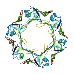
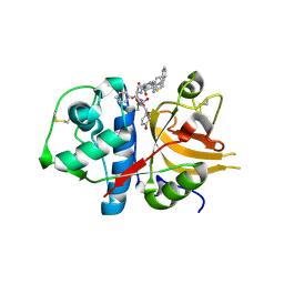
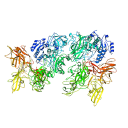
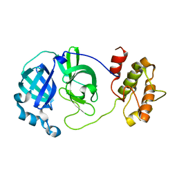
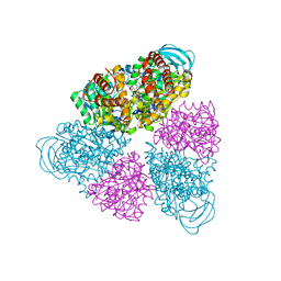
5JYP
 
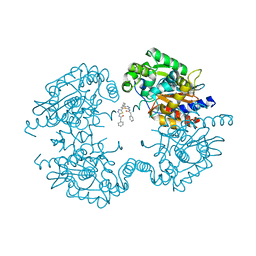 | | Allosteric inhibition of Kidney Isoform of Glutaminase | | 分子名称: | 2-phenyl-~{N}-[5-[(1~{S},3~{S})-3-[5-(2-phenylethanoylamino)-1,3,4-thiadiazol-2-yl]cyclohexyl]-1,3,4-thiadiazol-2-yl]ethanamide, Glutaminase kidney isoform, mitochondrial | | 著者 | Ramachandran, S, Sivaraman, J. | | 登録日 | 2016-05-15 | | 公開日 | 2016-08-03 | | 最終更新日 | 2023-11-08 | | 実験手法 | X-RAY DIFFRACTION (2.74 Å) | | 主引用文献 | Structural basis for exploring the allosteric inhibition of human kidney type glutaminase.
Oncotarget, 7, 2016
|
|
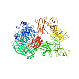
7EQX
 | | Crystal structure of an Aedes aegypti procarboxypeptidase B1 | | 分子名称: | Carboxypeptidase B, ZINC ION | | 著者 | Choong, Y.K, Gavor, E, Jobichen, C, Sivaraman, J. | | 登録日 | 2021-05-05 | | 公開日 | 2021-11-10 | | 最終更新日 | 2023-11-29 | | 実験手法 | X-RAY DIFFRACTION (2.08 Å) | | 主引用文献 | Structure of Aedes aegypti procarboxypeptidase B1 and its binding with Dengue virus for controlling infection.
Life Sci Alliance, 5, 2022
|
|
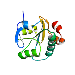
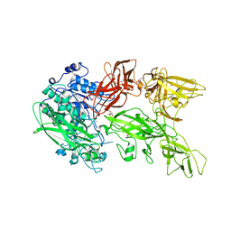
5XWR
 | | Crystal Structure of RBBP4-peptide complex | | 分子名称: | Histone-binding protein RBBP4, MET-SER-ARG-ARG-LYS-GLN-ALA-LYS-PRO-GLN-HIS-ILE | | 著者 | Jobichen, C, Lui, B.H, Daniel, G.T, Sivaraman, J. | | 登録日 | 2017-06-30 | | 公開日 | 2018-07-11 | | 最終更新日 | 2023-11-22 | | 実験手法 | X-RAY DIFFRACTION (2.69 Å) | | 主引用文献 | Targeting cancer addiction for SALL4 by shifting its transcriptome with a pharmacologic peptide.
Proc. Natl. Acad. Sci. U.S.A., 115, 2018
|
|
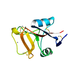
2GML
 | | Crystal Structure of Catalytic Domain of E.coli RluF | | 分子名称: | Ribosomal large subunit pseudouridine synthase F | | 著者 | Sunita, S, Zhenxing, H, Swaathi, J, Cygler, M, Matte, A, Sivaraman, J. | | 登録日 | 2006-04-06 | | 公開日 | 2006-07-18 | | 最終更新日 | 2011-07-13 | | 実験手法 | X-RAY DIFFRACTION (2.6 Å) | | 主引用文献 | Domain Organization and Crystal Structure of the Catalytic Domain of E.coli RluF, a Pseudouridine Synthase that Acts on 23S rRNA
J.Mol.Biol., 359, 2006
|
|
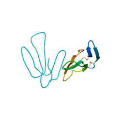
5XX9
 | |
6A2V
 | | Crystal structure of Hcp protein | | 分子名称: | Type VI secretion system tube protein Hcp | | 著者 | Jobichen, C, Sivaraman, J. | | 登録日 | 2018-06-13 | | 公開日 | 2018-09-19 | | 最終更新日 | 2023-11-22 | | 実験手法 | X-RAY DIFFRACTION (2.588 Å) | | 主引用文献 | Structural basis for the pathogenesis of Campylobacter jejuni Hcp1, a structural and effector protein of the Type VI Secretion System.
FEBS J., 285, 2018
|
|
1PVJ
 | | Crystal structure of the Streptococcal pyrogenic exotoxin B (SpeB)- inhibitor complex | | 分子名称: | (3R)-3-{[(BENZYLOXY)CARBONYL]AMINO}-2-OXO-4-PHENYLBUTANE-1-DIAZONIUM, pyrogenic exotoxin B | | 著者 | Ziomek, E, Sivaraman, J, Doran, J, Menard, R, Cygler, M. | | 登録日 | 2003-06-27 | | 公開日 | 2004-09-28 | | 最終更新日 | 2017-10-11 | | 実験手法 | X-RAY DIFFRACTION (3 Å) | | 主引用文献 | Inhibition of autoprocessing of the streptococcal pyrogenic exotoxin B (speB). Crystal structure of the proenzyme-inhibitor complex
To be published
|
|
5H7R
 | |
5H7S
 | |
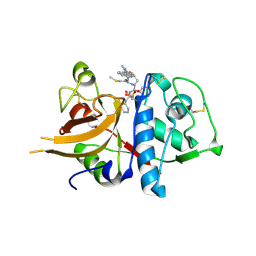
3BC3
 | | Exploring inhibitor binding at the S subsites of cathepsin L | | 分子名称: | Cathepsin L heavy and light chains, S-benzyl-N-(biphenyl-4-ylacetyl)-L-cysteinyl-N~5~-(diaminomethyl)-D-ornithyl-N-(2-phenylethyl)-L-tyrosinamide | | 著者 | Chowdhury, S.F, Joseph, L, Kumar, S, Tulsidas, S.R, Bhat, S, Ziomek, E, Nard, R.M, Sivaraman, J, Purisima, E.O. | | 登録日 | 2007-11-12 | | 公開日 | 2008-03-18 | | 最終更新日 | 2023-08-30 | | 実験手法 | X-RAY DIFFRACTION (2.2 Å) | | 主引用文献 | Exploring inhibitor binding at the S' subsites of cathepsin L
J.Med.Chem., 51, 2008
|
|
2LUS
 | |
3H8C
 | | A combined crystallographic and molecular dynamics study of cathepsin-L retro-binding inhibitors (compound 14) | | 分子名称: | Cathepsin L1, N-(biphenyl-4-ylacetyl)-S-methyl-L-cysteinyl-D-arginyl-N-(2-phenylethyl)-L-phenylalaninamide | | 著者 | Tulsidas, S.R, Chowdhury, S.F, Kumar, S, Joseph, L, Purisima, E.O, Sivaraman, J. | | 登録日 | 2009-04-29 | | 公開日 | 2009-10-20 | | 最終更新日 | 2014-11-12 | | 実験手法 | X-RAY DIFFRACTION (2.5 Å) | | 主引用文献 | A Combined Crystallographic and Molecular Dynamics Study of Cathepsin L Retrobinding Inhibitors
J.Med.Chem., 2009
|
|
3HH7
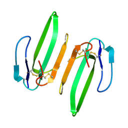 | | Structural and Functional Characterization of a Novel Homodimeric Three-finger Neurotoxin from the Venom of Ophiophagus hannah (King Cobra) | | 分子名称: | Muscarinic toxin-like protein 3 homolog | | 著者 | Roy, A, Zhou, X, Chong, M.Z, D'hoedt, D, Foo, C.S, Rajagopalan, N, Nirthanan, S, Bertrand, D, Sivaraman, J, Kini, R.M. | | 登録日 | 2009-05-15 | | 公開日 | 2010-01-12 | | 最終更新日 | 2017-11-01 | | 実験手法 | X-RAY DIFFRACTION (1.55 Å) | | 主引用文献 | Structural and Functional Characterization of a Novel Homodimeric Three-finger Neurotoxin from the Venom of Ophiophagus hannah (King Cobra)
J.Biol.Chem., 285, 2010
|
|
5XXZ
 | |
5XYA
 | | Crystal structure of a serine protease from Streptococcus species | | 分子名称: | 4-(2-AMINOETHYL)BENZENESULFONYL FLUORIDE, CALCIUM ION, Chemokine protease C, ... | | 著者 | Jobichen, C, Sivaraman, J. | | 登録日 | 2017-07-06 | | 公開日 | 2018-08-08 | | 最終更新日 | 2023-11-22 | | 実験手法 | X-RAY DIFFRACTION (3 Å) | | 主引用文献 | Structure of ScpC, a virulence protease fromStreptococcus pyogenes, reveals the functional domains and maturation mechanism.
Biochem. J., 475, 2018
|
|
5XYR
 | | Crystal structure of a serine protease from Streptococcus species | | 分子名称: | CALCIUM ION, CHLORIDE ION, Chemokine protease C, ... | | 著者 | Jobichen, C, Sivaraman, J. | | 登録日 | 2017-07-10 | | 公開日 | 2018-08-08 | | 最終更新日 | 2023-11-22 | | 実験手法 | X-RAY DIFFRACTION (2.8 Å) | | 主引用文献 | Structure of ScpC, a virulence protease fromStreptococcus pyogenes, reveals the functional domains and maturation mechanism.
Biochem. J., 475, 2018
|
|
2PY2
 | | Structure of Herring Type II Antifreeze Protein | | 分子名称: | Antifreeze protein type II, CALCIUM ION | | 著者 | Liu, Y, Li, Z, Lin, Q, Seetharaman, J, Sivaraman, J, Hew, C.-L. | | 登録日 | 2007-05-15 | | 公開日 | 2007-06-26 | | 最終更新日 | 2011-07-13 | | 実験手法 | X-RAY DIFFRACTION (1.7 Å) | | 主引用文献 | Structure and Evolutionary Origin of Ca-Dependent Herring Type II Antifreeze Protein.
PLoS ONE, 2, 2007
|
|
2QCY
 | |
4ZQY
 | |
5BN5
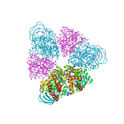 | | Structural basis for a unique ATP synthase core complex from Nanoarcheaum equitans | | 分子名称: | NEQ263, SULFATE ION, V-type ATP synthase alpha chain | | 著者 | Mohanty, S, Jobichen, C, Chichili, V.P.R, Sivaraman, J. | | 登録日 | 2015-05-25 | | 公開日 | 2015-09-16 | | 最終更新日 | 2023-11-08 | | 実験手法 | X-RAY DIFFRACTION (2.997 Å) | | 主引用文献 | Structural Basis for a Unique ATP Synthase Core Complex from Nanoarcheaum equitans
J.Biol.Chem., 290, 2015
|
|
5BN3
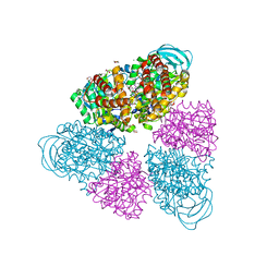 | | Structure of a unique ATP synthase NeqA-NeqB in complex with ADP from Nanoarcheaum equitans | | 分子名称: | 1,4-DIETHYLENE DIOXIDE, ADENOSINE-5'-DIPHOSPHATE, GLYCEROL, ... | | 著者 | Mohanty, S, Jobichen, C, Chichili, V.P.R, Sivaraman, J. | | 登録日 | 2015-05-25 | | 公開日 | 2015-09-16 | | 最終更新日 | 2023-11-08 | | 実験手法 | X-RAY DIFFRACTION (2 Å) | | 主引用文献 | Structural Basis for a Unique ATP Synthase Core Complex from Nanoarcheaum equitans
J.Biol.Chem., 290, 2015
|
|
5BN4
 | | Structure of a unique ATP synthase NeqA-NeqB in complex with ANP from Nanoarcheaum equitans | | 分子名称: | MAGNESIUM ION, NEQ263, PHOSPHOAMINOPHOSPHONIC ACID-ADENYLATE ESTER, ... | | 著者 | Mohanty, S, Jobichen, C, Chichili, V.P.R, Sivaraman, J. | | 登録日 | 2015-05-25 | | 公開日 | 2015-09-16 | | 最終更新日 | 2023-11-08 | | 実験手法 | X-RAY DIFFRACTION (2.699 Å) | | 主引用文献 | Structural Basis for a Unique ATP Synthase Core Complex from Nanoarcheaum equitans
J.Biol.Chem., 290, 2015
|
|
2ZQT
 | |
5BO5
 | | Structure of a unique ATP synthase subunit NeqB from Nanoarcheaum equitans | | 分子名称: | MAGNESIUM ION, NEQ263, SULFATE ION | | 著者 | Mohanty, S, Jobichen, C, Chichili, V.P.R, Sivaraman, J. | | 登録日 | 2015-05-27 | | 公開日 | 2015-09-16 | | 最終更新日 | 2023-11-08 | | 実験手法 | X-RAY DIFFRACTION (2.808 Å) | | 主引用文献 | Structural Basis for a Unique ATP Synthase Core Complex from Nanoarcheaum equitans
J.Biol.Chem., 290, 2015
|
|
2ZR6
 | |


