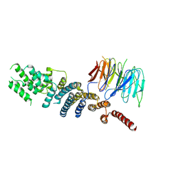
4O6M
 
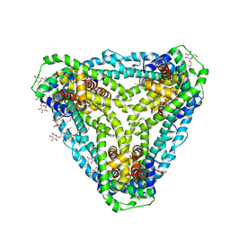
 | | Structure of AF2299, a CDP-alcohol phosphotransferase (CMP-bound) | | 分子名称: | AF2299, a CDP-alcohol phosphotransferase, CALCIUM ION, ... | | 著者 | Clarke, O.B, Sciara, G, Tomasek, D, Banerjee, S, Rajashankar, K.R, Shapiro, L, Mancia, F, New York Consortium on Membrane Protein Structure (NYCOMPS) | | 登録日 | 2013-12-22 | | 公開日 | 2014-05-14 | | 最終更新日 | 2024-02-28 | | 実験手法 | X-RAY DIFFRACTION (1.901 Å) | | 主引用文献 | Structural basis for catalysis in a CDP-alcohol phosphotransferase.
Nat Commun, 5, 2014
|
|
4O6N
 | | Structure of AF2299, a CDP-alcohol phosphotransferase (CDP-bound) | | 分子名称: | AF2299, a CDP-alcohol phosphotransferase, CALCIUM ION, ... | | 著者 | Clarke, O.B, Sciara, G, Tomasek, D, Banerjee, S, Rajashankar, K.R, Shapiro, L, Mancia, F, New York Consortium on Membrane Protein Structure (NYCOMPS) | | 登録日 | 2013-12-22 | | 公開日 | 2014-05-14 | | 最終更新日 | 2024-02-28 | | 実験手法 | X-RAY DIFFRACTION (2.1 Å) | | 主引用文献 | Structural basis for catalysis in a CDP-alcohol phosphotransferase.
Nat Commun, 5, 2014
|
|
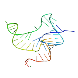
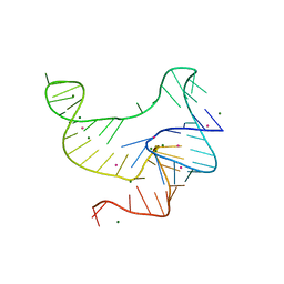
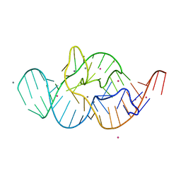
6CIH
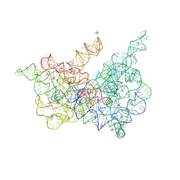 | | Crystal structure of a group II intron lariat in the post-catalytic state | | 分子名称: | IRIDIUM HEXAMMINE ION, MAGNESIUM ION, RNA (5'-R(P*UP*GP*UP*UP*UP*AP*UP*UP*AP*AP*AP*AP*AP*C*-3'), ... | | 著者 | Chan, R.T, Peters, J.K, Robart, A.R, Wiryaman, T, Rajashankar, K.R, Toor, N. | | 登録日 | 2018-02-23 | | 公開日 | 2018-11-21 | | 最終更新日 | 2024-03-13 | | 実験手法 | X-RAY DIFFRACTION (3.676 Å) | | 主引用文献 | Structural basis for the second step of group II intron splicing.
Nat Commun, 9, 2018
|
|
6D9Z
 | | Structure of CysZ, a sulfate permease from Pseudomonas Denitrificans | | 分子名称: | Sulfate transporter CysZ, octyl beta-D-glucopyranoside | | 著者 | Sanghai, Z.A, Clarke, O.B, Liu, Q, Banerjee, S, Rajashankar, K.R, Hendrickson, W.A, Mancia, F. | | 登録日 | 2018-04-30 | | 公開日 | 2018-05-23 | | 最終更新日 | 2023-10-04 | | 実験手法 | X-RAY DIFFRACTION (3.4021318 Å) | | 主引用文献 | Structure-based analysis of CysZ-mediated cellular uptake of sulfate.
Elife, 7, 2018
|
|
6D79
 | | Structure of CysZ, a sulfate permease from Pseudomonas Fragi | | 分子名称: | Sulfate transporter CysZ | | 著者 | Sanghai, Z.A, Liu, Q, Clarke, O.B, Banerjee, S, Rajashankar, K.R, Hendrickson, W.A, Mancia, F. | | 登録日 | 2018-04-24 | | 公開日 | 2018-05-16 | | 最終更新日 | 2024-10-16 | | 実験手法 | X-RAY DIFFRACTION (3.501 Å) | | 主引用文献 | Structure-based analysis of CysZ-mediated cellular uptake of sulfate.
Elife, 7, 2018
|
|
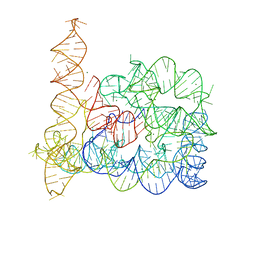
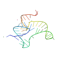
6CHR
 | | Crystal structure of a group II intron lariat with an intact 3' splice site (pre-2s state) | | 分子名称: | MAGNESIUM ION, RNA (5'-R(P*UP*GP*UP*UP*UP*AP*UP*UP*AP*AP*AP*AP*A)-3'), RNA (621-MER), ... | | 著者 | Chan, R.T, Peters, J.K, Robart, A.R, Wiryaman, T, Rajashankar, K.R, Toor, N. | | 登録日 | 2018-02-22 | | 公開日 | 2018-11-21 | | 最終更新日 | 2024-03-13 | | 実験手法 | X-RAY DIFFRACTION (3.7 Å) | | 主引用文献 | Structural basis for the second step of group II intron splicing.
Nat Commun, 9, 2018
|
|
4ENB
 | | Crystal structure of fluoride riboswitch, bound to Iridium | | 分子名称: | FLUORIDE ION, Fluoride riboswitch, IRIDIUM HEXAMMINE ION, ... | | 著者 | Ren, A.M, Rajashankar, K.R, Patel, D.J. | | 登録日 | 2012-04-12 | | 公開日 | 2012-05-09 | | 最終更新日 | 2024-02-28 | | 実験手法 | X-RAY DIFFRACTION (2.302 Å) | | 主引用文献 | Fluoride ion encapsulation by Mg2+ ions and phosphates in a fluoride riboswitch.
Nature, 486, 2012
|
|
4ENC
 | | Crystal structure of fluoride riboswitch | | 分子名称: | FLUORIDE ION, Fluoride riboswitch, MAGNESIUM ION, ... | | 著者 | Ren, A.M, Rajashankar, K.R, Patel, D.J. | | 登録日 | 2012-04-12 | | 公開日 | 2012-05-09 | | 最終更新日 | 2023-09-13 | | 実験手法 | X-RAY DIFFRACTION (2.272 Å) | | 主引用文献 | Fluoride ion encapsulation by Mg2+ ions and phosphates in a fluoride riboswitch.
Nature, 486, 2012
|
|
4DS6
 | | Crystal structure of a group II intron in the pre-catalytic state | | 分子名称: | MAGNESIUM ION, Mutant Group IIC Intron | | 著者 | Chan, R.T, Robart, A.R, Rajashankar, K.R, Pyle, A.M, Toor, N. | | 登録日 | 2012-02-17 | | 公開日 | 2012-04-18 | | 最終更新日 | 2023-09-13 | | 実験手法 | X-RAY DIFFRACTION (3.644 Å) | | 主引用文献 | Crystal structure of a group II intron in the pre-catalytic state.
Nat.Struct.Mol.Biol., 19, 2012
|
|
3EWE
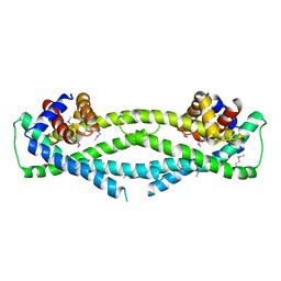
 | | Crystal Structure of the Nup85/Seh1 Complex | | 分子名称: | Nucleoporin NUP85, Nucleoporin SEH1 | | 著者 | Brohawn, S.G, Leksa, N.C, Rajashankar, K.R, Schwartz, T.U. | | 登録日 | 2008-10-14 | | 公開日 | 2008-11-11 | | 最終更新日 | 2024-10-09 | | 実験手法 | X-RAY DIFFRACTION (3.5 Å) | | 主引用文献 | Structural evidence for common ancestry of the nuclear pore complex and vesicle coats.
Science, 322, 2008
|
|
3FEF
 | | Crystal structure of putative glucosidase lplD from bacillus subtilis | | 分子名称: | MAGNESIUM ION, Putative glucosidase lplD, ALPHA-GALACTURONIDASE, ... | | 著者 | Ramagopal, U.A, Rajashankar, K.R, Toro, R, Burley, S.K, Almo, S.C, New York SGX Research Center for Structural Genomics (NYSGXRC) | | 登録日 | 2008-11-28 | | 公開日 | 2008-12-30 | | 最終更新日 | 2024-11-20 | | 実験手法 | X-RAY DIFFRACTION (2.2 Å) | | 主引用文献 | Crystal structure of putative glucosidase lplD from bacillus subtilis.
To be published
|
|
4EN5
 | | Crystal structure of fluoride riboswitch, Tl-Acetate soaked | | 分子名称: | FLUORIDE ION, Fluoride riboswitch, MAGNESIUM ION, ... | | 著者 | Ren, A.M, Rajashankar, K.R, Patel, D.J. | | 登録日 | 2012-04-12 | | 公開日 | 2012-05-09 | | 最終更新日 | 2023-09-13 | | 実験手法 | X-RAY DIFFRACTION (2.957 Å) | | 主引用文献 | Fluoride ion encapsulation by Mg2+ ions and phosphates in a fluoride riboswitch.
Nature, 486, 2012
|
|
4ENA
 | | Crystal structure of fluoride riboswitch, soaked in Cs+ | | 分子名称: | CESIUM ION, FLUORIDE ION, Fluoride riboswitch, ... | | 著者 | Ren, A.M, Rajashankar, K.R, Patel, D.J. | | 登録日 | 2012-04-12 | | 公開日 | 2012-05-09 | | 最終更新日 | 2023-09-13 | | 実験手法 | X-RAY DIFFRACTION (2.85 Å) | | 主引用文献 | Fluoride ion encapsulation by Mg2+ ions and phosphates in a fluoride riboswitch.
Nature, 486, 2012
|
|
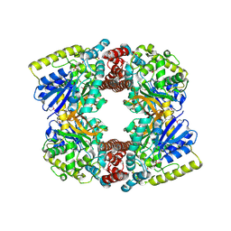
1HA7
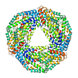 | | STRUCTURE OF A LIGHT-HARVESTING PHYCOBILIPROTEIN, C-PHYCOCYANIN FROM SPIRULINA PLATENSIS AT 2.2A RESOLUTION | | 分子名称: | C-PHYCOCYANIN ALPHA CHAIN, C-PHYCOCYANIN BETA CHAIN, PHYCOCYANOBILIN | | 著者 | Padyana, A.K, Rajashankar, K.R, Ramakumar, S. | | 登録日 | 2001-03-29 | | 公開日 | 2002-03-28 | | 最終更新日 | 2023-12-13 | | 実験手法 | X-RAY DIFFRACTION (2.2 Å) | | 主引用文献 | Crystal Structure of a Light-Harvesting Protein C-Phycocyanin from Spirulina Platensis
Biochem.Biophys.Res.Commun., 282, 2001
|
|
1GQB
 | | HUMAN MIR-RECEPTOR, REPEAT 11 | | 分子名称: | BROMIDE ION, CATION-INDEPENDENT MANNOSE-6-PHOSPHATE RECEPTOR | | 著者 | Von Buelow, R, Dauter, M, Dauter, Z, Rajashankar, K.R, Grimme, S, Schmidt, B, Von Figura, K, Uson, I. | | 登録日 | 2001-11-22 | | 公開日 | 2002-12-05 | | 最終更新日 | 2024-11-06 | | 実験手法 | X-RAY DIFFRACTION (1.8 Å) | | 主引用文献 | Locating the Anomalous Scatterer Substructures in Halide and Sulfur Phasing
Acta Crystallogr.,Sect.D, 59, 2003
|
|
1XBA
 | | Crystal structure of apo syk tyrosine kinase domain | | 分子名称: | Tyrosine-protein kinase SYK | | 著者 | Atwell, S, Adams, J.M, Badger, J, Buchanan, M.D, Feil, I.K, Froning, K.J, Gao, X, Hendle, J, Keegan, K, Leon, B.C, Muller-Deickmann, H.J, Nienaber, V.L, Noland, B.W, Post, K, Rajashankar, K.R, Ramos, A, Russell, M, Burley, S.K, Buchanan, S.G. | | 登録日 | 2004-08-30 | | 公開日 | 2004-11-02 | | 最終更新日 | 2024-02-14 | | 実験手法 | X-RAY DIFFRACTION (2 Å) | | 主引用文献 | A novel mode of Gleevec binding is revealed by the structure of spleen tyrosine kinase.
J.Biol.Chem., 279, 2004
|
|
1JLT
 | | Vipoxin Complex | | 分子名称: | (4R)-2-METHYLPENTANE-2,4-DIOL, (4S)-2-METHYL-2,4-PENTANEDIOL, PHOSPHOLIPASE A2, ... | | 著者 | Banumathi, S, Rajashankar, K.R, Notzel, C, Aleksiev, B, Singh, T.P, Genov, N, Betzel, C. | | 登録日 | 2001-07-16 | | 公開日 | 2001-10-31 | | 最終更新日 | 2024-11-13 | | 実験手法 | X-RAY DIFFRACTION (1.4 Å) | | 主引用文献 | Structure of the neurotoxic complex vipoxin at 1.4 A resolution.
Acta Crystallogr.,Sect.D, 57, 2001
|
|
1IIN
 | | thymidylyltransferase complexed with UDP-glucose | | 分子名称: | URIDINE-5'-DIPHOSPHATE-GLUCOSE, glucose-1-phosphate thymidylyltransferase | | 著者 | Barton, W.A, Lesniak, J, Biggins, J.B, Jeffrey, P.D, Jiang, J, Rajashankar, K.R, Thorson, J.S, Nikolov, D.B. | | 登録日 | 2001-04-23 | | 公開日 | 2001-05-09 | | 最終更新日 | 2024-02-07 | | 実験手法 | X-RAY DIFFRACTION (2.1 Å) | | 主引用文献 | Structure, mechanism and engineering of a nucleotidylyltransferase as a first step toward glycorandomization.
Nat.Struct.Biol., 8, 2001
|
|
1XBB
 | | Crystal structure of the syk tyrosine kinase domain with Gleevec | | 分子名称: | 4-(4-METHYL-PIPERAZIN-1-YLMETHYL)-N-[4-METHYL-3-(4-PYRIDIN-3-YL-PYRIMIDIN-2-YLAMINO)-PHENYL]-BENZAMIDE, Tyrosine-protein kinase SYK | | 著者 | Nienaber, V.L, Atwell, S, Adams, J.M, Badger, J, Buchanan, M.D, Feil, I.K, Froning, K.J, Gao, X, Hendle, J, Keegan, K, Leon, B.C, Muller-Deickmann, H.J, Noland, B.W, Post, K, Rajashankar, K.R, Ramos, A, Russell, M, Burley, S.K, Buchanan, S.G. | | 登録日 | 2004-08-30 | | 公開日 | 2004-11-02 | | 最終更新日 | 2024-02-14 | | 実験手法 | X-RAY DIFFRACTION (1.57 Å) | | 主引用文献 | A Novel Mode of Gleevec Binding Is Revealed by the Structure of Spleen Tyrosine Kinase
J.Biol.Chem., 279, 2004
|
|
1XBC
 | | Crystal structure of the syk tyrosine kinase domain with Staurosporin | | 分子名称: | STAUROSPORINE, Tyrosine-protein kinase SYK | | 著者 | Badger, J, Atwell, S, Adams, J.M, Buchanan, M.D, Feil, I.K, Froning, K.J, Gao, X, Hendle, J, Keegan, K, Leon, B.C, Muller-Deickmann, H.J, Nienaber, V.L, Noland, B.W, Post, K, Rajashankar, K.R, Ramos, A, Russell, M, Burley, S.K, Buchanan, S.G. | | 登録日 | 2004-08-30 | | 公開日 | 2004-11-02 | | 最終更新日 | 2024-02-14 | | 実験手法 | X-RAY DIFFRACTION (2 Å) | | 主引用文献 | A novel mode of Gleevec binding is revealed by the structure of spleen tyrosine kinase
J.Biol.Chem., 279, 2004
|
|
1IIM
 | | thymidylyltransferase complexed with TTP | | 分子名称: | THYMIDINE-5'-TRIPHOSPHATE, glucose-1-phosphate thymidylyltransferase | | 著者 | Barton, W.A, Lesniak, J, Biggins, J.B, Jeffrey, P.D, Jiang, J, Rajashankar, K.R, Thorson, J.S, Nikolov, D.B. | | 登録日 | 2001-04-23 | | 公開日 | 2001-05-09 | | 最終更新日 | 2024-02-07 | | 実験手法 | X-RAY DIFFRACTION (2.1 Å) | | 主引用文献 | Structure, mechanism and engineering of a nucleotidylyltransferase as a first step toward glycorandomization.
Nat.Struct.Biol., 8, 2001
|
|
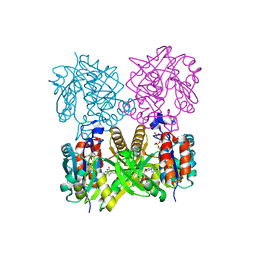
1KGY
 | | Crystal Structure of the EphB2-ephrinB2 complex | | 分子名称: | EPHRIN TYPE-B RECEPTOR 2, EPHRIN-B2 | | 著者 | Himanen, J.P, Rajashankar, K.R, Lackmann, M, Cowan, C.A, Henkemeyer, M, Nikolov, D.B. | | 登録日 | 2001-11-28 | | 公開日 | 2002-05-28 | | 最終更新日 | 2024-10-30 | | 実験手法 | X-RAY DIFFRACTION (2.7 Å) | | 主引用文献 | Crystal structure of an Eph receptor-ephrin complex.
Nature, 414, 2001
|
|
3MX0
 | | Crystal Structure of EphA2 ectodomain in complex with ephrin-A5 | | 分子名称: | 2-acetamido-2-deoxy-beta-D-glucopyranose, 2-acetamido-2-deoxy-beta-D-glucopyranose-(1-4)-2-acetamido-2-deoxy-beta-D-glucopyranose-(1-4)-2-acetamido-2-deoxy-beta-D-glucopyranose, Ephrin type-A receptor 2, ... | | 著者 | Himanen, J.P, Yermekbayeva, L, Janes, P.W, Walker, J.R, Xu, K, Atapattu, L, Rajashankar, K.R, Mensinga, A, Lackmann, M, Nikolov, D.B, Dhe-Paganon, S. | | 登録日 | 2010-05-06 | | 公開日 | 2010-06-30 | | 最終更新日 | 2024-10-30 | | 実験手法 | X-RAY DIFFRACTION (3.506 Å) | | 主引用文献 | Architecture of Eph receptor clusters.
Proc.Natl.Acad.Sci.USA, 107, 2010
|
|
4Q7C
 | | Structure of AF2299, a CDP-alcohol phosphotransferase | | 分子名称: | AF2299, a CDP-alcohol phosphotransferase, CALCIUM ION, ... | | 著者 | Clarke, O.B, Sciara, G, Tomasek, D, Banerjee, S, Rajashankar, K.R, Shapiro, L, Mancia, F, New York Consortium on Membrane Protein Structure (NYCOMPS) | | 登録日 | 2014-04-24 | | 公開日 | 2014-05-28 | | 最終更新日 | 2023-09-20 | | 実験手法 | X-RAY DIFFRACTION (3.102 Å) | | 主引用文献 | Structural basis for catalysis in a CDP-alcohol phosphotransferase.
Nat Commun, 5, 2014
|
|
4RGF
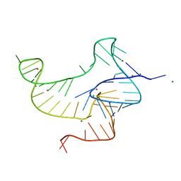
 | | Crystal structure of the in-line aligned env22 twister ribozyme soaked with Mn2+ | | 分子名称: | MAGNESIUM ION, MANGANESE (II) ION, POTASSIUM ION, ... | | 著者 | Ren, A, Rajashankar, K.R, Simanshu, D, Patel, D. | | 登録日 | 2014-09-30 | | 公開日 | 2014-12-03 | | 最終更新日 | 2024-02-28 | | 実験手法 | X-RAY DIFFRACTION (3.2008 Å) | | 主引用文献 | In-line alignment and Mg(2+) coordination at the cleavage site of the env22 twister ribozyme.
Nat Commun, 5, 2014
|
|


