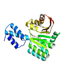
4R9P
 
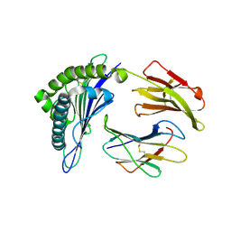
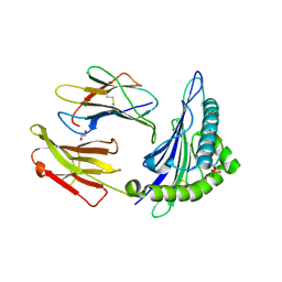
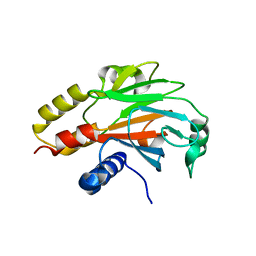 | | An Expansion to the Smad MH2-family: The structure of the N-MH2 expanded domain | | 分子名称: | RE28239p | | 著者 | Beich-Frandsen, M, Aragon, E, Llimargas, M, Benach, J, Riera, A, Macias, M.J. | | 登録日 | 2014-09-06 | | 公開日 | 2015-04-08 | | 最終更新日 | 2024-02-28 | | 実験手法 | X-RAY DIFFRACTION (1.592 Å) | | 主引用文献 | Structure of the N-terminal domain of the protein Expansion: an 'Expansion' to the Smad MH2
Acta Crystallogr.,Sect.D, 71, 2015
|
|
5NP6
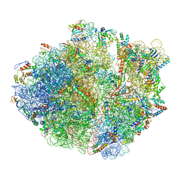 | | 70S structure prior to bypassing | | 分子名称: | 16S ribosomal RNA, 23S ribosomal RNA, 30S ribosomal protein S10, ... | | 著者 | Agirrezabala, X, Samatova, E, Klimova, M, Zamora, M, Gil-Carton, D, Rodnina, M, Valle, M. | | 登録日 | 2017-04-13 | | 公開日 | 2017-06-14 | | 最終更新日 | 2024-04-24 | | 実験手法 | ELECTRON MICROSCOPY (3.6 Å) | | 主引用文献 | Ribosome rearrangements at the onset of translational bypassing.
Sci Adv, 3, 2017
|
|
8A23
 | | Crystal structure of SARS-CoV-2 nsp10/nsp16 methyltransferase in complex with TO383 | | 分子名称: | (2R,3R,4S,5R)-2-[4-azanyl-5-(2-quinolin-3-ylethynyl)pyrrolo[2,3-d]pyrimidin-7-yl]-5-(hydroxymethyl)oxolane-3,4-diol, 2'-O-methyltransferase nsp16, GLYCEROL, ... | | 著者 | Hanigovsky, M, Krafcikova, P, Klima, M, Boura, E. | | 登録日 | 2022-06-02 | | 公開日 | 2023-06-14 | | 最終更新日 | 2024-02-07 | | 実験手法 | X-RAY DIFFRACTION (2.8 Å) | | 主引用文献 | Crystal structure of SARS-CoV-2 nsp10/nsp16 methyltransferase in
complex with TO383
To Be Published
|
|
6Q69
 | |
6Q68
 | |
6Q67
 | |
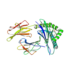
6R35
 | | Structure of the LecB lectin from Pseudomonas aeruginosa strain PAO1 in complex with lewis x tetrasaccharide | | 分子名称: | 1,2-ETHANEDIOL, CALCIUM ION, Fucose-binding lectin PA-IIL, ... | | 著者 | Lepsik, M, Sommer, R, Kuhaudomlarp, S, Lelimousin, M, Varrot, A, Titz, A, Imberty, A. | | 登録日 | 2019-03-19 | | 公開日 | 2019-06-12 | | 最終更新日 | 2024-01-24 | | 実験手法 | X-RAY DIFFRACTION (1.8 Å) | | 主引用文献 | Induction of rare conformation of oligosaccharide by binding to calcium-dependent bacterial lectin: X-ray crystallography and modelling study.
Eur.J.Med.Chem., 177, 2019
|
|
7R50
 | |
3FRH
 | | Structure of the 16S rRNA methylase RmtB, P21 | | 分子名称: | 16S rRNA methylase, S-ADENOSYL-L-HOMOCYSTEINE | | 著者 | Schmitt, E, Galimand, M, Panvert, M, Dupechez, M, Courvalin, P, Mechulam, Y. | | 登録日 | 2009-01-08 | | 公開日 | 2009-08-11 | | 最終更新日 | 2023-11-01 | | 実験手法 | X-RAY DIFFRACTION (1.2 Å) | | 主引用文献 | Structural bases for 16 S rRNA methylation catalyzed by ArmA and RmtB methyltransferases
J.Mol.Biol., 388, 2009
|
|
3FRI
 | | Structure of the 16S rRNA methylase RmtB, I222 | | 分子名称: | 16S rRNA methylase, S-ADENOSYL-L-HOMOCYSTEINE | | 著者 | Schmitt, E, Galimand, M, Panvert, M, Dupechez, M, Courvalin, P, Mechulam, Y. | | 登録日 | 2009-01-08 | | 公開日 | 2009-08-11 | | 最終更新日 | 2023-11-01 | | 実験手法 | X-RAY DIFFRACTION (1.8 Å) | | 主引用文献 | Structural bases for 16 S rRNA methylation catalyzed by ArmA and RmtB methyltransferases
J.Mol.Biol., 388, 2009
|
|
4RZD
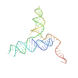 | | Crystal Structure of a PreQ1 Riboswitch | | 分子名称: | 7-DEAZA-7-AMINOMETHYL-GUANINE, PreQ1-III Riboswitch (Class 3) | | 著者 | Wedekind, J.E, Liberman, J.A, Salim, M. | | 登録日 | 2014-12-20 | | 公開日 | 2015-07-01 | | 最終更新日 | 2024-02-28 | | 実験手法 | X-RAY DIFFRACTION (2.75 Å) | | 主引用文献 | Structural analysis of a class III preQ1 riboswitch reveals an aptamer distant from a ribosome-binding site regulated by fast dynamics.
Proc.Natl.Acad.Sci.USA, 112, 2015
|
|
7ZIU
 | |
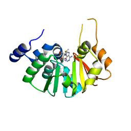
4B5Y
 | | X-ray structure of the cyan fluorescent protein mTurquoise-GL (K206A mutant) in space group C222(1) | | 分子名称: | GREEN FLUORESCENT PROTEIN | | 著者 | von Stetten, D, Lelimousin, M, Oost, K, Noirclerc-Savoye, M, Gadella, T.W.J, Goedhart, J, Royant, A. | | 登録日 | 2012-08-08 | | 公開日 | 2013-08-28 | | 最終更新日 | 2024-10-09 | | 実験手法 | X-RAY DIFFRACTION (1.45 Å) | | 主引用文献 | Influence of the H148G Mutation on Fluorescence Properties of Cyan Fluorescent Proteins
To be Published
|
|
3D25
 | | Crystal structure of HA-1 minor histocompatibility antigen bound to human class I MHC HLA-A2 | | 分子名称: | Beta-2-microglobulin, HLA class I histocompatibility antigen, A-2 alpha chain, ... | | 著者 | Nicholls, S, Piper, K.P, Mohammed, F, Dafforn, T.R, Tenzer, S, Salim, M, Mahendra, P, Craddock, C, van Endert, P, Schild, H, Cobbold, M, Engelhard, V.H, Moss, P.A.H, Willcox, B.E. | | 登録日 | 2008-05-07 | | 公開日 | 2009-02-10 | | 最終更新日 | 2024-10-09 | | 実験手法 | X-RAY DIFFRACTION (1.3 Å) | | 主引用文献 | Secondary anchor polymorphism in the HA-1 minor histocompatibility antigen critically affects MHC stability and TCR recognition
Proc.Natl.Acad.Sci.USA, 106, 2009
|
|
2YE0
 | | X-ray structure of the cyan fluorescent protein mTurquoise (K206A mutant) | | 分子名称: | GREEN FLUORESCENT PROTEIN | | 著者 | von Stetten, D, Goedhart, J, Noirclerc-Savoye, M, Lelimousin, M, Joosen, L, Hink, M.A, van Weeren, L, Gadella, T.W.J, Royant, A. | | 登録日 | 2011-03-25 | | 公開日 | 2012-03-21 | | 最終更新日 | 2023-12-20 | | 実験手法 | X-RAY DIFFRACTION (1.47 Å) | | 主引用文献 | Structure-Guided Evolution of Cyan Fluorescent Proteins Towards a Quantum Yield of 93%
Nat.Commun, 3, 2012
|
|
3FZG
 | | Structure of the 16S rRNA methylase ArmA | | 分子名称: | 16S rRNA methylase, S-ADENOSYLMETHIONINE | | 著者 | Schmitt, E, Galimand, M, Panvert, M, Courvalin, P, Mechulam, Y. | | 登録日 | 2009-01-26 | | 公開日 | 2009-08-11 | | 最終更新日 | 2024-03-20 | | 実験手法 | X-RAY DIFFRACTION (2 Å) | | 主引用文献 | Structural bases for 16 S rRNA methylation catalyzed by ArmA and RmtB methyltransferases
J.Mol.Biol., 388, 2009
|
|
2YDZ
 | | X-ray structure of the cyan fluorescent protein SCFP3A (K206A mutant) | | 分子名称: | GREEN FLUORESCENT PROTEIN | | 著者 | von Stetten, D, Goedhart, J, Noirclerc-Savoye, M, Lelimousin, M, Joosen, L, Hink, M.A, van Weeren, L, Gadella, T.W.J, Royant, A. | | 登録日 | 2011-03-25 | | 公開日 | 2012-03-21 | | 最終更新日 | 2024-10-09 | | 実験手法 | X-RAY DIFFRACTION (1.59 Å) | | 主引用文献 | Structure-Guided Evolution of Cyan Fluorescent Proteins Towards a Quantum Yield of 93%
Nat.Commun, 3, 2012
|
|
3BH9
 | | Crystal Structure of RTY Phosphopeptide Bound to Human Class I MHC HLA-A2 | | 分子名称: | 1,2-ETHANEDIOL, Beta-2-microglobulin, HLA class I histocompatibility antigen, ... | | 著者 | Mohammed, F, Cobbold, M, Zarling, A.L, Salim, M, Barrett-Wilt, G.A, Shabanowitz, J, Hunt, D.F, Engelhard, V.H, Willcox, B.E. | | 登録日 | 2007-11-28 | | 公開日 | 2008-10-21 | | 最終更新日 | 2017-10-11 | | 実験手法 | X-RAY DIFFRACTION (1.7 Å) | | 主引用文献 | Phosphorylation-dependent interaction between antigenic peptides and MHC class I: a molecular basis for the presentation of transformed self
Nat.Immunol., 9, 2008
|
|
3BH8
 | | Crystal Structure of RQA_M Phosphopeptide Bound to HUMAN Class I MHC HLA-A2 | | 分子名称: | 1,2-ETHANEDIOL, Beta-2-microglobulin, HLA class I histocompatibility antigen, ... | | 著者 | Mohammed, F, Cobbold, M, Zarling, A.L, Salim, M, Barrett-Wilt, G.A, Shabanowitz, J, Hunt, D.F, Engelhard, V.H, Willcox, B.E. | | 登録日 | 2007-11-28 | | 公開日 | 2008-10-21 | | 最終更新日 | 2024-10-09 | | 実験手法 | X-RAY DIFFRACTION (1.65 Å) | | 主引用文献 | Phosphorylation-dependent interaction between antigenic peptides and MHC class I: a molecular basis for the presentation of transformed self
Nat.Immunol., 9, 2008
|
|
3BHB
 | | Crystal Structure of KMD Phosphopeptide Bound to Human Class I MHC HLA-A2 | | 分子名称: | 1,2-ETHANEDIOL, Beta-2-microglobulin, HLA class I histocompatibility antigen, ... | | 著者 | Mohammed, F, Cobbold, M, Zarling, A.L, Salim, M, Barrett-Wilt, G.A, Shabanowitz, J, Hunt, D.F, Engelhard, V.H, Willcox, B.E. | | 登録日 | 2007-11-28 | | 公開日 | 2008-10-21 | | 最終更新日 | 2011-07-13 | | 実験手法 | X-RAY DIFFRACTION (2.2 Å) | | 主引用文献 | Phosphorylation-dependent interaction between antigenic peptides and MHC class I: a molecular basis for the presentation of transformed self
Nat.Immunol., 9, 2008
|
|
3BGM
 | | Crystal Structure of PKD2 Phosphopeptide Bound to Human Class I MHC HLA-A2 | | 分子名称: | 1,2-ETHANEDIOL, Beta-2-microglobulin, HLA class I histocompatibility antigen, ... | | 著者 | Mohammed, F, Cobbold, M, Zarling, A.L, Salim, M, Barrett-Wilt, G.A, Shabanowitz, J, Hunt, D.F, Engelhard, V.H, Willcox, B.E. | | 登録日 | 2007-11-27 | | 公開日 | 2008-10-21 | | 最終更新日 | 2011-07-13 | | 実験手法 | X-RAY DIFFRACTION (1.6 Å) | | 主引用文献 | Phosphorylation-dependent interaction between antigenic peptides and MHC class I: a molecular basis for the presentation of transformed self
Nat.Immunol., 9, 2008
|
|
3ZTF
 | | X-ray Structure of the Cyan Fluorescent Protein mTurquoise2 (K206A mutant) | | 分子名称: | GREEN FLUORESCENT PROTEIN | | 著者 | von Stetten, D, Goedhart, J, Noirclerc-Savoye, M, Lelimousin, M, Joosen, L, Hink, M.A, van Weeren, L, Gadella, T.W.J, Royant, A. | | 登録日 | 2011-07-07 | | 公開日 | 2012-03-21 | | 最終更新日 | 2023-12-20 | | 実験手法 | X-RAY DIFFRACTION (1.31 Å) | | 主引用文献 | Structure-Guided Evolution of Cyan Fluorescent Proteins Towards a Quantum Yield of 93%
Nat.Commun, 3, 2012
|
|
6YHU
 | |
8OIV
 | | Monkeypox virus VP39 in complex with SAH and cap0 | | 分子名称: | 7-METHYL-GUANOSINE-5'-TRIPHOSPHATE-5'-GUANOSINE, Cap-specific mRNA (nucleoside-2'-O-)-methyltransferase, S-ADENOSYL-L-HOMOCYSTEINE, ... | | 著者 | Skvara, P, Chalupska, D, Silhan, J, Klima, M, Boura, E. | | 登録日 | 2023-03-23 | | 公開日 | 2023-08-23 | | 実験手法 | X-RAY DIFFRACTION (2.12 Å) | | 主引用文献 | Structural basis for RNA-cap recognition and methylation by the mpox methyltransferase VP39.
Antiviral Res., 216, 2023
|
|
8P7A
 | |


