5WPY
 
 | |
5WPX
 | |
3TYW
 | |
3QZ1
 | |
3TZO
 | |
2MY5
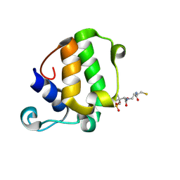
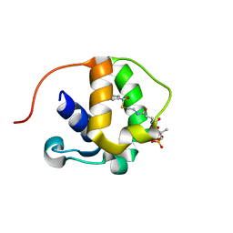
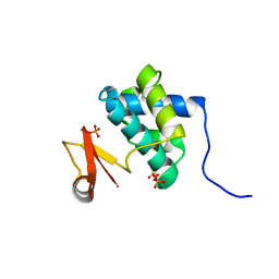
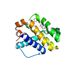
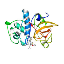
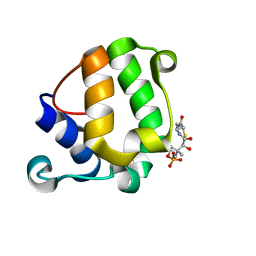 | | Solution Structure of KstB-PCP in kosinostatin biosynthesis | | 分子名称: | N~3~-[(2S)-2-hydroxy-3,3-dimethyl-4-(phosphonooxy)butanoyl]-N-(2-sulfanylethyl)-beta-alaninamide, Peptidyl carrier protein | | 著者 | Zhao, B, Lan, W, Wang, C, Tang, G, Cao, C. | | 登録日 | 2015-01-20 | | 公開日 | 2016-01-20 | | 最終更新日 | 2023-06-14 | | 実験手法 | SOLUTION NMR | | 主引用文献 | 1H and 15N Assigned Chemical Shifts for KstB-PCP
To be Published
|
|
2MY6
 | |
7JJ1
 | |
4F55
 | |
4HRO
 | |
4HRS
 | |
7DLY
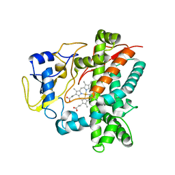
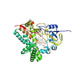
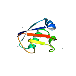
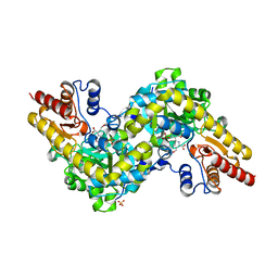
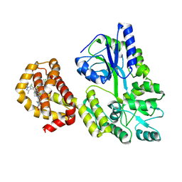
 | | Crystal structure of Arabidopsis ACS7 mutant in complex with PPG | | 分子名称: | (2E,3E)-4-(2-aminoethoxy)-2-[({3-hydroxy-2-methyl-5-[(phosphonooxy)methyl]pyridin-4-yl}methyl)imino]but-3-enoic acid, 1-aminocyclopropane-1-carboxylate synthase 7 | | 著者 | Hao, B, Zhang, Y, Li, X, Rao, Z. | | 登録日 | 2020-11-30 | | 公開日 | 2021-09-29 | | 最終更新日 | 2023-11-29 | | 実験手法 | X-RAY DIFFRACTION (2.94 Å) | | 主引用文献 | Dual activities of ACC synthase: Novel clues regarding the molecular evolution of ACS genes.
Sci Adv, 7, 2021
|
|
7DLW
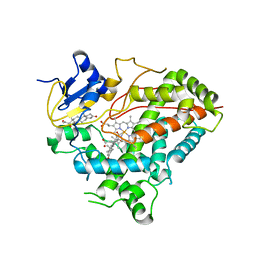
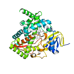
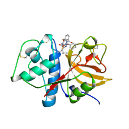
 | | Crystal structure of Arabidopsis ACS7 in complex with PPG | | 分子名称: | (2E,3E)-4-(2-aminoethoxy)-2-[({3-hydroxy-2-methyl-5-[(phosphonooxy)methyl]pyridin-4-yl}methyl)imino]but-3-enoic acid, 1-aminocyclopropane-1-carboxylate synthase 7, SULFATE ION | | 著者 | Hao, B, Zhang, Y, Li, X, Rao, Z. | | 登録日 | 2020-11-30 | | 公開日 | 2021-09-29 | | 最終更新日 | 2023-11-29 | | 実験手法 | X-RAY DIFFRACTION (2.19 Å) | | 主引用文献 | Dual activities of ACC synthase: Novel clues regarding the molecular evolution of ACS genes.
Sci Adv, 7, 2021
|
|
4X1N
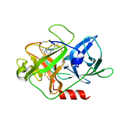 | | The crystal structure of mupain-1-16 in complex with murinised human uPA at pH7.4 | | 分子名称: | Urokinase-type plasminogen activator, mupain-1-16, piperidine-1-carboximidamide | | 著者 | Jiang, L, Zhao, B, Xu, P, Andreasen, P, Huang, M. | | 登録日 | 2014-11-25 | | 公開日 | 2015-03-25 | | 最終更新日 | 2023-11-08 | | 実験手法 | X-RAY DIFFRACTION (1.8 Å) | | 主引用文献 | A cyclic peptidic serine protease inhibitor: increasing affinity by increasing peptide flexibility.
Plos One, 9, 2014
|
|
4X1Q
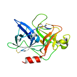 | | The crystal structure of mupain-1 in complex with murinised human uPA at pH7.4 | | 分子名称: | Urokinase-type plasminogen activator, mupain-1 | | 著者 | Jiang, L, Zhao, B, Xu, P, Andreasen, P, Huang, M. | | 登録日 | 2014-11-25 | | 公開日 | 2015-03-25 | | 最終更新日 | 2015-11-04 | | 実験手法 | X-RAY DIFFRACTION (2.28 Å) | | 主引用文献 | A cyclic peptidic serine protease inhibitor: increasing affinity by increasing peptide flexibility.
Plos One, 9, 2014
|
|
4X1S
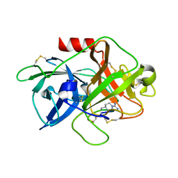 | | The crystal structure of mupain-1-16-D9A in complex with murinised human uPA at pH7.4 | | 分子名称: | Urokinase-type plasminogen activator, mupain-1-16, piperidine-1-carboximidamide | | 著者 | Jiang, L, Zhao, B, Xu, P, Andreasen, P, Huang, M. | | 登録日 | 2014-11-25 | | 公開日 | 2015-10-28 | | 最終更新日 | 2023-11-08 | | 実験手法 | X-RAY DIFFRACTION (1.9 Å) | | 主引用文献 | A cyclic peptidic serine protease inhibitor: increasing affinity by increasing peptide flexibility.
Plos One, 9, 2014
|
|
6NE5
 | |
6O8C
 | | Crystal structure of STING CTT in complex with TBK1 | | 分子名称: | N-(3-{[5-iodo-4-({3-[(thiophen-2-ylcarbonyl)amino]propyl}amino)pyrimidin-2-yl]amino}phenyl)pyrrolidine-1-carboxamide, Serine/threonine-protein kinase TBK1, Stimulator of interferon genes protein | | 著者 | Li, P, Zhao, B, Du, F. | | 登録日 | 2019-03-09 | | 公開日 | 2019-05-22 | | 最終更新日 | 2023-10-11 | | 実験手法 | X-RAY DIFFRACTION (3.17 Å) | | 主引用文献 | A conserved PLPLRT/SD motif of STING mediates the recruitment and activation of TBK1.
Nature, 569, 2019
|
|
6O8B
 | | Crystal structure of STING CTD in complex with TBK1 | | 分子名称: | N-(3-{[5-iodo-4-({3-[(thiophen-2-ylcarbonyl)amino]propyl}amino)pyrimidin-2-yl]amino}phenyl)pyrrolidine-1-carboxamide, Serine/threonine-protein kinase TBK1, Stimulator of interferon genes protein | | 著者 | Li, P, Zhao, B, Du, F. | | 登録日 | 2019-03-09 | | 公開日 | 2019-05-22 | | 最終更新日 | 2023-10-11 | | 実験手法 | X-RAY DIFFRACTION (3.4 Å) | | 主引用文献 | A conserved PLPLRT/SD motif of STING mediates the recruitment and activation of TBK1.
Nature, 569, 2019
|
|
5KH8
 | |
9BCG
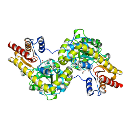
 | | Myeloid cell leukemia-1 (Mcl-1) complexed with compound | | 分子名称: | 7-[(4R,5S,6P)-7-chloro-10-[3-(4-chloro-3,5-dimethylphenoxy)propyl]-4-methyl-1-oxo-6-(1,3,5-trimethyl-1H-pyrazol-4-yl)-3,4-dihydropyrazino[1,2-a]indol-2(1H)-yl]-4,5-dimethoxy-1-methyl-1H-indole-2-carboxylic acid, Maltose/maltodextrin-binding periplasmic protein,Induced myeloid leukemia cell differentiation protein Mcl-1, alpha-D-glucopyranose-(1-4)-alpha-D-glucopyranose | | 著者 | Zhao, B, Fesik, S.W. | | 登録日 | 2024-04-09 | | 公開日 | 2024-08-07 | | 実験手法 | X-RAY DIFFRACTION (1.898 Å) | | 主引用文献 | Discovery of a Myeloid Cell Leukemia 1 (Mcl-1) Inhibitor that Demonstrates Potent in Vivo Activities in Mouse Models of Hematological and Solid Tumors
J.Med.Chem., 2024
|
|
1AYW
 | | CRYSTAL STRUCTURE OF CYSTEINE PROTEASE HUMAN CATHEPSIN K IN COMPLEX WITH A COVALENT BENZYLOXYBENZOYLCARBOHYDRAZIDE INHIBITOR | | 分子名称: | 1-(N-BENZYLOXYCARBONYL-L-LEUCINYL)-5-(3-BENZYLOXY BENZOYL)CARBOHYDRAZIDE, CATHEPSIN K | | 著者 | Zhao, B, Smith, W.W, Janson, C.A, Abdel-Meguid, S.S. | | 登録日 | 1997-11-10 | | 公開日 | 1998-11-25 | | 最終更新日 | 2023-08-02 | | 実験手法 | X-RAY DIFFRACTION (2.4 Å) | | 主引用文献 | Design of potent and selective human cathepsin K inhibitors that span the active site.
Proc.Natl.Acad.Sci.USA, 94, 1997
|
|
1AYV
 | | CRYSTAL STRUCTURE OF CYSTEINE PROTEASE HUMAN CATHEPSIN K IN COMPLEX WITH A COVALENT THIAZOLHYDRAZIDE INHIBITOR | | 分子名称: | CATHEPSIN K, N-[2-[1-(N-BENZYLOXYCARBONYLAMINO)-3-METHYLBUTYL]THIAZOL-4-YLCARBONYL]-N'-(BENZYLOXYCARBONYL-L-LEUCINYL)HYDRAZIDE | | 著者 | Zhao, B, Smith, W.W, Janson, C.A, Abdel-Meguid, S.S. | | 登録日 | 1997-11-10 | | 公開日 | 1998-11-25 | | 最終更新日 | 2023-08-02 | | 実験手法 | X-RAY DIFFRACTION (2.3 Å) | | 主引用文献 | Design of potent and selective human cathepsin K inhibitors that span the active site.
Proc.Natl.Acad.Sci.USA, 94, 1997
|
|
1AU3
 | | CRYSTAL STRUCTURE OF THE CYSTEINE PROTEASE HUMAN CATHEPSIN K IN COMPLEX WITH A COVALENT PYRROLIDINONE INHIBITOR | | 分子名称: | 1-[N[(PHENYLMETHOXY)CARBONYL]-L-LEUCYL-4-[[N/N-[(PHENYLMETHOXY)CARBONYL]-/NL-LEUCYL]AMINO]-3-PYRROLIDINONE/N, CATHEPSIN K | | 著者 | Zhao, B, Smith, W.W, Janson, C.A, Abdel-Meguid, S.S. | | 登録日 | 1997-09-10 | | 公開日 | 1998-10-14 | | 最終更新日 | 2023-08-02 | | 実験手法 | X-RAY DIFFRACTION (2.5 Å) | | 主引用文献 | Conformationally constrained 1,3-diamino ketones: a series of potent inhibitors of the cysteine protease cathepsin K.
J.Med.Chem., 41, 1998
|
|
1AYU
 | | CRYSTAL STRUCTURE OF CYSTEINE PROTEASE HUMAN CATHEPSIN K IN COMPLEX WITH A COVALENT SYMMETRIC BISCARBOHYDRAZIDE INHIBITOR | | 分子名称: | 1,5-BIS(N-BENZYLOXYCARBONYL-L-LEUCINYL)CARBOHYDRAZIDE, CATHEPSIN K | | 著者 | Zhao, B, Smith, W.W, Janson, C.A, Abdel-Meguid, S.S. | | 登録日 | 1997-11-10 | | 公開日 | 1998-11-25 | | 最終更新日 | 2023-08-02 | | 実験手法 | X-RAY DIFFRACTION (2.2 Å) | | 主引用文献 | Design of potent and selective human cathepsin K inhibitors that span the active site.
Proc.Natl.Acad.Sci.USA, 94, 1997
|
|


