5VED
 
 | |
3RQE
 | |
5VEF
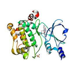
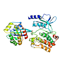
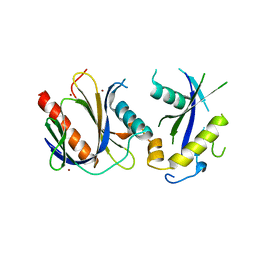
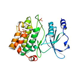
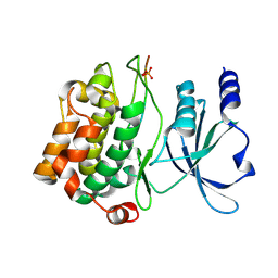
 | | PAK4 kinase domain in complex with fasudil | | 分子名称: | 5-(1,4-DIAZEPAN-1-SULFONYL)ISOQUINOLINE, ACETATE ION, Serine/threonine-protein kinase PAK 4 | | 著者 | Zhang, E.Y, Ha, B.H, Boggon, T.J. | | 登録日 | 2017-04-04 | | 公開日 | 2017-10-18 | | 最終更新日 | 2023-10-04 | | 実験手法 | X-RAY DIFFRACTION (1.752 Å) | | 主引用文献 | PAK4 crystal structures suggest unusual kinase conformational movements.
Biochim. Biophys. Acta, 1866, 2018
|
|
5U4U
 | |
5UPL
 | |
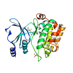
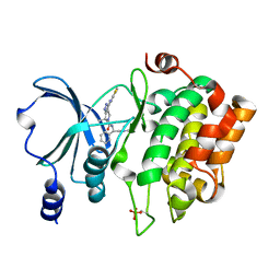
3TH5
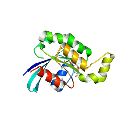 | | Crystal structure of wild-type RAC1 | | 分子名称: | MAGNESIUM ION, PHOSPHOAMINOPHOSPHONIC ACID-GUANYLATE ESTER, Ras-related C3 botulinum toxin substrate 1 | | 著者 | Ha, B.H, Boggon, T.J. | | 登録日 | 2011-08-18 | | 公開日 | 2012-07-18 | | 最終更新日 | 2023-09-13 | | 実験手法 | X-RAY DIFFRACTION (2.3 Å) | | 主引用文献 | Exome sequencing identifies recurrent somatic RAC1 mutations in melanoma.
Nat.Genet., 44, 2012
|
|
4DX9
 | | ICAP1 in complex with integrin beta 1 cytoplasmic tail | | 分子名称: | Integrin beta-1, Integrin beta-1-binding protein 1 | | 著者 | Liu, W, Draheim, K, Zhang, R, Calderwood, D.A, Boggon, T.J. | | 登録日 | 2012-02-27 | | 公開日 | 2013-01-09 | | 最終更新日 | 2020-09-02 | | 実験手法 | X-RAY DIFFRACTION (2.99 Å) | | 主引用文献 | Mechanism for KRIT1 Release of ICAP1-Mediated Suppression of Integrin Activation.
Mol.Cell, 49, 2013
|
|
4DXA
 | | Co-crystal structure of Rap1 in complex with KRIT1 | | 分子名称: | 5'-GUANOSINE-DIPHOSPHATE-MONOTHIOPHOSPHATE, Krev interaction trapped protein 1, MAGNESIUM ION, ... | | 著者 | Li, X, Zhang, R, Boggon, T.J. | | 登録日 | 2012-02-27 | | 公開日 | 2012-05-16 | | 最終更新日 | 2023-09-13 | | 実験手法 | X-RAY DIFFRACTION (1.95 Å) | | 主引用文献 | Structural Basis for Small G Protein Effector Interaction of Ras-related Protein 1 (Rap1) and Adaptor Protein Krev Interaction Trapped 1 (KRIT1).
J.Biol.Chem., 287, 2012
|
|
4DX8
 | | ICAP1 in complex with KRIT1 N-terminus | | 分子名称: | BROMIDE ION, Integrin beta-1-binding protein 1, Krev interaction trapped protein 1 | | 著者 | Liu, W, Draheim, K, Zhang, R, Calderwood, D.A, Boggon, T.J. | | 登録日 | 2012-02-27 | | 公開日 | 2013-01-09 | | 最終更新日 | 2024-02-28 | | 実験手法 | X-RAY DIFFRACTION (2.54 Å) | | 主引用文献 | Mechanism for KRIT1 Release of ICAP1-Mediated Suppression of Integrin Activation.
Mol.Cell, 49, 2013
|
|
4EDL
 | | Crystal structure of beta-parvin CH2 domain | | 分子名称: | 1,2-ETHANEDIOL, Beta-parvin | | 著者 | Stiegler, A.L, Draheim, K.M, Li, X, Chayen, N.E, Calderwood, D.A, Boggon, T.J. | | 登録日 | 2012-03-27 | | 公開日 | 2012-08-08 | | 最終更新日 | 2024-02-28 | | 実験手法 | X-RAY DIFFRACTION (2.1 Å) | | 主引用文献 | Structural basis for paxillin binding and focal adhesion targeting of beta-parvin.
J.Biol.Chem., 287, 2012
|
|
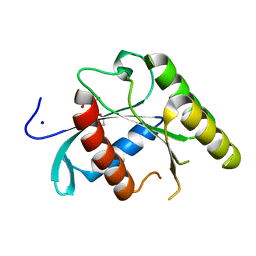
4EIH
 | | Crystal structure of Arg SH2 domain | | 分子名称: | Abelson tyrosine-protein kinase 2, CHLORIDE ION | | 著者 | Liu, W, MacGrath, S.M, Koleske, A.J, Boggon, T.J. | | 登録日 | 2012-04-05 | | 公開日 | 2013-04-10 | | 最終更新日 | 2023-09-13 | | 実験手法 | X-RAY DIFFRACTION (1.2 Å) | | 主引用文献 | Two Amino Acid Residues Confer Different Binding Affinities of Abelson Family Kinase Src Homology 2 Domains for Phosphorylated Cortactin.
J.Biol.Chem., 289, 2014
|
|
4EDN
 | | Crystal structure of beta-parvin CH2 domain in complex with paxillin LD1 motif | | 分子名称: | Beta-parvin, Paxillin, SULFATE ION | | 著者 | Stiegler, A.L, Draheim, K.M, Li, X, Chayen, N.E, Calderwood, D.A, Boggon, T.J. | | 登録日 | 2012-03-27 | | 公開日 | 2012-08-08 | | 最終更新日 | 2013-06-19 | | 実験手法 | X-RAY DIFFRACTION (2.9 Å) | | 主引用文献 | Structural basis for paxillin binding and focal adhesion targeting of beta-parvin.
J.Biol.Chem., 287, 2012
|
|
4EDM
 | | Crystal structure of beta-parvin CH2 domain | | 分子名称: | 1,2-ETHANEDIOL, Beta-parvin | | 著者 | Stiegler, A.L, Draheim, K.M, Li, X, Chayen, N.E, Calderwood, D.A, Boggon, T.J. | | 登録日 | 2012-03-27 | | 公開日 | 2012-08-08 | | 最終更新日 | 2024-02-28 | | 実験手法 | X-RAY DIFFRACTION (2 Å) | | 主引用文献 | Structural basis for paxillin binding and focal adhesion targeting of beta-parvin.
J.Biol.Chem., 287, 2012
|
|
1BWJ
 | | THE 1.8 A STRUCTURE OF MICROGRAVITY GROWN TETRAGONAL HEN EGG WHITE LYSOZYME | | 分子名称: | PROTEIN (LYSOZYME) | | 著者 | Dong, J, Boggon, T.J, Chayen, N.E, Raftery, J, Bi, R.C. | | 登録日 | 1998-09-18 | | 公開日 | 1998-09-30 | | 最終更新日 | 2023-08-09 | | 実験手法 | X-RAY DIFFRACTION (1.8 Å) | | 主引用文献 | Bound-solvent structures for microgravity-, ground control-, gel- and microbatch-grown hen egg-white lysozyme crystals at 1.8 A resolution.
Acta Crystallogr.,Sect.D, 55, 1999
|
|
1BWH
 | | THE 1.8 A STRUCTURE OF GROUND CONTROL GROWN TETRAGONAL HEN EGG WHITE LYSOZYME | | 分子名称: | PROTEIN (LYSOZYME) | | 著者 | Dong, J, Boggon, T.J, Chayen, N.E, Raftery, J, Bi, R.C. | | 登録日 | 1998-09-24 | | 公開日 | 1998-09-30 | | 最終更新日 | 2023-08-09 | | 実験手法 | X-RAY DIFFRACTION (1.8 Å) | | 主引用文献 | Bound-solvent structures for microgravity-, ground control-, gel- and microbatch-grown hen egg-white lysozyme crystals at 1.8 A resolution.
Acta Crystallogr.,Sect.D, 55, 1999
|
|
1BWI
 | | THE 1.8 A STRUCTURE OF MICROBATCH OIL DROP GROWN TETRAGONAL HEN EGG WHITE LYSOZYME | | 分子名称: | PROTEIN (LYSOZYME) | | 著者 | Dong, J, Boggon, T.J, Chayen, N.E, Raftery, J, Bi, R.C. | | 登録日 | 1998-09-24 | | 公開日 | 1998-09-30 | | 最終更新日 | 2023-08-09 | | 実験手法 | X-RAY DIFFRACTION (1.8 Å) | | 主引用文献 | Bound-solvent structures for microgravity-, ground control-, gel- and microbatch-grown hen egg-white lysozyme crystals at 1.8 A resolution.
Acta Crystallogr.,Sect.D, 55, 1999
|
|
1BVX
 | | THE 1.8 A STRUCTURE OF GEL GROWN TETRAGONAL HEN EGG WHITE LYSOZYME | | 分子名称: | PROTEIN (LYSOZYME) | | 著者 | Dong, J, Boggon, T.J, Chayen, N.E, Raftery, J, Bi, R.C, Helliwell, J.R. | | 登録日 | 1998-09-18 | | 公開日 | 1998-09-23 | | 最終更新日 | 2023-08-09 | | 実験手法 | X-RAY DIFFRACTION (1.8 Å) | | 主引用文献 | Bound-solvent structures for microgravity-, ground control-, gel- and microbatch-grown hen egg-white lysozyme crystals at 1.8 A resolution.
Acta Crystallogr.,Sect.D, 55, 1999
|
|
4JDK
 | |
4JIF
 | |
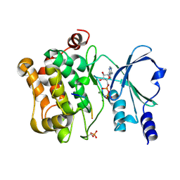
4KS8
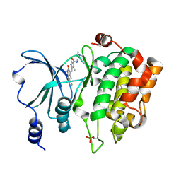 | | PAK6 kinase domain in complex with sunitinib | | 分子名称: | N-[2-(diethylamino)ethyl]-5-[(Z)-(5-fluoro-2-oxo-1,2-dihydro-3H-indol-3-ylidene)methyl]-2,4-dimethyl-1H-pyrrole-3-carbo xamide, Serine/threonine-protein kinase PAK 6 | | 著者 | Gao, J, Boggon, T.J. | | 登録日 | 2013-05-17 | | 公開日 | 2013-09-11 | | 最終更新日 | 2023-09-20 | | 実験手法 | X-RAY DIFFRACTION (1.95 Å) | | 主引用文献 | Substrate and Inhibitor Specificity of the Type II p21-Activated Kinase, PAK6.
Plos One, 8, 2013
|
|
4JDH
 | |
4KS7
 | |
4JDI
 | |
4JDJ
 | |
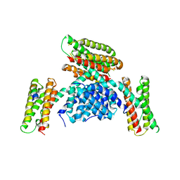
4K94
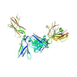 | | Crystal structure of KIT D4D5 fragment in complex with anti-Kit antibody Fab19 | | 分子名称: | 2-acetamido-2-deoxy-beta-D-glucopyranose, Fab19 heavy chain, Fab19 light chain, ... | | 著者 | Resheynyak, A.V, Boggon, T.J, Lax, I, Schlessinger, J. | | 登録日 | 2013-04-19 | | 公開日 | 2013-10-16 | | 最終更新日 | 2020-07-29 | | 実験手法 | X-RAY DIFFRACTION (2.4 Å) | | 主引用文献 | Structural basis for KIT receptor tyrosine kinase inhibition by antibodies targeting the D4 membrane-proximal region.
Proc.Natl.Acad.Sci.USA, 110, 2013
|
|


