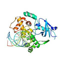
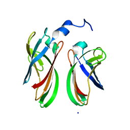
1VRL
 
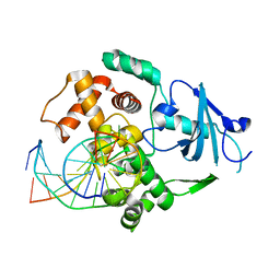
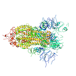
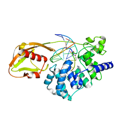 | | MutY adenine glycosylase in complex with DNA and soaked adenine free base | | 分子名称: | 5'-D(*AP*AP*GP*AP*CP*(8OG)P*TP*GP*GP*AP*C)-3', 5'-D(*TP*GP*TP*CP*CP*AP*(HPD)P*GP*TP*CP*T)-3', ADENINE, ... | | 著者 | Fromme, J.C, Banerjee, A, Huang, S.J, Verdine, G.L. | | 登録日 | 2005-03-08 | | 公開日 | 2005-03-22 | | 最終更新日 | 2023-12-27 | | 実験手法 | X-RAY DIFFRACTION (2.5 Å) | | 主引用文献 | Structural basis for removal of adenine mispaired with 8-oxoguanine by MutY adenine DNA glycosylase
Nature, 427, 2004
|
|
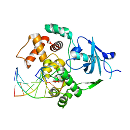
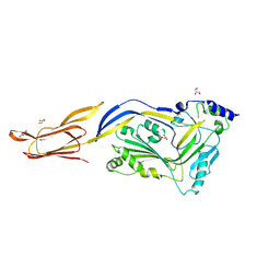
6BMN
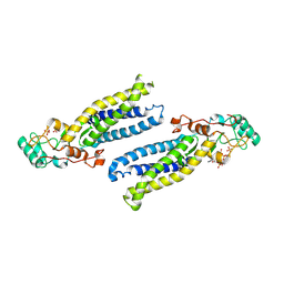 | | Structure of human DHHC20 palmitoyltransferase, space group P63 | | 分子名称: | 3'-PHOSPHATE-ADENOSINE-5'-DIPHOSPHATE, PHOSPHATE ION, ZINC ION, ... | | 著者 | Rana, M.S, Lee, C.-J, Banerjee, A. | | 登録日 | 2017-11-15 | | 公開日 | 2018-01-24 | | 最終更新日 | 2018-03-28 | | 実験手法 | X-RAY DIFFRACTION (2.25 Å) | | 主引用文献 | Fatty acyl recognition and transfer by an integral membraneS-acyltransferase.
Science, 359, 2018
|
|
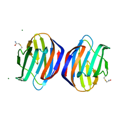
6BML
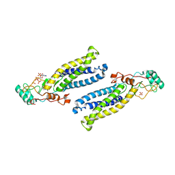 | | Structure of human DHHC20 palmitoyltransferase, irreversibly inhibited by 2-bromopalmitate | | 分子名称: | 3'-PHOSPHATE-ADENOSINE-5'-DIPHOSPHATE, PALMITIC ACID, PHOSPHATE ION, ... | | 著者 | Rana, M.S, Lee, C.-J, Banerjee, A. | | 登録日 | 2017-11-15 | | 公開日 | 2018-01-24 | | 最終更新日 | 2018-03-28 | | 実験手法 | X-RAY DIFFRACTION (2.95 Å) | | 主引用文献 | Fatty acyl recognition and transfer by an integral membraneS-acyltransferase.
Science, 359, 2018
|
|
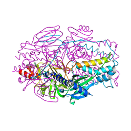
6BMM
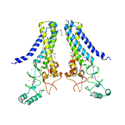 | | Structure of human DHHC20 palmitoyltransferase, space group P21 | | 分子名称: | (2S)-2,3-dihydroxypropyl (9Z)-octadec-9-enoate, (2S,5S)-hexane-2,5-diol, PHOSPHATE ION, ... | | 著者 | Rana, M.S, Lee, C.-J, Banerjee, A. | | 登録日 | 2017-11-15 | | 公開日 | 2018-01-24 | | 最終更新日 | 2018-03-28 | | 実験手法 | X-RAY DIFFRACTION (2.35 Å) | | 主引用文献 | Fatty acyl recognition and transfer by an integral membraneS-acyltransferase.
Science, 359, 2018
|
|
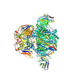
8P8M
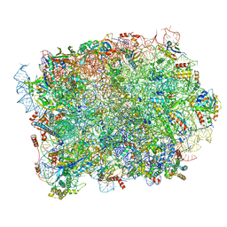 | | Yeast 60S ribosomal subunit, RPL39 deletion | | 分子名称: | 25S rRNA, 5.8S rRNA, 5S rRNA, ... | | 著者 | Rabl, J, Banerjee, A, Boehringer, D, Zavolan, M. | | 登録日 | 2023-06-01 | | 公開日 | 2024-06-12 | | 実験手法 | ELECTRON MICROSCOPY (2.66 Å) | | 主引用文献 | Yeast 60S ribosomal subunit, RPL39 deletion
To Be Published
|
|
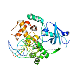
2NOH
 | | Structure of catalytically inactive Q315A human 8-oxoguanine glycosylase complexed to 8-oxoguanine DNA | | 分子名称: | 5'-D(*GP*CP*GP*TP*CP*CP*AP*(G42)P*GP*TP*CP*TP*AP*CP*C)-3', 5'-D(*GP*GP*TP*AP*GP*AP*CP*CP*TP*GP*GP*AP*CP*GP*C)-3', CALCIUM ION, ... | | 著者 | Radom, C.T, Banerjee, A, Verdine, G.L. | | 登録日 | 2006-10-25 | | 公開日 | 2006-11-21 | | 最終更新日 | 2023-12-27 | | 実験手法 | X-RAY DIFFRACTION (2.01 Å) | | 主引用文献 | Structural characterization of human 8-oxoguanine DNA glycosylase variants bearing active site mutations.
J.Biol.Chem., 282, 2007
|
|
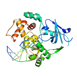
2NOZ
 | | Structure of Q315F human 8-oxoguanine glycosylase distal crosslink to 8-oxoguanine DNA | | 分子名称: | 5'-D(*G*CP*GP*TP*CP*CP*AP*(G42)P*GP*TP*CP*TP*AP*CP*C)-3', 5'-D(*GP*G*TP*AP*GP*AP*CP*CP*TP*GP*GP*AP*CP*GP*C)-3', CALCIUM ION, ... | | 著者 | Radom, C.T, Banerjee, A, Verdine, G.L. | | 登録日 | 2006-10-26 | | 公開日 | 2006-11-21 | | 最終更新日 | 2023-12-27 | | 実験手法 | X-RAY DIFFRACTION (2.43 Å) | | 主引用文献 | Structural characterization of human 8-oxoguanine DNA glycosylase variants bearing active site mutations.
J.Biol.Chem., 282, 2007
|
|
2NOF
 | | Structure of Q315F human 8-oxoguanine glycosylase proximal crosslink to 8-oxoguanine DNA | | 分子名称: | 5'-D(*GP*CP*GP*TP*C*CP*AP*(G42)P*GP*TP*CP*TP*AP*CP*C)-3', 5'-D(*GP*GP*TP*AP*GP*AP*CP*CP*TP*GP*GP*AP*CP*GP*C)-3', CALCIUM ION, ... | | 著者 | Radom, C.T, Banerjee, A, Verdine, G.L. | | 登録日 | 2006-10-25 | | 公開日 | 2006-11-21 | | 最終更新日 | 2023-12-27 | | 実験手法 | X-RAY DIFFRACTION (2.35 Å) | | 主引用文献 | Structural characterization of human 8-oxoguanine DNA glycosylase variants bearing active site mutations.
J.Biol.Chem., 282, 2007
|
|
2NOB
 | | Structure of catalytically inactive H270A human 8-oxoguanine glycosylase crosslinked to 8-oxoguanine DNA | | 分子名称: | 5'-D(*G*CP*GP*TP*CP*CP*AP*(G42)P*GP*TP*CP*TP*AP*CP*C)-3', 5'-D(*T*GP*GP*TP*AP*GP*AP*CP*CP*TP*GP*GP*AP*CP*GP*C)-3', CALCIUM ION, ... | | 著者 | Radom, C.T, Banerjee, A, Verdine, G.L. | | 登録日 | 2006-10-25 | | 公開日 | 2006-11-21 | | 最終更新日 | 2023-12-27 | | 実験手法 | X-RAY DIFFRACTION (2.1 Å) | | 主引用文献 | Structural characterization of human 8-oxoguanine DNA glycosylase variants bearing active site mutations.
J.Biol.Chem., 282, 2007
|
|
2NOI
 | | Structure of G42A human 8-oxoguanine glycosylase crosslinked to undamaged G-containing DNA | | 分子名称: | 5'-D(*GP*CP*GP*TP*C*CP*AP*GP*GP*TP*CP*TP*AP*CP*C)-3', 5'-D(*GP*GP*TP*AP*GP*AP*CP*CP*TP*GP*GP*AP*CP*GP*C)-3', CALCIUM ION, ... | | 著者 | Radom, C.T, Banerjee, A, Verdine, G.L. | | 登録日 | 2006-10-25 | | 公開日 | 2006-11-21 | | 最終更新日 | 2023-12-27 | | 実験手法 | X-RAY DIFFRACTION (2.35 Å) | | 主引用文献 | Structural characterization of human 8-oxoguanine DNA glycosylase variants bearing active site mutations.
J.Biol.Chem., 282, 2007
|
|
2NOE
 | | Structure of catalytically inactive G42A human 8-oxoguanine glycosylase complexed to 8-oxoguanine DNA | | 分子名称: | 5'-D(*G*CP*GP*TP*CP*CP*AP*(G42)P*GP*TP*CP*TP*AP*CP*C)-3', 5'-D(*G*GP*TP*AP*GP*AP*CP*CP*TP*GP*GP*AP*CP*GP*C)-3', CALCIUM ION, ... | | 著者 | Radom, C.T, Banerjee, A, Verdine, G.L. | | 登録日 | 2006-10-25 | | 公開日 | 2006-11-21 | | 最終更新日 | 2023-12-27 | | 実験手法 | X-RAY DIFFRACTION (2.2 Å) | | 主引用文献 | Structural characterization of human 8-oxoguanine DNA glycosylase variants bearing active site mutations.
J.Biol.Chem., 282, 2007
|
|
7UR4
 | |
3M4D
 | |
3M3R
 | |
3M2L
 | |
3M4E
 | |
4ZBH
 | |
5AHW
 | | Crystal structure of universal stress protein MSMEG_3811 in complex with cAMP | | 分子名称: | (20S)-2,5,8,11,14,17-HEXAMETHYL-3,6,9,12,15,18-HEXAOXAHENICOSANE-1,20-DIOL, ADENOSINE-3',5'-CYCLIC-MONOPHOSPHATE, CHLORIDE ION, ... | | 著者 | Adolph, R.S, Kleinboelting, S, Weyand, M, Steegborn, C. | | 登録日 | 2015-02-10 | | 公開日 | 2015-04-01 | | 最終更新日 | 2024-01-10 | | 実験手法 | X-RAY DIFFRACTION (2.15 Å) | | 主引用文献 | A Universal Stress Protein (Usp) in Mycobacteria Binds Camp
J.Biol.Chem., 290, 2015
|
|
4P94
 | | The crystal structure of the soluble domain of Sulfolobus acidocaldarius FlaF (residues 35-164) | | 分子名称: | Conserved flagellar protein F, SODIUM ION | | 著者 | Tsai, C.-L, Arvai, A.S, Ishida, J.P, Tainer, J.A. | | 登録日 | 2014-04-02 | | 公開日 | 2015-04-22 | | 最終更新日 | 2023-09-27 | | 実験手法 | X-RAY DIFFRACTION (1.651 Å) | | 主引用文献 | FlaF Is a beta-Sandwich Protein that Anchors the Archaellum in the Archaeal Cell Envelope by Binding the S-Layer Protein.
Structure, 23, 2015
|
|
6JMP
 | |
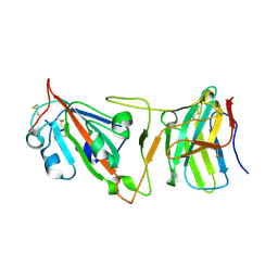
6E20
 | | Crystal structure of the Dario rerio galectin-1-L2 | | 分子名称: | Galectin, MAGNESIUM ION, beta-D-galactopyranose-(1-4)-2-acetamido-2-deoxy-alpha-D-glucopyranose | | 著者 | Ghosh, A, Bianchet, M.A. | | 登録日 | 2018-07-10 | | 公開日 | 2019-03-20 | | 最終更新日 | 2023-10-11 | | 実験手法 | X-RAY DIFFRACTION (2 Å) | | 主引用文献 | Structure of the zebrafish galectin-1-L2 and model of its interaction with the infectious hematopoietic necrosis virus (IHNV) envelope glycoprotein.
Glycobiology, 29, 2019
|
|
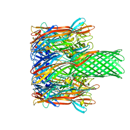
8E15
 | | A computationally stabilized hMPV F protein | | 分子名称: | 2-acetamido-2-deoxy-beta-D-glucopyranose, F1 protein with Fibritin peptide, F2 protein, ... | | 著者 | Huang, J, Gonzalez, K, Mousa, J, Strauch, E. | | 登録日 | 2022-08-09 | | 公開日 | 2023-04-12 | | 最終更新日 | 2024-07-24 | | 実験手法 | X-RAY DIFFRACTION (2.41 Å) | | 主引用文献 | A general computational design strategy for stabilizing viral class I fusion proteins.
Nat Commun, 15, 2024
|
|
8FEZ
 | |
8DI5
 | | Cryo-EM structure of SARS-CoV-2 Beta (B.1.351) spike protein in complex with VH domain F6 (focused refinement of RBD and VH F6) | | 分子名称: | 2-acetamido-2-deoxy-beta-D-glucopyranose, Spike glycoprotein, VH F6 | | 著者 | Zhu, X, Saville, J.W, Mannar, D, Berezuk, A.M, Subramaniam, S. | | 登録日 | 2022-06-28 | | 公開日 | 2022-08-24 | | 実験手法 | ELECTRON MICROSCOPY (3.04 Å) | | 主引用文献 | Potent and broad neutralization of SARS-CoV-2 variants of concern (VOCs) including omicron sub-lineages BA.1 and BA.2 by biparatopic human VH domains.
Iscience, 25, 2022
|
|
5W6L
 | | Crystal Structure of RRSP, a MARTX Toxin Effector Domain from Vibrio vulnificus CMCP6 | | 分子名称: | CHLORIDE ION, GLYCEROL, RTX repeat-containing cytotoxin, ... | | 著者 | Minasov, G, Wawrzak, Z, Biancucci, M, Shuvalova, L, Dubrovska, I, Satchell, K.J, Anderson, W.F, Center for Structural Genomics of Infectious Diseases (CSGID) | | 登録日 | 2017-06-16 | | 公開日 | 2018-06-27 | | 最終更新日 | 2018-10-10 | | 実験手法 | X-RAY DIFFRACTION (3.45 Å) | | 主引用文献 | The bacterial Ras/Rap1 site-specific endopeptidase RRSP cleaves Ras through an atypical mechanism to disrupt Ras-ERK signaling.
Sci Signal, 11, 2018
|
|


