7MDW
 
 | |
7ME7
 | |
7N9T
 | |
6UP7
 | | neurotensin receptor and arrestin2 complex | | 分子名称: | ARG-ARG-PRO-TYR-ILE-LEU, Beta-arrestin-1, Neurotensin receptor type 1, ... | | 著者 | Qu, Q.H, Huang, W, Masureel, M, Janetzko, J, Kobilka, B.K, Skiniotis, G. | | 登録日 | 2019-10-16 | | 公開日 | 2020-02-26 | | 最終更新日 | 2020-06-17 | | 実験手法 | ELECTRON MICROSCOPY (4.2 Å) | | 主引用文献 | Structure of the neurotensin receptor 1 in complex with beta-arrestin 1.
Nature, 579, 2020
|
|
1FC5
 | |
6NTS
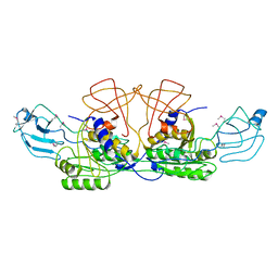
 | | Protein Phosphatase 2A (Aalpha-B56alpha-Calpha) holoenzyme in complex with a Small Molecule Activator of PP2A (SMAP) | | 分子名称: | MANGANESE (II) ION, N-[(1R,2R,3S)-2-hydroxy-3-(10H-phenoxazin-10-yl)cyclohexyl]-4-(trifluoromethoxy)benzene-1-sulfonamide, Serine/threonine-protein phosphatase 2A 56 kDa regulatory subunit alpha isoform, ... | | 著者 | Huang, W, Taylor, D. | | 登録日 | 2019-01-30 | | 公開日 | 2020-05-06 | | 最終更新日 | 2023-11-15 | | 実験手法 | ELECTRON MICROSCOPY (3.63 Å) | | 主引用文献 | Selective PP2A Enhancement through Biased Heterotrimer Stabilization.
Cell, 181, 2020
|
|
7SBF
 | | PZM21 bound Mu Opioid Receptor-Gi Protein Complex | | 分子名称: | Guanine nucleotide-binding protein G(I)/G(S)/G(O) subunit gamma-2, Guanine nucleotide-binding protein G(I)/G(S)/G(T) subunit beta-1, Guanine nucleotide-binding protein G(i) subunit alpha-1, ... | | 著者 | Huang, W, Qu, Q, Wang, H, Skiniotis, G, Kobilka, B. | | 登録日 | 2021-09-24 | | 公開日 | 2022-04-20 | | 最終更新日 | 2022-07-06 | | 実験手法 | ELECTRON MICROSCOPY (2.9 Å) | | 主引用文献 | Structure-Based Evolution of G Protein-Biased mu-Opioid Receptor Agonists.
Angew.Chem.Int.Ed.Engl., 61, 2022
|
|
6JM5
 | | Crystal structure of TBC1D23 C terminal domain | | 分子名称: | SODIUM ION, TBC1 domain family member 23 | | 著者 | Sun, Q, Huang, W. | | 登録日 | 2019-03-07 | | 公開日 | 2019-10-16 | | 最終更新日 | 2024-03-27 | | 実験手法 | X-RAY DIFFRACTION (1.6 Å) | | 主引用文献 | Structural and functional studies of TBC1D23 C-terminal domain provide a link between endosomal trafficking and PCH.
Proc.Natl.Acad.Sci.USA, 116, 2019
|
|
8INV
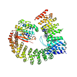
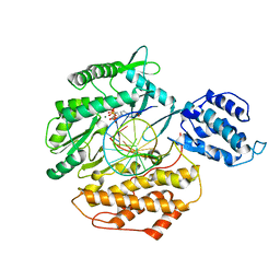
 | | Crystal structure of UGT74AN3-UDP-BUF | | 分子名称: | 2-AMINO-2-HYDROXYMETHYL-PROPANE-1,3-DIOL, Glycosyltransferase, URIDINE-5'-DIPHOSPHATE, ... | | 著者 | Long, F, Huang, W. | | 登録日 | 2023-03-10 | | 公開日 | 2024-01-24 | | 実験手法 | X-RAY DIFFRACTION (1.85 Å) | | 主引用文献 | Substrate Promiscuity, Crystal Structure, and Application of a Plant UDP-Glycosyltransferase UGT74AN3
Acs Catalysis, 14, 2024
|
|
8INA
 | | Crystal structure of UGT74AN3-UDP | | 分子名称: | GLYCEROL, Glycosyltransferase, URIDINE-5'-DIPHOSPHATE | | 著者 | Long, F, Huang, W. | | 登録日 | 2023-03-09 | | 公開日 | 2024-01-24 | | 実験手法 | X-RAY DIFFRACTION (1.86 Å) | | 主引用文献 | Substrate Promiscuity, Crystal Structure, and Application of a Plant UDP-Glycosyltransferase UGT74AN3
Acs Catalysis, 14, 2024
|
|
8INO
 | | Crystal structure of UGT74AN3 in complex UDP and PER | | 分子名称: | 2-AMINO-2-HYDROXYMETHYL-PROPANE-1,3-DIOL, 3-[(3S,5S,8S,9S,10R,13R,14S,17R)-10,13-dimethyl-3,5,14-tris(oxidanyl)-2,3,4,6,7,8,9,11,12,15,16,17-dodecahydro-1H-cyclopenta[a]phenanthren-17-yl]-2H-furan-5-one, Glycosyltransferase, ... | | 著者 | Long, F, Huang, W. | | 登録日 | 2023-03-10 | | 公開日 | 2024-01-24 | | 実験手法 | X-RAY DIFFRACTION (2.3 Å) | | 主引用文献 | Substrate Promiscuity, Crystal Structure, and Application of a Plant UDP-Glycosyltransferase UGT74AN3
Acs Catalysis, 14, 2024
|
|
8IR4
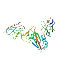
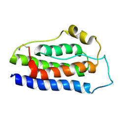
 | | Crystal structure of the SLF1 BRCT domain in complex with a Rad18 peptide containing pS442 | | 分子名称: | CHLORIDE ION, ISOPROPYL ALCOHOL, MAGNESIUM ION, ... | | 著者 | Xiang, S, Huang, W, Qiu, F. | | 登録日 | 2023-03-17 | | 公開日 | 2023-11-15 | | 実験手法 | X-RAY DIFFRACTION (1.62 Å) | | 主引用文献 | Structural insights into Rad18 targeting by the SLF1 BRCT domains.
J.Biol.Chem., 299, 2023
|
|
8IR2
 | | Crystal structure of the SLF1 BRCT domain in complex with a Rad18 peptide containing pS442 and pS444 | | 分子名称: | CHLORIDE ION, ISOPROPYL ALCOHOL, MAGNESIUM ION, ... | | 著者 | Xiang, S, Huang, W, Qiu, F. | | 登録日 | 2023-03-17 | | 公開日 | 2023-11-15 | | 実験手法 | X-RAY DIFFRACTION (1.75 Å) | | 主引用文献 | Structural insights into Rad18 targeting by the SLF1 BRCT domains.
J.Biol.Chem., 299, 2023
|
|
7UO0
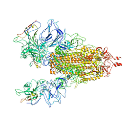
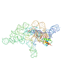 | | E.coli RNaseP Holoenzyme with Mg2+ | | 分子名称: | CALCIUM ION, Precursor tRNA substrate G(-1) G(-2), RNase P RNA, ... | | 著者 | Huang, W, Taylor, D.J. | | 登録日 | 2022-04-12 | | 公開日 | 2022-09-28 | | 最終更新日 | 2024-06-12 | | 実験手法 | ELECTRON MICROSCOPY (3.4 Å) | | 主引用文献 | Structural and mechanistic basis for recognition of alternative tRNA precursor substrates by bacterial ribonuclease P.
Nat Commun, 13, 2022
|
|
7UO1
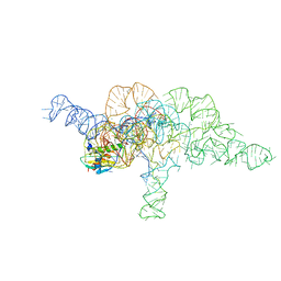 | | E.coli RNaseP Holoenzyme with Mg2+ | | 分子名称: | CALCIUM ION, E.coli RNase P RNA, Precursor tRNA substrate U(-1) and A(-2), ... | | 著者 | Huang, W, Taylor, D.J. | | 登録日 | 2022-04-12 | | 公開日 | 2022-09-28 | | 最終更新日 | 2024-06-12 | | 実験手法 | ELECTRON MICROSCOPY (3.2 Å) | | 主引用文献 | Structural and mechanistic basis for recognition of alternative tRNA precursor substrates by bacterial ribonuclease P.
Nat Commun, 13, 2022
|
|
7UO2
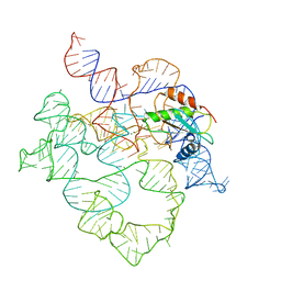 | | E.coli RNaseP Holoenzyme with Mg2+ | | 分子名称: | MAGNESIUM ION, RNase P RNA, Ribonuclease P protein component | | 著者 | Huang, W, Taylor, D.J. | | 登録日 | 2022-04-12 | | 公開日 | 2022-09-28 | | 最終更新日 | 2024-06-12 | | 実験手法 | ELECTRON MICROSCOPY (3.1 Å) | | 主引用文献 | Structural and mechanistic basis for recognition of alternative tRNA precursor substrates by bacterial ribonuclease P.
Nat Commun, 13, 2022
|
|
7UO5
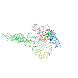 | | E.coli RNaseP Holoenzyme with Mg2+ | | 分子名称: | CALCIUM ION, RNase P RNA, Ribonuclease P protein component | | 著者 | Huang, W, Taylor, D.J. | | 登録日 | 2022-04-12 | | 公開日 | 2022-09-28 | | 最終更新日 | 2024-06-12 | | 実験手法 | ELECTRON MICROSCOPY (3.1 Å) | | 主引用文献 | Structural and mechanistic basis for recognition of alternative tRNA precursor substrates by bacterial ribonuclease P.
Nat Commun, 13, 2022
|
|
6E53
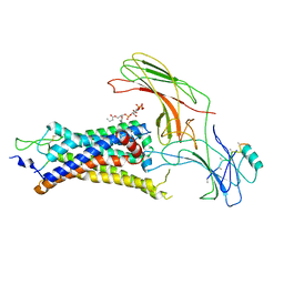
 | | Structure of TERT in complex with a novel telomerase inhibitor | | 分子名称: | MAGNESIUM ION, RNA/DNA hairpin, Telomerase reverse transcriptase, ... | | 著者 | Hernandez-Sanchez, W, Huang, W, Plucinsky, B, Garcia-Vazquez, N, Berdis, A.J, Skordalakes, E, Taylor, D.J. | | 登録日 | 2018-07-19 | | 公開日 | 2019-03-27 | | 最終更新日 | 2023-10-11 | | 実験手法 | X-RAY DIFFRACTION (2.8 Å) | | 主引用文献 | A non-natural nucleotide uses a specific pocket to selectively inhibit telomerase activity.
Plos Biol., 17, 2019
|
|
1AFR
 | |
2B34
 | | Structure of MAR1 Ribonuclease from Caenorhabditis elegans | | 分子名称: | MAR1 Ribonuclease | | 著者 | Schormann, N, Karpova, E, Li, S, Symersky, J, Zhang, Y, Lu, S, Zhou, Q, Lin, G, Cao, Z, Luo, M, Qiu, S, Luan, C.-H, Luo, D, Huang, W, Shang, Q, McKinstry, A, An, J, Tsao, J, Carson, M, Stinnett, M, Chen, Y, Johnson, D, Gary, R, Arabshahi, A, Bunzel, R, Bray, T, DeLucas, L, Southeast Collaboratory for Structural Genomics (SECSG) | | 登録日 | 2005-09-19 | | 公開日 | 2005-09-27 | | 最終更新日 | 2023-08-23 | | 実験手法 | X-RAY DIFFRACTION (2.141 Å) | | 主引用文献 | Structure of MAR1 Ribonuclease from Caenorhabditis elegans
To be Published
|
|
1EAP
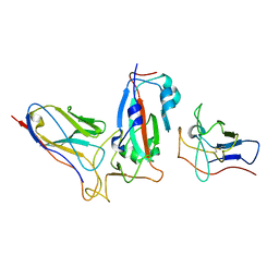
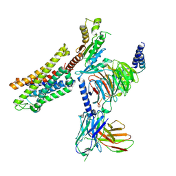
 | | CRYSTAL STRUCTURE OF A CATALYTIC ANTIBODY WITH A SERINE PROTEASE ACTIVE SITE | | 分子名称: | IGG2B-KAPPA 17E8 FAB (HEAVY CHAIN), IGG2B-KAPPA 17E8 FAB (LIGHT CHAIN), PHENYL[1-(N-SUCCINYLAMINO)PENTYL]PHOSPHONATE | | 著者 | Zhou, G.W, Guo, J, Huang, W, Scanlan, T.S, Fletterick, R.J. | | 登録日 | 1994-08-10 | | 公開日 | 1994-12-20 | | 最終更新日 | 2024-06-05 | | 実験手法 | X-RAY DIFFRACTION (2.4 Å) | | 主引用文献 | Crystal structure of a catalytic antibody with a serine protease active site.
Science, 265, 1994
|
|
8K6Z
 | | NMR structure of human leptin | | 分子名称: | Leptin | | 著者 | Fan, X, Qin, R, Yuan, W, Fan, J, Huang, W, Lin, Z. | | 登録日 | 2023-07-26 | | 公開日 | 2024-02-07 | | 最終更新日 | 2024-05-15 | | 実験手法 | SOLUTION NMR | | 主引用文献 | The solution structure of human leptin reveals a conformational plasticity important for receptor recognition.
Structure, 32, 2024
|
|
1XDO
 | |
1XDP
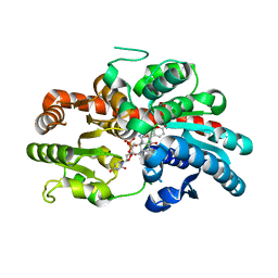
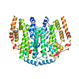
 | | Crystal Structure of the E.coli Polyphosphate Kinase in complex with AMPPNP | | 分子名称: | ADENOSINE-5'-TRIPHOSPHATE, MAGNESIUM ION, Polyphosphate kinase | | 著者 | Zhu, Y, Huang, W, Lee, S.S, Xu, W. | | 登録日 | 2004-09-07 | | 公開日 | 2005-06-21 | | 最終更新日 | 2024-02-14 | | 実験手法 | X-RAY DIFFRACTION (2.5 Å) | | 主引用文献 | Crystal structure of a polyphosphate kinase and its implications for polyphosphate synthesis
Embo Rep., 6, 2005
|
|
1ONR
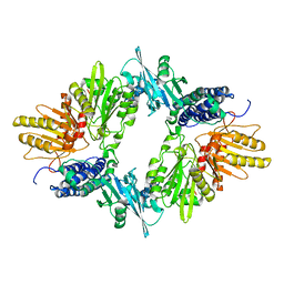
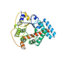 | | STRUCTURE OF TRANSALDOLASE B | | 分子名称: | TRANSALDOLASE B | | 著者 | Jia, J, Huang, W, Lindqvist, Y, Schneider, G. | | 登録日 | 1996-08-13 | | 公開日 | 1997-03-12 | | 最終更新日 | 2024-02-14 | | 実験手法 | X-RAY DIFFRACTION (1.87 Å) | | 主引用文献 | Crystal structure of transaldolase B from Escherichia coli suggests a circular permutation of the alpha/beta barrel within the class I aldolase family.
Structure, 4, 1996
|
|


