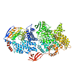
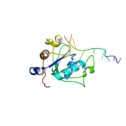
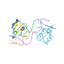
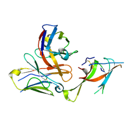
8H3T
 
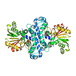 | | The crystal structure of AlpH | | 分子名称: | AlpH, GLYCEROL | | 著者 | Zhao, Y, Li, M, Jiang, M, Pan, L.F. | | 登録日 | 2022-10-09 | | 公開日 | 2023-09-13 | | 最終更新日 | 2024-02-14 | | 実験手法 | X-RAY DIFFRACTION (1.866 Å) | | 主引用文献 | O-methyltransferase-like enzyme catalyzed diazo installation in polyketide biosynthesis.
Nat Commun, 14, 2023
|
|
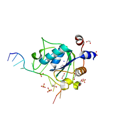
6LCV
 | |
6LCU
 | |
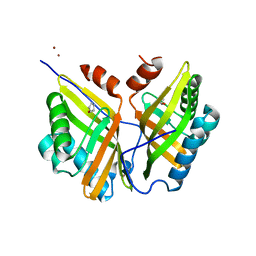
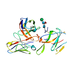
4OBA
 | | Co-crystal structure of MDM2 with Inhibitor Compound 4 | | 分子名称: | E3 ubiquitin-protein ligase Mdm2, [(2R,5R,6R)-4-[(1S)-2-(tert-butylsulfonyl)-1-cyclopropylethyl]-6-(3-chlorophenyl)-5-(4-chlorophenyl)-3-oxomorpholin-2-yl]acetic acid | | 著者 | Shaffer, P.L, Huang, X, Yakowec, P, Long, A.M. | | 登録日 | 2014-01-07 | | 公開日 | 2014-03-19 | | 最終更新日 | 2023-09-20 | | 実験手法 | X-RAY DIFFRACTION (1.602 Å) | | 主引用文献 | Selective and Potent Morpholinone Inhibitors of the MDM2-p53 Protein-Protein Interaction.
J.Med.Chem., 57, 2014
|
|
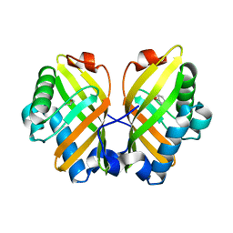
4OCC
 | | co-crystal structure of MDM2(17-111) in complex with compound 48 | | 分子名称: | E3 ubiquitin-protein ligase Mdm2, [(2R,5R,6R)-4-[(2S)-1-(tert-butylsulfonyl)butan-2-yl]-6-(3-chlorophenyl)-5-(4-chlorophenyl)-3-oxomorpholin-2-yl]acetic acid | | 著者 | Huang, X. | | 登録日 | 2014-01-08 | | 公開日 | 2014-04-02 | | 最終更新日 | 2024-02-28 | | 実験手法 | X-RAY DIFFRACTION (1.8 Å) | | 主引用文献 | Novel Inhibitors of the MDM2-p53 Interaction Featuring Hydrogen Bond Acceptors as Carboxylic Acid Isosteres.
J.Med.Chem., 57, 2014
|
|
4ODF
 | | Co-Crystal Structure of MDM2 with Inhibitor Compound 47 | | 分子名称: | 6-{[(2S,5R,6R)-4-[(1S)-2-(tert-butylsulfonyl)-1-cyclopropylethyl]-6-(3-chlorophenyl)-5-(4-chlorophenyl)-2-methyl-3-oxomorpholin-2-yl]methyl}pyridine-3-carboxylic acid, E3 ubiquitin-protein ligase Mdm2 | | 著者 | Shaffer, P.L, Huang, X, Yakowec, P, Long, A.M. | | 登録日 | 2014-01-10 | | 公開日 | 2014-04-02 | | 最終更新日 | 2023-09-20 | | 実験手法 | X-RAY DIFFRACTION (2.2006 Å) | | 主引用文献 | Novel Inhibitors of the MDM2-p53 Interaction Featuring Hydrogen Bond Acceptors as Carboxylic Acid Isosteres.
J.Med.Chem., 57, 2014
|
|
4QO4
 | |
8VXC
 | |
8VX9
 | |
6WE8
 | | YTH domain of human YTHDC1 | | 分子名称: | 1,2-ETHANEDIOL, GLYCEROL, SULFATE ION, ... | | 著者 | Horton, J.R, Cheng, X. | | 登録日 | 2020-04-01 | | 公開日 | 2020-07-15 | | 最終更新日 | 2023-10-18 | | 実験手法 | X-RAY DIFFRACTION (1.18 Å) | | 主引用文献 | Biochemical and structural basis for YTH domain of human YTHDC1 binding to methylated adenine in DNA.
Nucleic Acids Res., 48, 2020
|
|
7ETK
 | |
7ETL
 | | The crystal structure of FtmOx1-Y68F | | 分子名称: | 1,2-ETHANEDIOL, 2-OXOGLUTARIC ACID, CHLORIDE ION, ... | | 著者 | Zhou, J.H, Wu, L. | | 登録日 | 2021-05-13 | | 公開日 | 2021-12-01 | | 最終更新日 | 2023-11-29 | | 実験手法 | X-RAY DIFFRACTION (1.992128 Å) | | 主引用文献 | Structural Insight into the Catalytic Mechanism of the Endoperoxide Synthase FtmOx1.
Angew.Chem.Int.Ed.Engl., 61, 2022
|
|
6WE9
 | |
4QOC
 | |
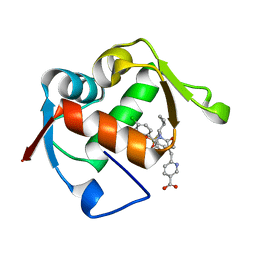
5YAO
 | | The complex structure of SZ529 and expoxid | | 分子名称: | (1R,5S)-6-oxabicyclo[3.1.0]hexane, Limonene-1,2-epoxide hydrolase, SODIUM ION | | 著者 | Lian, W, Sun, Z.T, Zhou, J.H, Reetz, M.T. | | 登録日 | 2017-09-01 | | 公開日 | 2018-06-27 | | 最終更新日 | 2023-11-22 | | 実験手法 | X-RAY DIFFRACTION (2.611 Å) | | 主引用文献 | Structural and Computational Insight into the Catalytic Mechanism of Limonene Epoxide Hydrolase Mutants in Stereoselective Transformations
J. Am. Chem. Soc., 140, 2018
|
|
7M10
 | |
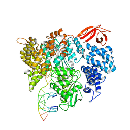
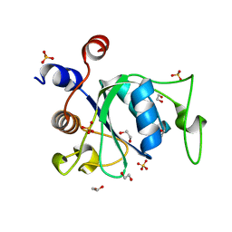
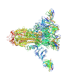
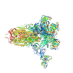
5H3D
 | | Helical structure of membrane tubules decorated by ACAP1 (BARPH doamin) protein by cryo-electron microscopy and MD simulation | | 分子名称: | Arf-GAP with coiled-coil, ANK repeat and PH domain-containing protein 1 | | 著者 | Chan, C, Pang, X.Y, Zhang, Y, Sun, F, Fan, J. | | 登録日 | 2016-10-22 | | 公開日 | 2019-01-16 | | 最終更新日 | 2024-03-20 | | 実験手法 | ELECTRON MICROSCOPY (14 Å) | | 主引用文献 | ACAP1 assembles into an unusual protein lattice for membrane deformation through multiple stages.
Plos Comput.Biol., 15, 2019
|
|
8TLL
 | | CDCA7 (Mouse) Binds Non-B-form 26-mer DNA oligo | | 分子名称: | 1,2-ETHANEDIOL, Cell division cycle-associated protein 7, DNA (26-MER), ... | | 著者 | Horton, J.R, Ren, R, Cheng, X. | | 登録日 | 2023-07-26 | | 公開日 | 2024-08-21 | | 最終更新日 | 2024-10-16 | | 実験手法 | X-RAY DIFFRACTION (2.58 Å) | | 主引用文献 | The ICF syndrome protein CDCA7 harbors a unique DNA binding domain that recognizes a CpG dyad in the context of a non-B DNA.
Sci Adv, 10, 2024
|
|
6WEA
 | |
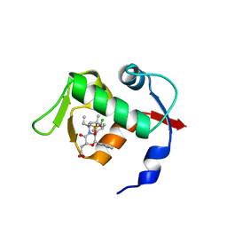
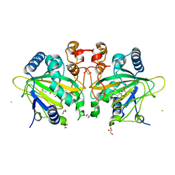
5YNG
 | | Crystal structure of SZ348 in complex with cyclopentene oxide | | 分子名称: | (1R,5S)-6-oxabicyclo[3.1.0]hexane, Limonene-1,2-epoxide hydrolase, NICKEL (II) ION, ... | | 著者 | Wu, L, Sun, Z.T, Reetz, M.T, Zhou, J.H. | | 登録日 | 2017-10-24 | | 公開日 | 2018-06-27 | | 最終更新日 | 2024-03-27 | | 実験手法 | X-RAY DIFFRACTION (2.497 Å) | | 主引用文献 | Structural and Computational Insight into the Catalytic Mechanism of Limonene Epoxide Hydrolase Mutants in Stereoselective Transformations.
J. Am. Chem. Soc., 140, 2018
|
|
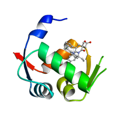
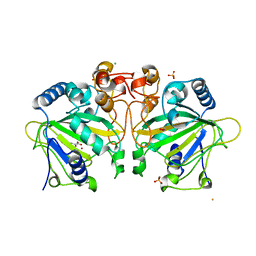
5YQT
 | | Crystal Structure of the L74F/M78V/I80V/L114F mutant of LEH complexed with cyclopentene oxide | | 分子名称: | (1R,5S)-6-oxabicyclo[3.1.0]hexane, Limonene-1,2-epoxide hydrolase | | 著者 | Kong, X.D, Sun, Z.T, Wu, L, Reetz, M.T, Zhou, J.H, Xu, J.H. | | 登録日 | 2017-11-07 | | 公開日 | 2018-06-27 | | 最終更新日 | 2023-11-22 | | 実験手法 | X-RAY DIFFRACTION (2.3 Å) | | 主引用文献 | Structural and Computational Insight into the Catalytic Mechanism of Limonene Epoxide Hydrolase Mutants in Stereoselective Transformations.
J. Am. Chem. Soc., 140, 2018
|
|
7LXW
 | |
7LY0
 | |
7LXZ
 | |
7LY2
 | |


