Loading
PDBj
メニューPDBj@FacebookPDBj@TwitterPDBj@YouTubewwPDB FoundationwwPDB
RCSB PDBPDBeBMRBAdv. SearchSearch help
Search by PDB author
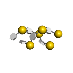
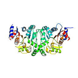
1CAR
DownloadVisualize
BU of 1car by Molmil
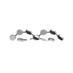
I-CARRAGEENAN. MOLECULAR STRUCTURE AND PACKING OF POLYSACCHARIDE DOUBLE HELICES IN ORIENTED FIBRES OF DIVALENT CATION SALTS
分子名称: 4-O-sulfo-beta-D-galactopyranose-(1-4)-3,6-anhydro-2-O-sulfo-alpha-D-galactopyranose-(1-3)-4-O-sulfo-beta-D-galactopyranose-(1-4)-3,6-anhydro-2-O-sulfo-alpha-D-galactopyranose-(1-3)-4-O-sulfo-beta-D-galactopyranose-(1-4)-3,6-anhydro-2-O-sulfo-alpha-D-galactopyranose
著者Arnott, S.
登録日1978-05-23
公開日1980-03-28
最終更新日2024-02-07
実験手法FIBER DIFFRACTION (3 Å)
主引用文献Iota-carrageenan: molecular structure and packing of polysaccharide double helices in oriented fibres of divalent cation salts.
J.Mol.Biol., 90, 1974
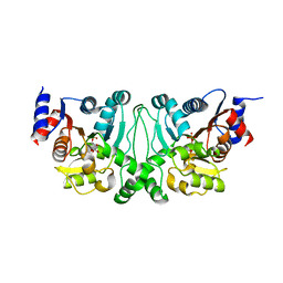
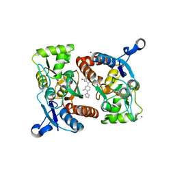
4L6B
DownloadVisualize
BU of 4l6b by Molmil
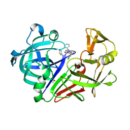
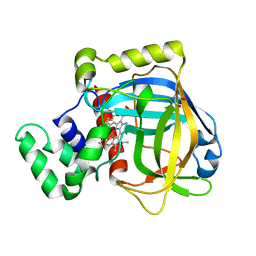
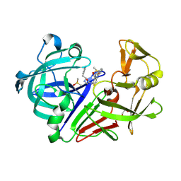
Endothiapepsin in complex with thiophen-based inhibitor SAP128
分子名称: DIMETHYL SULFOXIDE, Endothiapepsin, GLYCEROL, ...
著者Park, A, Heine, A, Klebe, G.
登録日2013-06-12
公開日2014-06-18
実験手法X-RAY DIFFRACTION (1.37 Å)
主引用文献Endothiapepsin in Complex with Thiophen based Inhibitors
To be Published
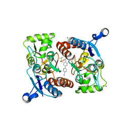
4L51
DownloadVisualize
BU of 4l51 by Molmil
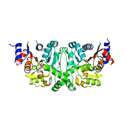
Crystal structures of the LsrR proteins complexed with phospho-AI-2 and its two different analogs reveal distinct mechanisms for ligand recognition
分子名称: 5-O-phosphono-alpha-D-ribofuranose, Transcriptional regulator LsrR
著者Ryu, K.S, Ha, J.H, Eo, Y.
登録日2013-06-10
公開日2013-11-06
最終更新日2024-02-28
実験手法X-RAY DIFFRACTION (1.9 Å)
主引用文献Crystal Structures of the LsrR Proteins Complexed with Phospho-AI-2 and Two Signal-Interrupting Analogues Reveal Distinct Mechanisms for Ligand Recognition.
J.Am.Chem.Soc., 135, 2013
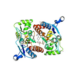
4L5J
DownloadVisualize
BU of 4l5j by Molmil
Crystal structures of the LsrR proteins complexed with phospho-AI-2 and its two different analogs reveal distinct mechanisms for ligand recognition
分子名称: 5-O-phosphono-alpha-D-ribofuranose, Transcriptional regulator LsrR
著者Ryu, K.S, Ha, J.H, Eo, Y.
登録日2013-06-11
公開日2013-11-06
最終更新日2024-02-28
実験手法X-RAY DIFFRACTION (2.6 Å)
主引用文献Crystal Structures of the LsrR Proteins Complexed with Phospho-AI-2 and Two Signal-Interrupting Analogues Reveal Distinct Mechanisms for Ligand Recognition.
J.Am.Chem.Soc., 135, 2013
4L5I
DownloadVisualize
BU of 4l5i by Molmil
Crystal structures of the LsrR proteins complexed with phospho-AI-2 and its two different analogs reveal distinct mechanisms for ligand recognition
分子名称: Transcriptional regulator LsrR
著者Ryu, K.S, Ha, J.H, Eo, Y.
登録日2013-06-11
公開日2013-11-06
最終更新日2024-02-28
実験手法X-RAY DIFFRACTION (3.21 Å)
主引用文献Crystal Structures of the LsrR Proteins Complexed with Phospho-AI-2 and Two Signal-Interrupting Analogues Reveal Distinct Mechanisms for Ligand Recognition.
J.Am.Chem.Soc., 135, 2013
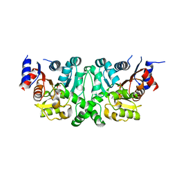
4LAP
DownloadVisualize
BU of 4lap by Molmil
Endothiapepsin in complex with thiophen-based inhibitor SAP114
分子名称: DIMETHYL SULFOXIDE, Endothiapepsin, GLYCEROL, ...
著者Park, A, Heine, A, Klebe, G.
登録日2013-06-20
公開日2014-12-24
実験手法X-RAY DIFFRACTION (1.12 Å)
主引用文献Endothiapepsin in Complex with Thiophen based Inhibitors
To be Published
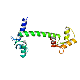
1CLL
DownloadVisualize
BU of 1cll by Molmil
CALMODULIN STRUCTURE REFINED AT 1.7 ANGSTROMS RESOLUTION
分子名称: CALCIUM ION, CALMODULIN, ETHANOL
著者Chattopadhyaya, R, Quiocho, F.A.
登録日1992-09-29
公開日1993-10-31
最終更新日2024-02-07
実験手法X-RAY DIFFRACTION (1.7 Å)
主引用文献Calmodulin structure refined at 1.7 A resolution.
J.Mol.Biol., 228, 1992
1AGA
DownloadVisualize
BU of 1aga by Molmil
THE AGAROSE DOUBLE HELIX AND ITS FUNCTION IN AGAROSE GEL STRUCTURE
分子名称: beta-D-galactopyranose-(1-4)-3,6-anhydro-alpha-L-galactopyranose-(1-3)-beta-D-galactopyranose-(1-4)-3,6-anhydro-alpha-L-galactopyranose-(1-3)-beta-D-galactopyranose-(1-4)-3,6-anhydro-alpha-L-galactopyranose
著者Arnott, S.
登録日1978-05-23
公開日1980-03-28
最終更新日2024-02-07
実験手法FIBER DIFFRACTION (3 Å)
主引用文献The agarose double helix and its function in agarose gel structure.
J.Mol.Biol., 90, 1974
4L4Z
DownloadVisualize
BU of 4l4z by Molmil
Crystal structures of the LsrR proteins complexed with phospho-AI-2 and its two different analogs reveal distinct mechanisms for ligand recognition
分子名称: (2S)-2,3,3-trihydroxy-4-oxopentyl dihydrogen phosphate, Transcriptional regulator LsrR
著者Ryu, K.S, Ha, J.H, Eo, Y.
登録日2013-06-10
公開日2013-11-06
最終更新日2024-02-28
実験手法X-RAY DIFFRACTION (2.3 Å)
主引用文献Crystal Structures of the LsrR Proteins Complexed with Phospho-AI-2 and Two Signal-Interrupting Analogues Reveal Distinct Mechanisms for Ligand Recognition.
J.Am.Chem.Soc., 135, 2013
4L50
DownloadVisualize
BU of 4l50 by Molmil
Crystal structures of the LsrR proteins complexed with phospho-AI-2 and its two different analogs reveal distinct mechanisms for ligand recognition
分子名称: (2S)-2,3,3-trihydroxy-6-methyl-4-oxoheptyl dihydrogen phosphate, Transcriptional regulator LsrR
著者Ryu, K.S, Ha, J.H, Eo, Y.
登録日2013-06-10
公開日2013-11-06
最終更新日2024-02-28
実験手法X-RAY DIFFRACTION (2.1 Å)
主引用文献Crystal Structures of the LsrR Proteins Complexed with Phospho-AI-2 and Two Signal-Interrupting Analogues Reveal Distinct Mechanisms for Ligand Recognition.
J.Am.Chem.Soc., 135, 2013
4L4Y
DownloadVisualize
BU of 4l4y by Molmil
Crystal structures of the LsrR proteins complexed with phospho-AI-2 and its two different analogs reveal distinct mechanisms for ligand recognition
分子名称: Transcriptional regulator LsrR
著者Ryu, K.S, Ha, J.H, Eo, Y.
登録日2013-06-10
公開日2013-11-06
最終更新日2024-02-28
実験手法X-RAY DIFFRACTION (1.9 Å)
主引用文献Crystal Structures of the LsrR Proteins Complexed with Phospho-AI-2 and Two Signal-Interrupting Analogues Reveal Distinct Mechanisms for Ligand Recognition.
J.Am.Chem.Soc., 135, 2013
4LZ7
DownloadVisualize
BU of 4lz7 by Molmil
Crystal structures of GLuR2 ligand-binding-domain in complex with glutamate and positive allosteric modulators
分子名称: GLUTAMIC ACID, Glutamate receptor 2, N-({(5S)-3-[3-fluoro-4-(pyrrolidin-1-yl)phenyl]-4,5-dihydro-1,2-oxazol-5-yl}methyl)acetamide, ...
著者Pandit, J.
登録日2013-07-31
公開日2013-12-04
最終更新日2023-09-20
実験手法X-RAY DIFFRACTION (2.1 Å)
主引用文献Discovery and Characterization of a Novel Dihydroisoxazole Class of alpha-Amino-3-hydroxy-5-methyl-4-isoxazolepropionic acid (AMPA) Receptor Potentiators.
J.Med.Chem., 56, 2013
4LZ5
DownloadVisualize
BU of 4lz5 by Molmil
Crystal structures of GLuR2 ligand-binding-domain in complex with glutamate and positive allosteric modulators
分子名称: GLUTAMIC ACID, Glutamate receptor 2, N-[(2R)-2-(4'-cyanobiphenyl-4-yl)propyl]propane-2-sulfonamide, ...
著者Pandit, J.
登録日2013-07-31
公開日2013-12-04
最終更新日2023-09-20
実験手法X-RAY DIFFRACTION (1.5 Å)
主引用文献Discovery and Characterization of a Novel Dihydroisoxazole Class of alpha-Amino-3-hydroxy-5-methyl-4-isoxazolepropionic acid (AMPA) Receptor Potentiators.
J.Med.Chem., 56, 2013
4LZ8
DownloadVisualize
BU of 4lz8 by Molmil
Crystal structures of GLuR2 ligand-binding-domain in complex with glutamate and positive allosteric modulators
分子名称: GLUTAMIC ACID, Glutamate receptor 2, N-[(3S)-1-{2-fluoro-4-[(5S)-5-{[(propan-2-ylsulfonyl)amino]methyl}-4,5-dihydro-1,2-oxazol-3-yl]phenyl}pyrrolidin-3-yl]acetamide, ...
著者Pandit, J.
登録日2013-07-31
公開日2013-12-04
最終更新日2023-09-20
実験手法X-RAY DIFFRACTION (1.85 Å)
主引用文献Discovery and Characterization of a Novel Dihydroisoxazole Class of alpha-Amino-3-hydroxy-5-methyl-4-isoxazolepropionic acid (AMPA) Receptor Potentiators.
J.Med.Chem., 56, 2013
4MLE
DownloadVisualize
BU of 4mle by Molmil
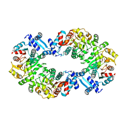
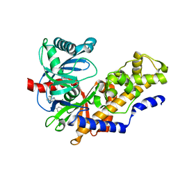
Human Glucokinase in Complex with Novel Amino Thiazole Activator
分子名称: 3-(benzyloxy)-N-(4-methyl-1,3-thiazol-2-yl)pyridin-2-amine, Glucokinase, alpha-D-glucopyranose
著者Voegtli, W.C.
登録日2013-09-06
公開日2013-09-25
最終更新日2024-02-28
実験手法X-RAY DIFFRACTION (2.6 Å)
主引用文献Identification of a New Class of Glucokinase Activators through Structure-Based Design.
J.Med.Chem., 56, 2013
4MLH
DownloadVisualize
BU of 4mlh by Molmil
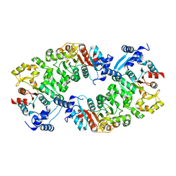
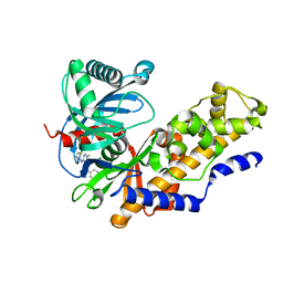
Human Glucokinase in Complex with a Novel Amino Thiazole Allosteric Activator
分子名称: 3-(benzyloxy)-5-methyl-N-(4-methyl-1,3-thiazol-2-yl)pyridin-2-amine, Glucokinase, alpha-D-glucopyranose
著者Voegtli, W.C.
登録日2013-09-06
公開日2013-09-25
最終更新日2024-02-28
実験手法X-RAY DIFFRACTION (2.9 Å)
主引用文献Identification of a New Class of Glucokinase Activators through Structure-Based Design.
J.Med.Chem., 56, 2013
6ZOD
DownloadVisualize
BU of 6zod by Molmil
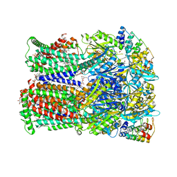
Fusidic acid binding to the allosteric deep transmembrane domain binding pocket, TM7/TM8 groove, and TM1/TM2 groove of the fully induced AcrB T protomer
分子名称: 1,2-ETHANEDIOL, DARPIN, DECANE, ...
著者Oswald, C, Tam, H.K, Pos, K.M.
登録日2020-07-07
公開日2021-05-19
最終更新日2024-01-31
実験手法X-RAY DIFFRACTION (2.85 Å)
主引用文献Allosteric drug transport mechanism of multidrug transporter AcrB.
Nat Commun, 12, 2021
4SGA
DownloadVisualize
BU of 4sga by Molmil
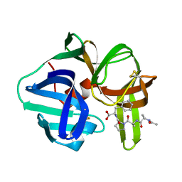
STRUCTURES OF PRODUCT AND INHIBITOR COMPLEXES OF STREPTOMYCES GRISEUS PROTEASE A AT 1.8 ANGSTROMS RESOLUTION. A MODEL FOR SERINE PROTEASE CATALYSIS
分子名称: PROTEINASE A (SGPA), TETRAPEPTIDE ACE-PRO-ALA-PRO-PHE
著者Sielecki, A.R, James, M.N.G.
登録日1990-05-29
公開日1991-10-15
最終更新日2024-06-05
実験手法X-RAY DIFFRACTION (1.8 Å)
主引用文献Structures of product and inhibitor complexes of Streptomyces griseus protease A at 1.8 A resolution. A model for serine protease catalysis.
J.Mol.Biol., 144, 1980
4PHV
DownloadVisualize
BU of 4phv by Molmil
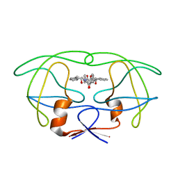
X-RAY CRYSTAL STRUCTURE OF THE HIV PROTEASE COMPLEX WITH L-700,417, AN INHIBITOR WITH PSEUDO C2 SYMMETRY
分子名称: HIV-1 PROTEASE, N,N-BIS(2-HYDROXY-1-INDANYL)-2,6- DIPHENYLMETHYL-4-HYDROXY-1,7-HEPTANDIAMIDE
著者Bone, R.
登録日1991-10-04
公開日1993-10-31
最終更新日2024-02-28
実験手法X-RAY DIFFRACTION (2.1 Å)
主引用文献X-Ray Crystal Structure of the HIV Protease Complex with L-700,417, an Inhibitor with Pseudo C2 Symmetry
J.Am.Chem.Soc., 113, 1991
5EZP
DownloadVisualize
BU of 5ezp by Molmil
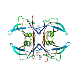
Human transthyretin (TTR) complexed with 4-hydroxy-chalcone
分子名称: 1,2-ETHANEDIOL, 4-hydroxy-chalcone, Transthyretin
著者Polsinelli, I, Nencetti, S, Shepard, W.E, Orlandini, E, Stura, E.A.
登録日2015-11-26
公開日2016-01-27
最終更新日2024-01-10
実験手法X-RAY DIFFRACTION (2.5 Å)
主引用文献A new crystal form of human transthyretin obtained with a curcumin derived ligand.
J.Struct.Biol., 194, 2016
2ADM
DownloadVisualize
BU of 2adm by Molmil
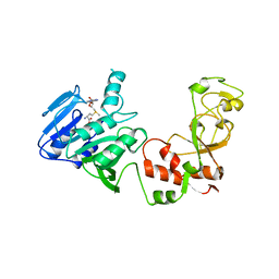
ADENINE-N6-DNA-METHYLTRANSFERASE TAQI
分子名称: ADENINE-N6-DNA-METHYLTRANSFERASE TAQI, S-ADENOSYLMETHIONINE
著者Schluckebier, G, Saenger, W.
登録日1996-07-15
公開日1997-01-27
最終更新日2024-02-14
実験手法X-RAY DIFFRACTION (2.6 Å)
主引用文献Differential binding of S-adenosylmethionine S-adenosylhomocysteine and Sinefungin to the adenine-specific DNA methyltransferase M.TaqI.
J.Mol.Biol., 265, 1997
3EKF
DownloadVisualize
BU of 3ekf by Molmil
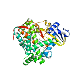
Crystal structure of the A264Q heme domain of cytochrome P450 BM3
分子名称: Cytochrome P450(BM-3), PROTOPORPHYRIN IX CONTAINING FE
著者Toogood, H.S, Leys, D.
登録日2008-09-19
公開日2008-12-30
最終更新日2023-08-30
実験手法X-RAY DIFFRACTION (2.1 Å)
主引用文献Novel haem co-ordination variants of flavocytochrome P450BM3.
Biochem.J., 417, 2009
3E4Y
DownloadVisualize
BU of 3e4y by Molmil
Crystal structure of a 33kDa catalase-related protein from Mycobacterium avium subsp. paratuberculosis. I2(1)2(1)2(1) crystal form
分子名称: GLYCEROL, PHOSPHATE ION, PROTOPORPHYRIN IX CONTAINING FE, ...
著者Pakhomova, S, Newcomer, M.E.
登録日2008-08-12
公開日2009-08-18
最終更新日2023-08-30
実験手法X-RAY DIFFRACTION (2.6 Å)
主引用文献The structure and peroxidase activity of a 33-kDa catalase-related protein from Mycobacterium avium ssp. paratuberculosis.
Protein Sci., 18, 2009
3EGK
DownloadVisualize
BU of 3egk by Molmil
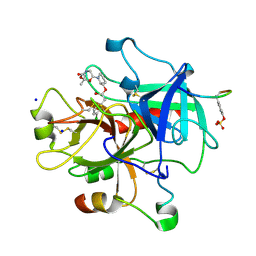
KNOBLE Inhibitor
分子名称: Hirudin variant-1, SODIUM ION, Thrombin heavy chain, ...
著者Baum, B, Heine, A, Klebe, G, Muenzel, M.
登録日2008-09-10
公開日2008-09-30
最終更新日2023-11-15
実験手法X-RAY DIFFRACTION (2.2 Å)
主引用文献KNOBLE: a knowledge-based approach for the design and synthesis of readily accessible small-molecule chemical probes to test protein binding
Angew.Chem.Int.Ed.Engl., 46, 2007
3VAC
DownloadVisualize
BU of 3vac by Molmil
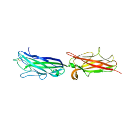
Crystal Structure of the CFA/I Enterotoxigenic E. coli adhesin CfaE mutant G168D
分子名称: CFA/I fimbrial subunit E
著者Liu, Y, Esser, L, Xia, D.
登録日2011-12-29
公開日2013-02-20
最終更新日2017-11-08
実験手法X-RAY DIFFRACTION (2.6 Å)
主引用文献Tight Conformational Coupling between the Domains of the Enterotoxigenic Escherichia coli Fimbrial Adhesin CfaE Regulates Binding State Transition.
J.Biol.Chem., 288, 2013

221051

件を2024-06-12に公開中

PDB statisticsPDBj update infoContact PDBjnumon