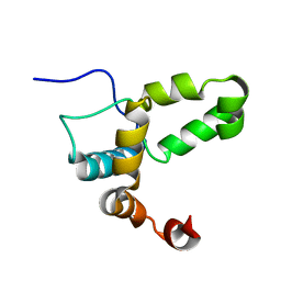
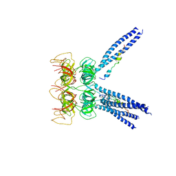
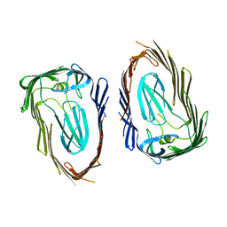
1UOS
 
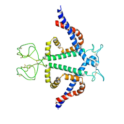
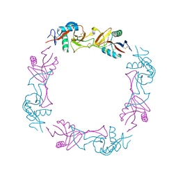 | | The Crystal Structure of the Snake Venom Toxin Convulxin | | 分子名称: | CONVULXIN ALPHA, CONVULXIN BETA | | 著者 | Batuwangala, T, Leduc, M, Gibbins, J.M, Bon, C, Jones, E.Y. | | 登録日 | 2003-09-22 | | 公開日 | 2003-10-14 | | 最終更新日 | 2023-12-13 | | 実験手法 | X-RAY DIFFRACTION (2.7 Å) | | 主引用文献 | Structure of the Snake-Venom Toxin Convulxin
Acta Crystallogr.,Sect.D, 60, 2004
|
|
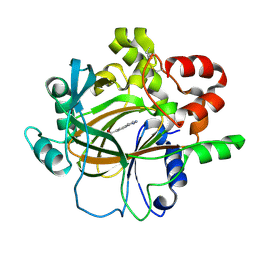
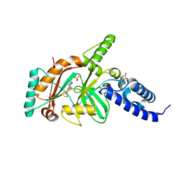
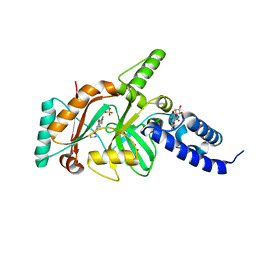
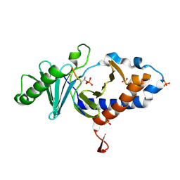
2H3B
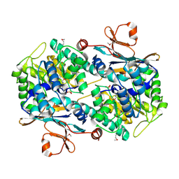 | | Crystal Structure of Mouse Nicotinamide Phosphoribosyltransferase/Visfatin/Pre-B Cell Colony Enhancing Factor 1 | | 分子名称: | Nicotinamide phosphoribosyltransferase, SULFATE ION | | 著者 | Wang, T, Zhang, X, Bheda, P, Revollo, J.R, Imai, S.I, Wolberger, C. | | 登録日 | 2006-05-22 | | 公開日 | 2006-06-20 | | 最終更新日 | 2021-10-20 | | 実験手法 | X-RAY DIFFRACTION (1.95 Å) | | 主引用文献 | Structure of Nampt/PBEF/visfatin, a mammalian NAD(+) biosynthetic enzyme.
Nat.Struct.Mol.Biol., 13, 2006
|
|
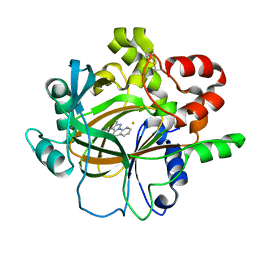
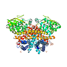
2H3D
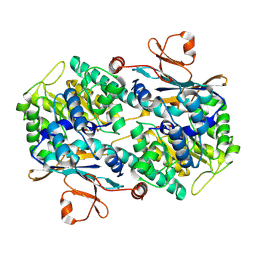 | | Crystal Structure of Mouse Nicotinamide Phosphoribosyltransferase/Visfatin/Pre-B Cell Colony Enhancing Factor in Complex with Nicotinamide Mononuleotide | | 分子名称: | BETA-NICOTINAMIDE RIBOSE MONOPHOSPHATE, Nicotinamide phosphoribosyltransferase | | 著者 | Wang, T, Zhang, X, Bheda, P, Revollo, J.R, Imai, S.I, Wolberger, C. | | 登録日 | 2006-05-22 | | 公開日 | 2006-06-20 | | 最終更新日 | 2011-07-13 | | 実験手法 | X-RAY DIFFRACTION (2.1 Å) | | 主引用文献 | Structure of Nampt/PBEF/visfatin, a mammalian NAD(+) biosynthetic enzyme.
Nat.Struct.Mol.Biol., 13, 2006
|
|
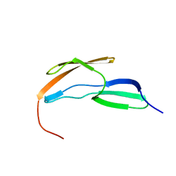
2L7Y
 | |
2LI6
 | |
7DYG
 | |
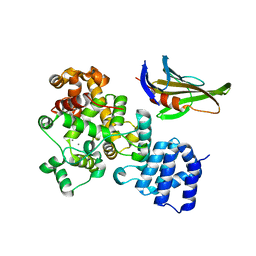
7DYQ
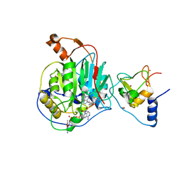
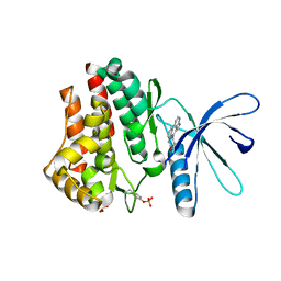
 | | Crystal structure of histone lysine demethylase 4D (KDM4D) in complex with the inhibitor 5-hydroxy-2-methylpyrazolo[1,5-a]pyrido[3,2-e]pyrimidine-3-carbonitrile | | 分子名称: | 5-hydroxy-2-methylpyrazolo[1,5-a]pyrido[3,2-e]pyrimidine-3-carbonitrile, FE (III) ION, Lysine-specific demethylase 4D | | 著者 | Wang, T, Yang, L. | | 登録日 | 2021-01-22 | | 公開日 | 2022-01-26 | | 最終更新日 | 2024-10-09 | | 実験手法 | X-RAY DIFFRACTION (1.998 Å) | | 主引用文献 | Crystal structure of histone lysine demethylase 4D (KDM4D) in complex with the inhibitor 5-hydroxy-2-methylpyrazolo[1,5-a]pyrido[3,2-e]pyrimidine-3-carbonitrile
To Be Published
|
|
4LUQ
 | |
6WKS
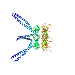
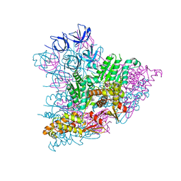
 | | Structure of SARS-CoV-2 nsp16/nsp10 in complex with RNA cap analogue (m7GpppA) and S-adenosylmethionine | | 分子名称: | 1,2-ETHANEDIOL, 2'-O-methyltransferase, ADENOSINE, ... | | 著者 | Gupta, Y.K, Viswanathan, T, Arya, S, Qi, S, Misra, A, Chan, S.-H. | | 登録日 | 2020-04-16 | | 公開日 | 2020-05-06 | | 最終更新日 | 2023-10-18 | | 実験手法 | X-RAY DIFFRACTION (1.8 Å) | | 主引用文献 | Structural basis of RNA cap modification by SARS-CoV-2.
Nat Commun, 11, 2020
|
|
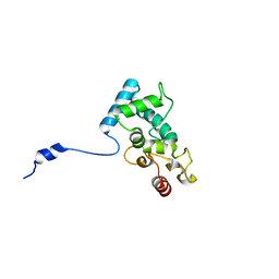
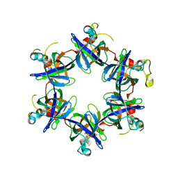
8YMK
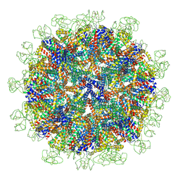
 | | Localized reconstruction of Hepatitis B virus surface antigen dimer in the subviral particle with D2 symmetry from dataset A0 | | 分子名称: | Isoform S of Large envelope protein | | 著者 | Wang, T, Cao, L, Mu, A, Wang, Q, Rao, Z.H. | | 登録日 | 2024-03-09 | | 公開日 | 2024-09-18 | | 最終更新日 | 2024-10-09 | | 実験手法 | ELECTRON MICROSCOPY (3.7 Å) | | 主引用文献 | Inherent symmetry and flexibility in hepatitis B virus subviral particles.
Science, 385, 2024
|
|
8YMJ
 | | Cryo-EM structure of Hepatitis B virus surface antigen subviral particle with D2 symmetry | | 分子名称: | Isoform S of Large envelope protein | | 著者 | Wang, T, Cao, L, Mu, A, Wang, Q, Rao, Z.H. | | 登録日 | 2024-03-09 | | 公開日 | 2024-09-18 | | 最終更新日 | 2024-10-09 | | 実験手法 | ELECTRON MICROSCOPY (6.6 Å) | | 主引用文献 | Inherent symmetry and flexibility in hepatitis B virus subviral particles.
Science, 385, 2024
|
|
3M9B
 | |
3M9D
 | |
3M9H
 | |
4EFE
 | | crystal structure of DNA ligase | | 分子名称: | BETA-NICOTINAMIDE RIBOSE MONOPHOSPHATE, DNA ligase, SULFATE ION, ... | | 著者 | Wei, Y, Wang, T, Charifson, P, Xu, W. | | 登録日 | 2012-03-29 | | 公開日 | 2013-04-03 | | 最終更新日 | 2024-02-28 | | 実験手法 | X-RAY DIFFRACTION (2 Å) | | 主引用文献 | crystal structure of DNA ligase
To be Published
|
|
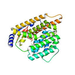
6ES9
 | | Methylsuccinyl-CoA dehydrogenase of Paracoccus denitrificans with bound flavin adenine dinucleotide | | 分子名称: | Acyl-CoA dehydrogenase, COENZYME A, FLAVIN-ADENINE DINUCLEOTIDE, ... | | 著者 | Zarzycki, J, Schwander, T, Erb, T.J. | | 登録日 | 2017-10-19 | | 公開日 | 2018-01-03 | | 最終更新日 | 2024-01-17 | | 実験手法 | X-RAY DIFFRACTION (1.37 Å) | | 主引用文献 | Structural basis for substrate specificity of methylsuccinyl-CoA dehydrogenase, an unusual member of the acyl-CoA dehydrogenase family.
J. Biol. Chem., 293, 2018
|
|
4EFB
 | | Crystal structure of DNA ligase | | 分子名称: | 4-amino-2-(cyclopentyloxy)-6-{[(1R,2S)-2-hydroxycyclopentyl]oxy}pyrimidine-5-carboxamide, BETA-NICOTINAMIDE RIBOSE MONOPHOSPHATE, DNA ligase, ... | | 著者 | Wei, Y, Wang, T, Charifson, P, Xu, W. | | 登録日 | 2012-03-29 | | 公開日 | 2013-04-03 | | 最終更新日 | 2024-02-28 | | 実験手法 | X-RAY DIFFRACTION (2.2 Å) | | 主引用文献 | Crystal structure of DNA ligase
To be Published
|
|
5KWA
 | |
4FQB
 | |
3KCK
 | | A Novel Chemotype of Kinase Inhibitors | | 分子名称: | 3-chloro-4-(4H-3,4,7-triazadibenzo[cd,f]azulen-6-yl)phenol, Tyrosine-protein kinase JAK2 | | 著者 | Zuccola, H.J, Wang, T, Ledeboer, M.W. | | 登録日 | 2009-10-21 | | 公開日 | 2009-11-24 | | 最終更新日 | 2011-07-13 | | 実験手法 | X-RAY DIFFRACTION (2.2 Å) | | 主引用文献 | A novel chemotype of kinase inhibitors: Discovery of 3,4-ring fused 7-azaindoles and deazapurines as potent JAK2 inhibitors.
Bioorg.Med.Chem.Lett., 20, 2010
|
|
4DOX
 | |
3OHN
 | |
4O56
 | | Structure of PLK1 in complex with peptide | | 分子名称: | PHOSPHATE ION, Serine/threonine-protein kinase PLK1, synthetic peptide | | 著者 | Wang, T. | | 登録日 | 2013-12-19 | | 公開日 | 2014-12-24 | | 最終更新日 | 2023-11-08 | | 実験手法 | X-RAY DIFFRACTION (1.8 Å) | | 主引用文献 | Integration of Charge-dipole Interaction and Intramolecular Hydrogen Bond in Ligand Design for the Polo-Box Domain of Polo-like Kinase 1
To be Published
|
|
3FP9
 | |
7W5G
 | | The apo structure of trichobrasilenol synthase TaTC6 with the space group of orthorhombic | | 分子名称: | GLYCEROL, SULFATE ION, Terpene cyclase 6 | | 著者 | Chen, C, Wang, T, Yang, Y, Zhang, L, Ko, T, Huang, J, Guo, R. | | 登録日 | 2021-11-30 | | 公開日 | 2022-06-01 | | 最終更新日 | 2023-11-29 | | 実験手法 | X-RAY DIFFRACTION (1.8 Å) | | 主引用文献 | Structural insights into the cyclization of unusual brasilane-type sesquiterpenes.
Int.J.Biol.Macromol., 209, 2022
|
|


