6NHX
 
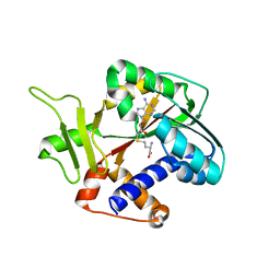
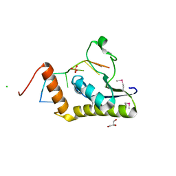
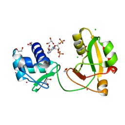
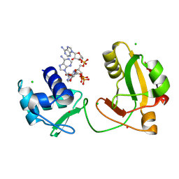
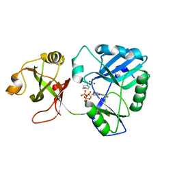 | | mycobacterial DNA ligase D complexed with ATP and MES | | 分子名称: | 2-(N-MORPHOLINO)-ETHANESULFONIC ACID, ADENOSINE-5'-TRIPHOSPHATE, ATP-dependent DNA ligase | | 著者 | Shuman, S, Unciuleac, M, Goldgur, Y. | | 登録日 | 2018-12-24 | | 公開日 | 2019-02-13 | | 最終更新日 | 2024-03-13 | | 実験手法 | X-RAY DIFFRACTION (1.4 Å) | | 主引用文献 | Structures of ATP-bound DNA ligase D in a closed domain conformation reveal a network of amino acid and metal contacts to the ATP phosphates.
J. Biol. Chem., 294, 2019
|
|
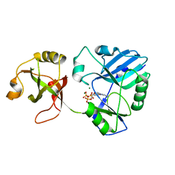
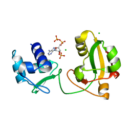
6NHZ
 | |
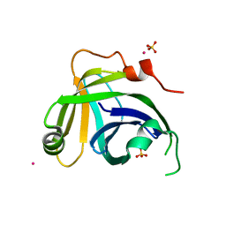
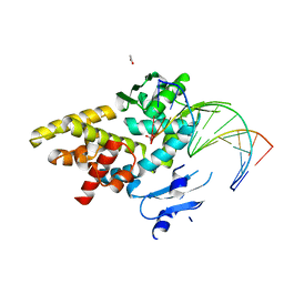
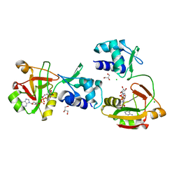
3KGD
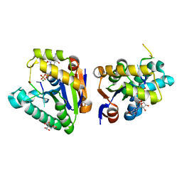
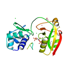
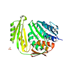 | | Crystal structure of E. coli RNA 3' cyclase | | 分子名称: | ADENOSINE MONOPHOSPHATE, GLYCEROL, RNA 3'-terminal phosphate cyclase, ... | | 著者 | Shuman, S, Tanaka, N, Smith, P. | | 登録日 | 2009-10-28 | | 公開日 | 2010-04-21 | | 最終更新日 | 2023-09-06 | | 実験手法 | X-RAY DIFFRACTION (1.68 Å) | | 主引用文献 | Structure of the RNA 3'-phosphate cyclase-adenylate intermediate illuminates nucleotide specificity and covalent nucleotidyl transfer.
Structure, 18, 2010
|
|
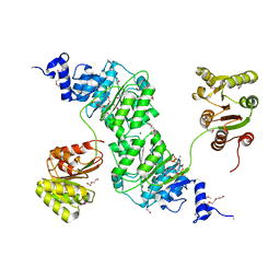
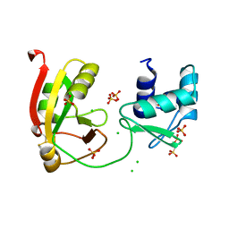
3N9B
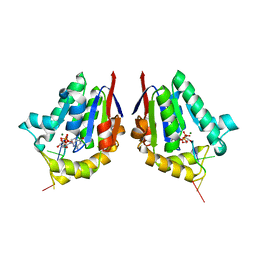
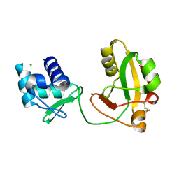
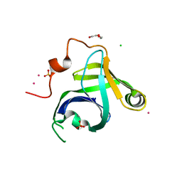 | | Crystal Structure of the P. aeruginosa LigD phosphoesterase domain | | 分子名称: | CHLORIDE ION, DI(HYDROXYETHYL)ETHER, MANGANESE (II) ION, ... | | 著者 | Shuman, S, Nair, P, Smith, P. | | 登録日 | 2010-05-28 | | 公開日 | 2010-08-11 | | 最終更新日 | 2024-02-21 | | 実験手法 | X-RAY DIFFRACTION (1.92 Å) | | 主引用文献 | Structure of bacterial LigD 3'-phosphoesterase unveils a DNA repair superfamily
Proc.Natl.Acad.Sci.USA, 107, 2010
|
|
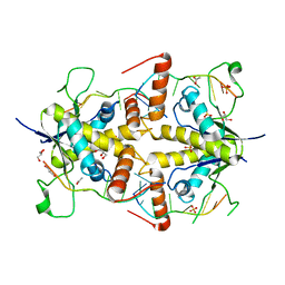
3N9D
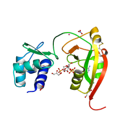
 | | Monoclinic Structure of P. aeruginosa LigD phosphoesterase domain | | 分子名称: | MANGANESE (II) ION, Probable ATP-dependent DNA ligase, SULFATE ION, ... | | 著者 | Shuman, S, Nair, P, Smith, P. | | 登録日 | 2010-05-28 | | 公開日 | 2010-08-11 | | 最終更新日 | 2023-09-06 | | 実験手法 | X-RAY DIFFRACTION (2.3 Å) | | 主引用文献 | Structure of bacterial LigD 3'-phosphoesterase unveils a DNA repair superfamily
Proc.Natl.Acad.Sci.USA, 107, 2010
|
|
5U32
 | |
2QY2
 | |
2OWO
 | | Last Stop on the Road to Repair: Structure of E.coli DNA Ligase Bound to Nicked DNA-Adenylate | | 分子名称: | 26-MER, 5'-D(*AP*CP*AP*AP*TP*TP*GP*CP*GP*AP*CP*(OMC)P*C)-3', 5'-D(*CP*AP*CP*TP*AP*TP*CP*GP*GP*AP*AP*TP*G)-3', ... | | 著者 | Shuman, S, Nandakumar, J, Nair, P.A. | | 登録日 | 2007-02-16 | | 公開日 | 2007-05-15 | | 最終更新日 | 2023-08-30 | | 実験手法 | X-RAY DIFFRACTION (2.3 Å) | | 主引用文献 | Last Stop on the Road to Repair: Structure of E. coli DNA Ligase Bound to Nicked DNA-Adenylate.
Mol.Cell, 26, 2007
|
|
4MDF
 | | Structure of bacterial polynucleotide kinase Michaelis complex bound to GTP and DNA | | 分子名称: | CITRIC ACID, DNA (5'-D(*CP*CP*TP*GP*T)-3'), GUANOSINE-5'-TRIPHOSPHATE, ... | | 著者 | Shuman, S, Das, U, Wang, L.K, Smith, P, Jacewicz, A. | | 登録日 | 2013-08-22 | | 公開日 | 2013-11-06 | | 最終更新日 | 2024-02-28 | | 実験手法 | X-RAY DIFFRACTION (1.727 Å) | | 主引用文献 | Structures of bacterial polynucleotide kinase in a Michaelis complex with GTP*Mg2+ and 5'-OH oligonucleotide and a product complex with GDP*Mg2+ and 5'-PO4 oligonucleotide reveal a mechanism of general acid-base catalysis and the determinants of phosphoacceptor recognition.
Nucleic Acids Res., 42, 2014
|
|
4MDE
 | | Structure of bacterial polynucleotide kinase product complex bound to GDP and DNA | | 分子名称: | DNA (5'-D(P*CP*CP*TP*GP*T)-3'), GUANOSINE-5'-DIPHOSPHATE, MAGNESIUM ION, ... | | 著者 | Shuman, S, Das, U, Wang, L.K, Smith, P, Jacewicz, A. | | 登録日 | 2013-08-22 | | 公開日 | 2013-11-06 | | 最終更新日 | 2024-02-28 | | 実験手法 | X-RAY DIFFRACTION (1.8 Å) | | 主引用文献 | Structures of bacterial polynucleotide kinase in a Michaelis complex with GTP*Mg2+ and 5'-OH oligonucleotide and a product complex with GDP*Mg2+ and 5'-PO4 oligonucleotide reveal a mechanism of general acid-base catalysis and the determinants of phosphoacceptor recognition.
Nucleic Acids Res., 42, 2014
|
|
5TC1
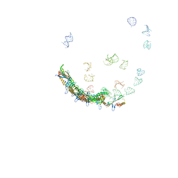 | | In situ structures of the genome and genome-delivery apparatus in ssRNA bacteriophage MS2 | | 分子名称: | Capsid protein, Maturation protein, phage MS2 genome | | 著者 | Dai, X.H, Li, Z.H, Lai, M, Shu, S, Du, Y.S, Zhou, Z.H, Sun, R. | | 登録日 | 2016-09-13 | | 公開日 | 2016-12-07 | | 最終更新日 | 2018-07-18 | | 実験手法 | ELECTRON MICROSCOPY (3.6 Å) | | 主引用文献 | In situ structures of the genome and genome-delivery apparatus in a single-stranded RNA virus.
Nature, 541, 2017
|
|
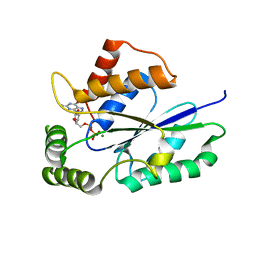
1RI1
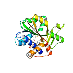 | | Structure and mechanism of mRNA cap (guanine N-7) methyltransferase | | 分子名称: | 7-METHYL-GUANOSINE-5'-TRIPHOSPHATE-5'-GUANOSINE, S-ADENOSYL-L-HOMOCYSTEINE, mRNA CAPPING ENZYME | | 著者 | Fabrega, C, Hausmann, S, Shen, V, Shuman, S, Lima, C.D. | | 登録日 | 2003-11-16 | | 公開日 | 2004-02-03 | | 最終更新日 | 2024-02-14 | | 実験手法 | X-RAY DIFFRACTION (2.5 Å) | | 主引用文献 | Structure and mechanism of mRNA cap (Guanine-n7) methyltransferase
Mol.Cell, 13, 2004
|
|
1RI3
 | | Structure and mechanism of mRNA cap (guanine N-7) methyltransferase | | 分子名称: | S-ADENOSYL-L-HOMOCYSTEINE, mRNA CAPPING ENZYME | | 著者 | Fabrega, C, Hausmann, S, Shen, V, Shuman, S, Lima, C.D. | | 登録日 | 2003-11-16 | | 公開日 | 2004-02-03 | | 最終更新日 | 2024-02-14 | | 実験手法 | X-RAY DIFFRACTION (2.5 Å) | | 主引用文献 | Structure and mechanism of mRNA cap (Guanine-n7) methyltransferase
Mol.Cell, 13, 2004
|
|
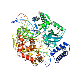
3M4A
 | | Crystal structure of a bacterial topoisomerase IB in complex with DNA reveals a secondary DNA binding site | | 分子名称: | ACETIC ACID, DNA (5'-D(*GP*AP*AP*TP*AP*AP*GP*GP*GP*CP*GP*C)-3'), DNA (5'-D(*GP*CP*GP*CP*CP*CP*TP*TP*AP*TP*TP*C)-3'), ... | | 著者 | Patel, A, Yakovleva, L, Shuman, S, Mondragon, A. | | 登録日 | 2010-03-10 | | 公開日 | 2010-07-14 | | 最終更新日 | 2023-09-06 | | 実験手法 | X-RAY DIFFRACTION (1.65 Å) | | 主引用文献 | Crystal structure of a bacterial topoisomerase IB in complex with DNA reveals a secondary DNA binding site.
Structure, 18, 2010
|
|
4W7S
 | | Crystal structure of the yeast DEAD-box splicing factor Prp28 at 2.54 Angstroms resolution | | 分子名称: | GLYCEROL, HEXAETHYLENE GLYCOL, MAGNESIUM ION, ... | | 著者 | Jacewicz, A, Smith, P, Schwer, B, Shuman, S. | | 登録日 | 2014-08-22 | | 公開日 | 2014-10-29 | | 最終更新日 | 2023-12-27 | | 実験手法 | X-RAY DIFFRACTION (2.542 Å) | | 主引用文献 | Crystal structure, mutational analysis and RNA-dependent ATPase activity of the yeast DEAD-box pre-mRNA splicing factor Prp28.
Nucleic Acids Res., 42, 2014
|
|
4WAN
 | | Crystal structure of Msl5 protein in complex with RNA at 1.8 A | | 分子名称: | ACETATE ION, Branchpoint-bridging protein, GLYCEROL, ... | | 著者 | Jacewicz, A, Smith, P, Chico, L, Schwer, B, Shuman, S. | | 登録日 | 2014-08-29 | | 公開日 | 2014-12-17 | | 最終更新日 | 2023-12-27 | | 実験手法 | X-RAY DIFFRACTION (1.8 Å) | | 主引用文献 | Structural basis for recognition of intron branchpoint RNA by yeast Msl5 and selective effects of interfacial mutations on splicing of yeast pre-mRNAs.
Rna, 21, 2015
|
|
4WAL
 | | Crystal structure of selenomethionine Msl5 protein in complex with RNA at 2.2 A | | 分子名称: | Branchpoint-bridging protein, CHLORIDE ION, GLYCEROL, ... | | 著者 | Jacewicz, A, Smith, P, Chico, L, Schwer, B, Shuman, S. | | 登録日 | 2014-08-29 | | 公開日 | 2014-12-17 | | 最終更新日 | 2023-12-27 | | 実験手法 | X-RAY DIFFRACTION (2.2 Å) | | 主引用文献 | Structural basis for recognition of intron branchpoint RNA by yeast Msl5 and selective effects of interfacial mutations on splicing of yeast pre-mRNAs.
Rna, 21, 2015
|
|
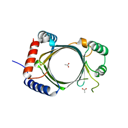
8TG4
 | | tRNA 2'-phosphotransferase (Tpt1) from Aeropyrum pernix in complex with ADP-ribose-2"-phosphate and 2'-OH RNA | | 分子名称: | 1,2-ETHANEDIOL, CHLORIDE ION, NITRATE ION, ... | | 著者 | Jacewicz, A, Dantuluri, S, Shuman, S. | | 登録日 | 2023-07-12 | | 公開日 | 2023-11-08 | | 実験手法 | X-RAY DIFFRACTION (1.37 Å) | | 主引用文献 | Structural basis for Tpt1-catalyzed 2'-PO 4 transfer from RNA and NADP(H) to NAD.
Proc.Natl.Acad.Sci.USA, 120, 2023
|
|
8TFI
 | |
8TG3
 | | tRNA 2'-phosphotransferase (Tpt1) from Aeropyrum pernix in complex with ADP-ribose-1" -phosphate | | 分子名称: | DI(HYDROXYETHYL)ETHER, GLYCEROL, NITRATE ION, ... | | 著者 | Jacewicz, A, Dantuluri, S, Shuman, S. | | 登録日 | 2023-07-12 | | 公開日 | 2023-11-08 | | 実験手法 | X-RAY DIFFRACTION (1.47 Å) | | 主引用文献 | Structural basis for Tpt1-catalyzed 2'-PO 4 transfer from RNA and NADP(H) to NAD.
Proc.Natl.Acad.Sci.USA, 120, 2023
|
|
8TFX
 | | tRNA 2'-phosphotransferase (Tpt1) from Pyrococcus horikoshii in complex with 2',5'-ADP | | 分子名称: | 1,2-ETHANEDIOL, ADENOSINE-2'-5'-DIPHOSPHATE, CHLORIDE ION, ... | | 著者 | Jacewicz, A, Dantuluri, S, Shuman, S. | | 登録日 | 2023-07-12 | | 公開日 | 2023-11-08 | | 実験手法 | X-RAY DIFFRACTION (1.7 Å) | | 主引用文献 | Structural basis for Tpt1-catalyzed 2'-PO 4 transfer from RNA and NADP(H) to NAD.
Proc.Natl.Acad.Sci.USA, 120, 2023
|
|
8TFZ
 | | tRNA 2'-phosphotransferase (Tpt1) from Pyrococcus horikoshii in complex with NAD | | 分子名称: | CHLORIDE ION, GLYCEROL, NICOTINAMIDE-ADENINE-DINUCLEOTIDE, ... | | 著者 | Jacewicz, A, Dantuluri, S, Shuman, S. | | 登録日 | 2023-07-12 | | 公開日 | 2023-11-08 | | 実験手法 | X-RAY DIFFRACTION (2.06 Å) | | 主引用文献 | Structural basis for Tpt1-catalyzed 2'-PO 4 transfer from RNA and NADP(H) to NAD.
Proc.Natl.Acad.Sci.USA, 120, 2023
|
|
8TFY
 | |
8TG6
 | |
8TG5
 | | tRNA 2'-phosphotransferase (Tpt1) from Pyrococcus horikoshii in complex with branched 2'-PO4 RNA | | 分子名称: | (2S,3R,4R,5S)-2-(6-amino-9H-purin-9-yl)-4-{[(S)-{[(2R,3S,4R,5S)-5-(6-amino-9H-purin-9-yl)-3,4-dihydroxyoxolan-2-yl]methoxy}(hydroxy)phosphoryl]oxy}-5-({[(S)-{[(2R,3R,4S,5R)-5-(6-amino-9H-purin-9-yl)-4-hydroxy-2-(hydroxymethyl)oxolan-3-yl]oxy}(hydroxy)phosphoryl]oxy}methyl)oxolan-3-yl dihydrogen phosphate (non-preferred name), CHLORIDE ION, POTASSIUM ION, ... | | 著者 | Jacewicz, A, Dantuluri, S, Shuman, S. | | 登録日 | 2023-07-12 | | 公開日 | 2024-06-05 | | 最終更新日 | 2024-06-26 | | 実験手法 | X-RAY DIFFRACTION (2 Å) | | 主引用文献 | Structural basis for Tpt1-catalyzed 2'-PO 4 transfer from RNA and NADP(H) to NAD.
Proc.Natl.Acad.Sci.USA, 120, 2023
|
|


